The request "neurite" is redirected here. A separate article should be created on this topic.
Axon of a 9-day-old mouse
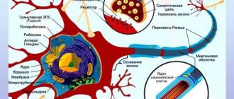
Axon
(ancient Greek ἄξων - “axis”) - a neurite (a long cylindrical process of a nerve cell), along which nerve impulses travel from the cell body (soma) to the innervated organs and other nerve cells.
Each neuron consists of one axon, a body (perikaryon) and several dendrites, depending on the number of which nerve cells are divided into unipolar, bipolar or multipolar. Transmission of a nerve impulse occurs from the dendrites (or cell body) to the axon, and then the generated action potential from the initial segment of the axon is transmitted back to the dendrites[1]. If an axon in nerve tissue connects to the body of the next nerve cell, such contact is called axo-somatic, with dendrites - axo-dendritic, with another axon - axo-axonal (a rare type of connection, found in the central nervous system).
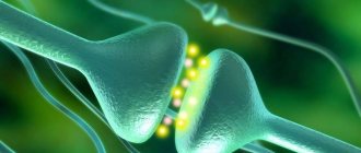
The ends of the axon - terminals - branch and contact other nerve, muscle or glandular cells. At the end of the axon there is a synaptic ending - the terminal portion of the terminal in contact with the target cell. Together with the postsynaptic membrane of the target cell, the synaptic ending forms a synapse. Excitation is transmitted through synapses.
Properties[edit | edit code]
The nutrition and growth of the axon depend on the body of the neuron: when the axon is cut, its peripheral part dies, while the central part remains viable.
With a diameter of several microns, the length of the axon can reach 1 meter or more in large animals (for example, axons coming from neurons in the spinal cord to the limb).
Many invertebrates (squid, annelids, phoronids, crustaceans) have giant axons hundreds of microns thick (in squids - up to 2-3 mm). Typically, such axons conduct signals to the muscles that provide the “flight reaction” (pulling into a hole, fast swimming, etc.). All other things being equal, as the diameter of the axon increases, the speed of nerve impulses along it increases.
Motor neurons
A person performs many actions and movements every day, and behind each of the simplest movements there is a huge mechanism of the motor-skeletal system.
We get up early in the morning, wash, someone does exercises, have breakfast and go to work, all this happens simply and routinely. But if we could look into what is happening behind the curtain of this performance, we would see that behind all these actions there are neurons of the brain, and, in particular, human motor neurons. What are these physiological mechanisms, where are they located, how do they work, where is the motor neuron located later in this article.
All physical actions that a person can perform are carried out according to the same principle: through contraction and stretching of muscles and tendons. These contractions occur due to the existence of communication between all muscles and tendons with a single coordination center - the brain. These messages consist of cells with different tasks - neurons.
Accordingly, special motor cells—motoneurons—participate in the implementation of motor functions.
Muscle contraction occurs by changing just two commands: relax and tense - that is, straighten and contract. A special motor neuron is responsible for each of these conditions. The motor neuron responsible for contraction is called a flexor, and the motor neuron responsible for relaxation is called an extensor.
Types of motor neurons
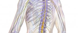
Motor neurons are divided into central and peripheral according to their location in the body. Accordingly, central motor cells are located in the spinal cord and brain, and peripheral ones are located directly in the muscles and are connected to them through the axons of neurons.
Central neurons are responsible for conscious and reflex movements; electrochemical impulses with commands diverge from them to the periphery and are transmitted to muscles, organs and other tissues.
The main accumulation of groups of motor cells of the somatic nervous system occurs in the region of the anterior horns of the spinal cord. Each group is responsible for contracting its own muscles.
For example, a group of motor neurons in the cervical region controls the muscles of the arms.
It is precisely because of the participation of the spinal cord and its motor neurons in the control of the motor system that the spine is dangerous to injure and there is a high risk of injury and disability. And even spinal massage should be trusted only to trusted professionals.
Classification of motor neurons:
- Renshaw cells
- Small alpha motor neurons.
- Large alpha motor neurons.
- Gamma motor neurons.
Large alphas form the trunk of the nerve chain, and small alpha and gamma with their small axons transmit signals to the most inaccessible areas. Renshaw cells perform a special function of signal switching. These are a kind of telephone operators who, in the last century, manually connected different telephone subscribers.
The entire nervous system, central and peripheral nerves, is a large and complex mechanism in which many elements work in concert. In fact, human upright posture is a unique and very costly function for the body, which requires a special kind of motor mechanism, and humans have it.
Any physical action comes down to the fact that a certain group of muscles flexes and extends, and for this there are special “flexor and extensor” cells.
A motor signal is formed in the corresponding part of the cerebral cortex. Another specialized cells are involved in this, which are called pyramidal for their shape. Pyramidal cells make up the pyramidal motor pathway through which the signal reaches the spinal cord.
Different areas of the cerebral cortex are responsible for the work of flexors and extensors, as a result of which muscle contraction occurs: a signal is formed in the area of the precentral gyrus, and the posterior regions of both hemispheres are already responsible for the work of flexors and extensors.
What muscles are motor neurons connected to?
All muscle fibers have their own motor neurons attached. Together, the motor cell and the muscle fiber to which it is attached are called a “motor unit.” Each such unit functions independently of other similar units. And each motor unit contains only one type of muscle fiber.
Types of muscle fibers:
- Slow oxidative fibers.
- Fast oxidative fibers.
- Fast glycolytic fibers.
Features of nerve cells
Neurons are somewhat reminiscent of a colony of ants - there are just as many of them and they are divided into various groups according to specialization. It is in the differences between these specializations that their specific features and differences lie.
Types of motor neurons, their characteristics and localization in the cerebral cortex:
- Central innervating flexors: localized in the area of the precentral gyrus and are responsible for compression (contraction) of skeletal muscles.
- Central innervating extensors: localized in the hindbrain region and are responsible for relaxing skeletal muscles.
- Peripheral alpha: cells that transmit commands to muscle fibers to contract. Localized in the anterior horns of the spinal cord.
- Peripheral gamma: cells responsible for muscle tone. They are localized in the same place, in the anterior horns of the spinal cord.
- Intercalary: present in all parts of the central nervous system, and carry out the role of communication of all signals in the central nervous system.
How many neurons are there in the body?
The number of nerve cells in the human brain alone is a magnitude of cosmic proportions. According to the results of recent studies conducted by Brazilian physiologists, there are about 86 billion of them in the human brain.
Structure of neurons
A motor nerve cell consists of three conventional parts: the body of the motor neuron, one axon and many dendrites.
Dendrites are active nerve endings of cells through which communication between neurons is established and electrochemical impulses pass. Nerves form connections with each other of varying degrees of stability.
And the axons are already connected to other cells and transmit command signals to them, forming the entire nervous system.
Some of the connections form a fully automated system for controlling many physiological processes that a person does not need to consciously control. These connections are called conditioned and unconditioned reflexes. Also, stable neural circuits are formed in the process of any activity, including thinking.
The more often a person performs the same action, thinks the same thoughts, reacts in the same way to the same stimuli, the more stable the connections that these events form become. This is how acquired reflexes, good and bad habits, physical and psychological dependencies are formed. Each person’s return to habitual behavior only strengthens the associated neural circuits, and any attempt to further change one’s character or behavior will encounter more and more resistance from the psyche (where the root of any addiction is located) and a feeling of discomfort. Reflex arc
The very majority of automated neural circuits that are responsible for the unconscious regulation of all processes in the body are, in fact, a reflex arc.
A “reflex arc” is a stable neural connection that is guaranteed to fire under certain identical conditions. For example, withdrawing your hand from a hot object is a reflex that makes a connection.
The reflex is triggered by an irritant - in this example, any hot object.
The general mechanism of reflexive activity is as follows:
- The signal about the presence of the stimulus is transmitted to the sensitive nerve endings and, through communication from the dendrites, is redirected for analysis to the brain. Each area of the cerebral cortex is responsible for a specific specialization. Accordingly, nerve endings throughout the body are tied to different areas of the brain, and each neuron sends signals exclusively to its own command center.
- After the dendrites are the first to react to the stimulus, this reaction passes to the cell.
- Information about the event is transformed into an electrochemical impulse, which is immediately transmitted throughout the nervous system to the corresponding parts of the cerebral cortex.
- The brain analyzes the received information and transmits the response impulse back along the entire chain with a set of mandatory instructions for the cells, how they should behave in the response phase and whether this phase is needed.
- The phase of physical response to a stimulus in which cells carry out instructions received.
Conclusion
The human body was, is and remains one of nature's greatest unsolved mysteries. And the structure of the human body is many times superior in its perfection to all our most advanced inventions and developments.
The main reason why humanity seeks to study the structure of the body is disease; the human body is as fragile as it is strong.
Another hundred, or even a thousand, years will pass before our science gets even a little closer to solving this mystery.
Source: https://mygenetics.ru/blog/nauka/dvigatelnye-neyrony/
Building[edit | edit code]
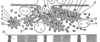
In the protoplasm of the axon - axoplasm - there are the thinnest fibers - neurofibrils, as well as microtubules, mitochondria and agranular (smooth) endoplasmic reticulum. Depending on whether the axons are covered with a myelin sheath or lack it, they form pulpy or non-myelinous nerve fibers.
The myelin sheath of axons is present only in vertebrates. It is formed by special Schwann cells (in the central nervous system - oligodendrocytes) that “wind” onto the axon, between which there are areas free from the myelin sheath - nodes of Ranvier. Only at the interceptions are voltage-gated sodium channels present and the action potential arises again. In this case, the nerve impulse spreads along the myelinated fibers in steps, which increases the speed of its propagation several times. The speed of signal transmission along myelin-covered axons reaches 100 meters per second [2].
Unmyelinated axons are smaller in size than axons covered with a myelin sheath, which compensates for the loss in signal propagation speed compared to myelin-sheathed axons.
At the junction of the axon with the neuron body, the largest pyramidal cells of the 5th layer of the cortex have an axon hillock. It was previously assumed that the conversion of the postsynaptic potential of the neuron into nerve impulses occurs here, but experimental data have not confirmed this. Registration of electrical potentials revealed that the nerve impulse is generated in the axon itself, namely in the initial segment at a distance of ~50 μm from the neuron body [3]. To generate an action potential in the initial segment of the axon, an increased concentration of sodium channels is required (up to a hundred times compared to the neuron body [4]).
Links
- Savelyev A.V.
Modeling the logic of self-organization of nerve bundle activity by ephaptic interactions at the axonal level // collection: Modeling of nonequilibrium systems. - Institute of Computational Modeling SB RAS, Krasnoyarsk, 2004. - P. 142-143.
| This is a draft article on histology. You can help the project by adding to it. |
| Afferent nerve/Sensory neuron | GSA GVA SSA SVA Nerve fibers (Muscle spindles (Ia), Neurotendon spindle (Ib), II or Aβ fibers, III or Aδ fibers, IV or C fibers) |
| Efferent nerve/Motor neuron | GSE GVE SVE Upper motor neuron Lower motor neuron (α motor neurons, γ motor neurons) |
| Synapse | Chemical synapse Neuromuscular synapse Ephaps (Electrical synapse) Neuropil Synaptic vesicle |
| Touch receptor | Meissner's corpuscle Merkel's corpuscle Pacini's corpuscle Ruffini's corpuscle Neuromuscular spindle Free nerve ending Olfactory neuron Photoreceptor cells Hair cells Taste bud |
| Neuroglia | Astrocytes (Radial glia) Oligodendrocytes Ependymal cells (Tanycytes) Microglia |
| Myelin (White matter) | CNS: Oligodendrocytes PNS: Schwann cells (Neurolemma · Node of Ranvier/Internodal segment · Myelin notch) |
| Connective tissue | Epineurium Perineurium Endoneurium Nerve fiber bundles Meninges: dura mater, arachnoid membrane, pia mater |