In the gray matter of the anterior horns of each spinal cord segment
there are several thousand neurons that are 50-100% larger than most other neurons. They are called anterior motor neurons. The axons of these motor neurons exit the spinal cord through the ventral roots and directly innervate skeletal muscle fibers. There are two types of these neurons: alpha motor neurons and gamma motor neurons.
Alpha motor neurons
. Alpha motor neurons give rise to large motor fibers of the A-alpha (Ace) type with an average diameter of 14 μm. After entering the skeletal muscle, these fibers branch repeatedly to innervate large muscle fibers. Stimulation of a single alpha fiber excites from three to several hundred skeletal muscle fibers, which, together with the motor neuron innervating them, constitute the so-called motor unit.
Gamma motor neurons
. Along with alpha motor neurons, the stimulation of which leads to contraction of skeletal muscle fibers, much smaller gamma motor neurons are localized in the anterior horns of the spinal cord, the number of which is approximately 2 times less. Gamma motor neurons transmit impulses along much thinner motor fibers of the A-gamma (Ay) type with an average diameter of about 5 microns.
They innervate small special fibers
skeletal muscles, called intrafusal muscle fibers. These fibers form the central part of the muscle spindles involved in the regulation of muscle tone.
Interneurons
. Interneurons are present in all areas of the gray matter of the spinal cord, in the dorsal and anterior horns, and in the space between them. These cells are approximately 30 times more numerous than anterior motor neurons. Interneurons are small in size and very excitable, often exhibit spontaneous activity and are capable of generating up to 1500 impulses/sec.
They have numerous connections
each other, and many also synapse directly with anterior motor neurons. Interconnections between interneurons and anterior motor neurons are responsible for most of the integrative functions of the spinal cord, as discussed later in this chapter.
Essentially the entire set of different types of neural circuits
, is found within a pool of spinal cord interneurons, including diverging, converging, rhythmically discharging, and other types of circuits. This chapter outlines the many ways in which these various circuits are involved in the spinal cord's execution of specific reflex actions.
Only a few sensory signals
, entering the spinal cord along the spinal nerves or descending from the brain, reach directly the anterior motor neurons. Instead, almost all signals are conducted first through interneurons, where they are processed accordingly. The corticospinal tract terminates almost entirely at the spinal interneurons, where signals from this tract combine with signals from other spinal tracts or spinal nerves before they converge on the anterior motor neurons to regulate muscle function.
A connecting neuron that lies between sensory (afferent) and motor (efferent) neurons. Located in the central nervous system. Also called an interneuron, and in older texts an association neuron.
See the meaning of Intercalary Neuron in other dictionaries
Intercalary Adj.
— 1. Intended for insertion, insertion. Explanatory Dictionary by Efremova
Neuron M.
— 1. Same as: neuron. Explanatory Dictionary by Efremova
Insertable
- (shn), insertion, insertion. Adj. to insert. Ushakov's Explanatory Dictionary
Neuron
- neuron, m. (Greek neuron - fiber, nerve) (anat.). Nerve cell. Ushakov's Explanatory Dictionary
Neuron
- -A; m. [from Greek. neuron - nerve] Special. A nerve cell with all the processes extending from it. Kuznetsov's Explanatory Dictionary
Insertion Disc
— (discus intercalatus, LNH) the general name of microscopic structures at the point of contact of adjacent muscle cells of the myocardium, ensuring their connection into muscle complexes and transmission…….. Big medical dictionary
Motor Neuron
— , a nerve cell that conducts information to EFFECTORS (usually muscles) from the CENTRAL NERVOUS SYSTEM (CNS), thus causing the appropriate response. Axons (processes,……..
Neuron
— (nerve cell), the main structural and functional unit of the NERVOUS SYSTEM, which carries out the rapid transmission of NERVE IMPULSES between various organs. Consists of…….. Scientific and technical encyclopedic dictionary
Sensory Neuron
- (sensitive neuron), a nerve cell that conducts information from RECEPTORS in any part of the body to the CENTRAL NERVOUS SYSTEM (CNS). Their nerve endings are located in…….. Scientific and technical encyclopedic dictionary
Neuron
— (neuronum, neurocytus, LNH; Greek neuron vein, nerve; synonym: nerve cell, neurocyte, neurocyte) a cell capable of perceiving irritation, becoming excited, producing…….. Big medical dictionary
Neuron Amacrine
- (n. amacrinum, LNH) N., located in the inner granular layer of the retina and providing communication between the neurons of this layer. Large medical dictionary
Neuron Associative
— see Intercalary neuron. Large medical dictionary
Neuron Afferent
- (n. afferens, n. sensorium: synonym: N. receptor, N. sensory, N. sensitive) N., which carries out the perception and transmission of excitation from receptors to other N. of the central nervous system. Large medical dictionary
Neuron Bipolar
- (n. bipolare, LNH) N., having two processes - an axon and a dendrite. Large medical dictionary
Neuron Vegetative
- the general name of N., which are part of the ganglia, plexuses and nerves of the autonomic nervous system. Large medical dictionary
Neuron Fusiform
- (n. fusiforme, LNH) multipolar intercalary N. of elongated shape, found in the molecular plate of the cerebral cortex. Large medical dictionary
Neuron Fusiform Horizontal
- (n. fusiforme horizontale, LNH) multipolar N. elongated, found mainly between the layer of piriform neurons and the granular layer of the cerebellar cortex. Large medical dictionary
Neuron Internal
— (n. internum, LNH) N. of the internal parts of the anterior horn of the spinal cord, the axon of which passes through the white commissure to the opposite half of the spinal cord. Large medical dictionary
Neuron Intercalary
- (n. intercalatum; synonym: N. associative, N. intermediate) N. involved in the transmission of excitation from afferent N. to efferent. Large medical dictionary
Neuron Input
- a formal neuron that performs an input function in a specific system of neurons (neural network), i.e., it perceives signals only from the environment external to this system. Large medical dictionary
Neuron Gigantopyramidal
- (n. gigantopyramidale, LNH; synonym: Betza cell, giant pyramidal cell) large pyramidal N. of the internal pyramidal plate of the cerebral cortex; axons of the N. g. form…….. Big medical dictionary
Neuron Horizontal
— (n. horizomale, LNH) 1) N. inner granular layer of the retina, the processes of which contact the central endings of photoreceptor cells, carrying out redistribution…….. Big medical dictionary
Neuron Piriform
- (n. piriforme, LNH; synonym Purkinje cell) efferent N. of the cerebellar cortex, located in its ganglion layer and having a pear-shaped shape. Large medical dictionary
Neuron Motor
— see Motor neuron. Large medical dictionary
Neuron Long axon
— (n. longiaxonicum, LNH; synonym for Dogel cell type I) multipolar vegetative N., the axon of which transmits impulses to smooth or cardiac muscle tissue. Large medical dictionary
Neuron Stellate
- (n. stellatum, LNH) intercalated N. star-shaped. Large medical dictionary
Neuron Stellate Long-axon
- (n. stellatum longiaxonicum, LNH) N. z., located in the granular layer of the cerebellar cortex, having an axon extending into the white matter. Large medical dictionary
Neuron Stellate Short Axon
- (n. stellatum breviaxonicum, LNH) N. z. granular layer of the cerebellar cortex, which has an axon going to the cerebellar glomeruli. Large medical dictionary
Neuron Granular
— (n. granulare, LNH) the general name of small N. round, angular and pyramidal in shape, located in the outer granular plate of the cerebral cortex, the dendrites of which rise…….. Big medical dictionary
Neuron Granular Large
— (granoneurocytus magnus, LNH) the general name of large N., located in the molecular layer of the cerebellar cortex, the dendrites of which spread in the molecular layer, and the axons go to the granular…….. Large medical dictionary
What are they needed for? Why are there so many of them? What is a sensory neuron? What function do intercalary and executive neurons perform? Let's take a closer look at these amazing cells.
Functions
Every second, many signals pass through our brain. The process does not stop even in sleep. The body needs to perceive the world around it, make movements, ensure the functioning of the heart, respiratory, digestive, genitourinary systems, etc. Two main groups of neurons are involved in organizing all this activity - sensory and motor.
When we touch cold or hot and feel the temperature of the object, this is the merit of the sensitive cells. They instantly transmit information received from the periphery of the body. This ensures reflex activity.
Neurons form our entire central nervous system. Their main tasks:
- get information;
- transmit it through the nervous system.
These unique cells are capable of instantly transmitting electrical impulses.
To ensure the process of life, the body must process a huge amount of information that comes to it from the outside world, and respond to any sign of changing environmental conditions. To make this process as efficient as possible, neurons are divided according to their functions into:
- Sensitive (afferent) are our guides to the world around us. They are the ones who perceive information from the outside, from the senses, and transmit it to the central nervous system. The peculiarity is that thanks to their contact activity, we feel temperature, pain, pressure, and have other feelings. Sensitive cells of narrow specialization transmit taste and smell.
- Motor (motor, efferent, motor neurons). Motor neurons transmit information through electrical impulses from the central nervous system to muscle groups and glands.
- Intermediate (associative, intercalary, intercalary). Now let’s take a closer look at what function interneurons perform, why they are needed, and what is their difference. They are located between sensory and motor neurons. Interneurons transmit nerve impulses from sensory fibers to motor fibers. They provide “communication” between efferent and afferent nerve cells. They should be treated as a kind of natural “extenders,” long cavities that help transmit a signal from a sensory neuron to a motor one. Without their participation this would not have been possible. This is their function.
The receptors themselves are cells of the skin, muscles, internal organs, and joints specially designated for this function. Receptors can begin in the cells of the epidermis and mucous membrane. They are able to accurately capture the smallest changes, both outside the body and inside it. Such changes may be physical or chemical. Then they are instantly transformed into special bioelectric impulses and sent directly to sensory neurons. This is how the signal travels from the periphery to the center of the body, where the brain deciphers its meaning.
Impulses from the organ to the brain are carried out by all three groups of neurons - motor, sensory and intermediate. The human nervous system consists of these groups of cells. This structure allows you to respond to signals from the outside world. They provide reflex activity of the body.
If a person ceases to feel taste, smell, hearing and vision decrease, this may indicate disorders in the central nervous system. Depending on which sense organs are affected, a neurologist can determine in which part of the brain the problems have arisen.
1) Somatic. This is conscious control of the skeletal muscles.
2) Vegetative (autonomous). This is control of internal organs uncontrolled by consciousness. The operation of this system occurs even if a person is in a state of sleep.
Sensory neurons are most often unipolar. This means that they are equipped with only one bifurcating process. It leaves the cell body (soma) and simultaneously performs the functions of both an axon and a dendrite. The axon is the input, and the dendrite of the sensory neuron is the output. After excitation of sensitive sensory cells, a bioelectric signal passes along the axon and dendrite.
There are also bipolar nerve cells that have two processes, respectively. They can be found, for example, in the retina and structures of the inner ear.
The body of the sensitive cell is shaped like a spindle. 1, and more often 2 processes (central and peripheral) extend from the body.
Peripheral in its shape is very similar to a thick long stick. It reaches the surface of the mucous membrane or skin. This process is similar to the dendrite of nerve cells.
The second, opposite process extends from the opposite part of the cell body and is shaped like a thin thread covered with swellings (they are called varicosities). This is an analogue of the nerve process of a neuron. This process is directed to a specific part of the central nervous system and branches out this way.
Sensitive cells are also called peripheral. Their peculiarity is that they are located directly behind the peripheral nervous system and the central nervous system, but without them the operation of these systems is unthinkable. For example, olfactory cells are located in the epithelium of the nasal mucosa.
Reviews of motor neuron treatment in China
Elena, 73 years old, Russia. Admission Treatment: Treatment of Motor Neurone Disease in China
A 73-year-old female patient diagnosed with amyotrophic lateral sclerosis was admitted to Dalian Military Hospital for treatment of motor neuron disease in very poor condition. She could not walk without assistance, could barely speak, and there was a complete lack of sensation in the left side of her body. I arrived with an accompanying person in a wheelchair. This condition is not often found in reviews of motor neuron treatment, as it is still very difficult for modern medicine.
Diagnosis was carried out at the Military Hospital
Motor neurone treatment was performed at Dalian Military Hospital. The patient underwent electroneuromyography, which identified specific areas of slowing of nerve fibers. Using transcranial magnetic stimulation, doctors were able to identify motor neurons with reduced excitability. An MRI was also performed, which helped to make an accurate diagnosis and select the correct treatment for the motor neuron. The examination was carried out under the supervision of Professor Zhou Xu Dong.
Treatment was carried out at the Military Hospital
The patient was prescribed specialized treatment for motor neuron disease involving herbal injections of a concentrated type of a rare formulation of drugs. This natural material can act as a repair system for various body tissues, including nerve fibers. The treatment lasted for three weeks, during which three injections of herbal compounds were made into the joints. The holistic therapy also included professional speech therapy, physical therapy and acupuncture to help the body prepare for active movement and recover faster.
The result of motor neuron treatment at the Military Hospital
Immediately after the correct diagnosis and treatment of motor neuron disease at Dalian Military Hospital, the left side of the patient's body began to feel faint electrical impulses. After three months, she could eat and bathe independently, walk unassisted by moving her left leg, and conduct conversations using her hands to write messages on the board. Although motor neurone disease is a terminal condition, after treatment in a hospital in China, the patient's quality of life significantly improved and, most importantly, she regained independence in movement.
Eduard, 49 years old, Dagestan. Diagnosis on admission: motor neuron treatment in China
A 49-year-old patient was admitted to Dalian Military Hospital with a rare type of motor neuron disease. The preliminary diagnosis is muscular progressive atrophy. Significant weight loss was observed, mainly due to abnormal decrease in muscle mass. The patient was very weak and could only move with outside help.
Examination carried out
After being examined by a professor at a Chinese clinic, the patient underwent an MRI. Based on a series of tests, a topography of muscle atrophy was formed. A family history was taken. Based on the results of modern diagnostics, it has been established that the disease has a secondary form with degeneration of peripheral nerve fibers.
Treatment carried out
Based on the diagnosis at Dalian Military Hospital, treatment for motor neuron disease consisted of a series of conservative procedures. The patient underwent complex acupuncture therapy. The professor selected pharmaceuticals based on natural ingredients based on the patient’s individual indicators. The medications were aimed not only at treating the motor neuron, but also at gradually cleansing the body of synthetic drugs, which by the time the patient was admitted to the Chinese clinic had already begun to cause the body’s dependence.
Result of motor neuron treatment in China
Two months after diagnosis at Dalian Military Hospital, patients in their reviews of the motor neuron treatment reported a number of improvements. Fatigue when moving decreased, the patient began to move without assistance. General health improved and, most importantly, the patient began to gain weight.
Nina, 62 years old, Russia
Diagnosis on admission
A 62-year-old female patient was admitted to Dalian Military Hospital with a diagnosis of lateral primary sclerosis. Previous treatment for motor neuron disease abroad and in a local hospital showed no improvement, and the disease progressed over 7 years. As a result, the patient experienced severe weakness in the muscles of her arms and face, which is why she could not speak clearly and could only eat with outside help.
Examination carried out
At Dalian Military Hospital, the patient underwent transcranial magnetic stimulation and electroneuromyography. Using MRI, suspicions of the development of third-party diseases, such as a tumor of the brain or spinal cord, were excluded. Based on the data obtained, the professor compiled a motor neuron treatment program.
Treatment carried out
Treatment for motor neuron disease at the Dalian Military Hospital lasted 3 courses. During this time, the patient took prescribed medications based on natural ingredients and underwent a course of acupuncture to activate the restoration processes of the motor cells of the central nervous system in the extremities. The patient also underwent individual physiotherapy using Chinese medicine methods.
Result of motor neuron treatment in China
Based on a personal review of motor neurone treatment in China, the patient gradually experienced improvement throughout the treatment. After 3 months, the trend towards increased quality of life and improvements in mobility continued. Acupuncture helped slow motor neuron degeneration and caused remission of the disease. The patient was able to eat on her own again.
David, 53 years old, Israel
Diagnosis on admission
A 53-year-old patient presented to Dalian Military Hospital with motor neuron disease. A more accurate diagnosis is amyotrophic lateral sclerosis. The patient had significant atrophy of the muscles of the limbs and paralysis of the left leg. The patient also complained of fatigue.
Examination carried out
To prescribe treatment for motor neuron disease, the Chinese clinic conducted a series of tests, including MRI, electroneuromyography and TMS (transcranial magnetic stimulation).
Treatment carried out
Based on the data obtained during the examination, a famous professor at the Dalian Military Hospital prescribed a series of drugs, individually selected and manufactured in the laboratory of a Chinese clinic. Acupuncture was also prescribed in the spinal cord and limbs. In parallel with the treatment of motor neuron disease using modern Chinese medicine, the patient underwent physical therapy to accelerate the recovery of muscle function.
Result of motor neuron treatment in Dalian
In the general treatment, there was a break of 3 weeks and a break of 3 months. After only two months spent in the Dalian military hospital, the patient reported long-awaited improvements in his review of the motor neuron treatment. The patient began to get less tired, the left leg began to feel touch, which indicates the start of restoration processes in the nervous tissues of the extremities. Physical therapy helped increase overall muscle mass. After six months of observation, the patient began to walk independently with the help of a small cane. His overall quality of life improved.
Maria, 59 years old, Kazakhstan
Diagnosis on admission
A 59-year-old female patient was admitted to a Chinese clinic with motor neuron disease. The medical history included a diagnosis of amyotrophic lateral sclerosis. The pathology was in an advanced form: the patient could not move independently, eat, and speak using long and short blinks.
Examination carried out
The patient was examined by a professor at the Dalian Military Hospital, and MRI, neuromyography and transcranial stimulation were also performed.
Treatment carried out
To ensure safe treatment of the extreme stage of motor neuron disease that the patient was diagnosed with, the doctor prescribed traditional Chinese medicine acupuncture and a number of natural-based medications. Acupuncture was supposed to trigger the generation of depleted neuron sheaths to increase the efficiency of nerve impulse transmission. The prescribed medications helped cleanse the body of previously taken synthetic drugs, which are used mainly for the treatment of motor neuron abroad and fought the cause of the disease.
The result of motor neuron treatment abroad
After the first course of motor neuron treatment, which lasted three weeks, the patient was able to move her mouth and began to chew food. Subsequent courses contributed to the restoration of speech function. A year later, the patient was able to move her hands and communicate using a notepad and pen. She wrote about her improvement in well-being in a review of motor neuron treatment in a clinic in China.
The State Military Hospital in Dalian invites patients to effective treatment of motor neurone in China, using the methods of the most ancient medicine in the world! We guarantee 100% quality of treatment, by highly qualified doctors at state prices!
How do they work
The function of a sensitive neuron is to receive a signal from special receptors located on the periphery of the body and determine its characteristics. The impulses are perceived by the peripheral processes of sensory neurons, then they are transmitted to their body, and then along the central processes they follow directly to the central nervous system.
The dendrites of sensory neurons connect to various receptors, and their axons connect to other neurons (interneurons). For a nerve impulse, the simplest path is the following - it must pass through three neurons: sensory, intercalary, motor.
The most typical example of the passage of an impulse is when a neurologist knocks on the knee joint with a hammer. In this case, a simple reflex is immediately triggered: the knee tendon, after a blow to it, sets in motion the muscle that is attached to it; Sensitive cells from the muscle transmit the signal through sensory neurons directly to the spinal cord. There, sensory neurons make contact with motor neurons, and they send impulses back to the muscle, causing it to contract, and the leg straightens.
By the way, in each section of the spinal cord (cervical, thoracic, lumbar, sacral, coccygeal) there is a pair of roots: sensory posterior, motor anterior. They form a single trunk. Each of these pairs controls its own specific part of the body and sends a centrifugal signal about what to do next, how to position a limb, torso, what to do to the gland, etc.
Sensory neurons take part in the work of the reflex arc. It consists of 5 elements:
- Receptor. Converts irritation into a nerve impulse.
- The impulse along the neuron follows from the receptor in the central nervous system.
- The interneuron, which is located in the brain, transmits a signal from the sensory neuron to the executive one.
- The motor (executive) neuron conducts the main impulse from the brain to the organ.
- An (executive) organ is a muscle, gland, etc. It reacts to the received signal by contraction, secretion, etc.
Conclusion
The biology of the human body is very thought out and perfect. Thanks to the activity of many sensitive neurons, we can interact with this wonderful world and respond to it. Our body is very receptive, the development of its receptors and sensitive nerve cells has reached the highest level. Thanks to such a thoughtful organization of the central nervous system, our senses can perceive and transmit the smallest shades of taste, smell, tactile sensations, sound, and color.
We often believe that the main thing in our consciousness and the functioning of the body is the cortex and hemispheres of the brain. At the same time, we forget what enormous capabilities the spinal cord provides. It is the functioning of the spinal cord that ensures the receipt of signals from all receptors.
It is difficult to name the limit of these possibilities. Our body is very plastic. The more a person develops, the more opportunities are placed at his disposal. This simple principle allows us to quickly adapt to changes in the world around us.
A neuron is a specific, electrically excitable cell in the human nervous system and has unique characteristics. Its functions are to process, store and transmit information. Neurons are characterized by a complex structure and narrow specialization. They are also divided into three types. This article describes in detail the interneuron and its role in the action of the central nervous system.
Symptoms
IHL can be divided into three stages: early, middle and advanced.
Early signs and symptoms
Symptoms develop slowly and may be confused with symptoms of some other unrelated neurological conditions.
Early symptoms depend on which body system is affected first. Typical symptoms begin in one of three areas: the arms and legs, the mouth (bulbar), or the respiratory system.
- weakening your grip, making it difficult to pick up and hold things.
- fatigue
- muscle pain, cramps and spasms.
- unclear and sometimes garbled speech.
- weakness of arms and legs
- increased clumsiness and stumbling.
- difficulty swallowing
- difficulty breathing or shortness of breath
Signs and symptoms of mid-stage disease
As the disease progresses, symptoms become more severe.
- Muscle pain and weakness increases, spasms and spasms become more severe.
- The limbs are becoming weaker.
- The limbus muscles begin to contract.
- Movement of the affected limbs becomes more difficult.
- The limbus muscles may become abnormally stiff.
- Joint pain is increasing.
- Eating, drinking and swallowing become more difficult.
- Salivation occurs due to problems with saliva control.
- Yawning occurs, sometimes during uncontrollable contractions.
- Pain in the jaws can be caused by a strong yawn.
- Speech problems get worse as the muscles in the throat and mouth weaken.
The person may show changes in personality and emotional state, with uncontrollable crying or laughing.
It was previously thought that MHD had no significant effect on brain function or memory, but research has shown that up to 50 percent of people with ALS now have some type of brain function involvement.
This includes difficulties with memory, planning, language, behavior and spatial relationships. Up to 15 percent of people with ALS have a form of dementia known as frontotemporal dementia (FTD).
Breathing problems can occur due to deterioration of the diaphragm, the main breathing muscle. Breathing may be difficult, even while sleeping or resting. Eventually you will need help with breathing.
Secondary symptoms are insomnia, anxiety and depression.
Signs and symptoms in advanced development
Eventually, the patient will be unable to move, eat, or breathe without assistance. Without supportive therapy, a person will die. Despite the best care currently available, the most common causes of death are complications of the respiratory system.
Classification of neurons
The human brain has approximately 65 billion neurons that constantly communicate with each other. These cells are divided into several types, each of which performs its own special functions.
The sensory neuron plays the role of a transmitter of information between the sense organs and the central parts of the human nervous system. It perceives a variety of irritations, which it converts into nerve impulses, and then transmits the signal to the human brain.
Motor - sends impulses to various organs and tissues. This type is mainly involved in the control of spinal cord reflexes.
The interneuron is responsible for processing and switching impulses. The functions of this type of cell are to receive and process information from the sensory and motor neurons between which they are located. Moreover, interneurons (or intermediate) neurons occupy 90% of the human central nervous system, and are also found in large quantities in all areas of the brain and spinal cord.
What causes the death of motor neurons?
According to experts, the main causes of MND are heredity and environmental factors that increase the risk of pathology.
A familial hereditary form has been identified in approximately 5-10% of patients with motor neuron disease. MND was mainly diagnosed (90-95% of cases) in a sporadic form that occurs for unknown reasons.
Epidemiological studies have revealed a possible connection between the effects of shock, mechanical injuries, high sports loads, and harmful substances on the development of the disease.
Currently, studies are being carried out on the mechanisms of damage to intracellular processes, due to which motor neurons and surrounding cells die.
Possible reasons include a strike and malfunction of “editors” in processing RNA, changes in chemical communication connections of the spinal cord, disruption of the transport of metabolic products, nutrients, accumulation of protein aggregates in neurons with disruption of normal functions and adhesion, the appearance of free oxygen radicals, disruption of the energy background cells and lack of nutrients for motor neurons. Also, the death of motor neurons can occur due to being surrounded by glial cells, which can be toxic.
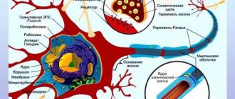
The structure of intermediate neurons
An interneuron consists of a body, an axon and dendrites. Each part has its own specific functions and is responsible for a specific action. Its body contains all the components from which cellular structures are created. The important role of this part of the neuron is to generate nerve impulses and perform trophic function. The elongated process that carries the signal from the cell body is called an axon. It is divided into two types: myelinated and non-myelinated. At the end of the axon there are various synapses. The third component of neurons is dendrites. They are short processes that branch in different directions. Their function is to deliver impulses to the neuron body, which ensures communication between different types of neurons in the central nervous system.
Sphere of influence
What determines the area of influence of an interneuron? First of all, his own structure. Basically, cells of this type have axons whose synapses end on neurons of the same center, which ensures their unification. Some interneurons are activated by others, from other centers, and then deliver information to their neural center. Such actions increase the impact of the signal, which is repeated in parallel paths, thereby extending the storage period of information data in the center. As a result, the location where the signal was delivered increases the reliability of the influence on the executive structure. Other interneurons can receive activation from connections of motor “brothers” from their center. Then they become transmitters of information back to their center, thereby creating feedback connections. Thus, the interneuron plays an important role in the formation of special closed networks that extend the storage period of information in the nerve center.
Excitatory type of interneurons
Interneurons are divided into two types: excitatory and inhibitory. When the former are activated, the transfer of data from one neural group to another is facilitated. This task is performed by “slow” neurons, which have the ability to activate for a long time. They transmit signals for quite a long time. In parallel with these actions, intermediate neurons activate their “fast” “colleagues”. When the activity of “slow” neurons increases, the reaction time of “fast” ones decreases. At the same time, the latter somewhat slow down the work of the “slow” ones.
The role of interneurons in the functioning of the spinal cord
In the functioning of the human spinal cord, an important role is played by the conduction pathways, which are located outside the bundles that perform the conduction function. It is along these paths that the impulses sent by the intercalary and sensory neurons move. Signals travel up and down these pathways, conveying various information to the corresponding parts of the brain. The interneurons of the spinal cord are located in the intermedial nucleus, which, in turn, is located in the dorsal horn. Interneurons are an important anterior part of the spinocerebellar tract. On the back of the spinal cord horn are fibers consisting of interneurons. They form the lateral spinothalamic tract, which performs a special function. It is a conductor, that is, it transmits signals about pain and temperature sensitivity first to the diencephalon, and then to the cerebral cortex itself.
Motor neurons are located in the anterior horn of the spinal cord
Motor neurons are located in the anterior (ventral) horns of the gray matter of the spinal cord. In the Rexed classification of layers, this is “layer” IX. In cross sections, this is not a layer, but several isolated zones, and in different illustrations, and at different levels of the spinal cord, the location of these zones varies quite a lot.
What is the reason? Already in 1888, Heinrich Waldeyer (the author of the term “neuron” and the neural doctrine) described that motor neurons form not so much layers, but areas extended over several segments. However, even after they were described in detail (for decades in the 40s and 50s by George Romanes - see Jessel 2011), many anatomists continued to call them "nuclei".
Very often you can also see attempts to apply some somatotopy to the areas where the “nuclei” of motor neurons are located. Although there are, of course, general principles for the relative location of motor neurons (detailed below), such “flat” pictures in cross sections are quite deceptive.