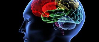
Functions and structure of the human brain
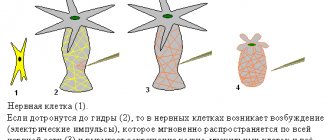
This organ is primarily made up of cells called neurons.
These nerve cells produce electrical impulses that enable the nervous system to function. The work of neurons is ensured by cells called neuroglia - they make up almost half of the total number of cells in the central nervous system.
Neurons, in turn, consist of a body and processes of two types: axons (transmitting impulses) and dendrites (receiving impulses). The bodies of nerve cells form a tissue mass, which is commonly called gray matter, and their axons are woven into nerve fibers and represent white matter.
To protect it, nature has created a whole arsenal of various means. Externally, the parts of the brain are protected by the cranium, under which there are three more membranes of the brain:
- Solid. It is a thin film, one side adjacent to the bone tissue of the skull, and the other directly to the cortex.
- Soft. It consists of loose tissue and tightly envelops the surface of the hemispheres, going into all the cracks and grooves. Its function is to supply blood to the organ.
- Arachnoid. It is located between the first and second membranes and exchanges cerebrospinal fluid (cerebrospinal fluid). Liquor is a natural shock absorber that protects the brain from damage when moving.
Next, let's take a closer look at how the human brain works. According to morpho-functional characteristics, the brain is also divided into three parts. The lowermost section is called rhomboid. Where the rhomboid part begins, the spinal cord ends - it passes into the medulla oblongata and posterior (pons and cerebellum).
Next comes the midbrain, which unites the lower parts with the main nerve center - the anterior section. The latter includes the telencephalon (cerebral hemispheres) and diencephalon. The key functions of the cerebral hemispheres are the organization of higher and lower nervous activity.
This small superficial layer of gray matter (up to 4.5 mm) is the youngest formation in the central nervous system. It is the cerebral cortex that is responsible for the work of higher nervous activity in humans.
Research has made it possible to determine which areas of the cortex were formed relatively recently during evolutionary development, and which were present in our prehistoric ancestors:
- neocortex - the new outer part of the cortex, which is its main part;
- archicortex - an older formation responsible for human instinctive behavior and emotions;
- The paleocortex is the most ancient area involved in the control of autonomic functions. In addition, it helps maintain the internal physiological balance of the body.
Frontal lobes
The largest lobes of the cerebral hemispheres, responsible for complex motor functions. In the frontal lobes of the brain, planning of voluntary movements occurs, and speech centers are also located here. It is in this part of the cortex that volitional control of behavior is exercised. If the frontal lobes are damaged, a person loses control over his actions, behaves antisocially and simply inappropriately.
Occipital lobes
Closely related to visual function, they are responsible for the processing and perception of optical information. That is, they transform the entire set of light signals that enter the retina into meaningful visual images.
Parietal lobes
They carry out spatial analysis and process most sensations (touch, pain, “muscle feeling”). In addition, it promotes the analysis and integration of various information into structured fragments - the ability to feel one’s own body and its sides, the ability to read, count and write.
Temporal lobes
In this department, audio information is analyzed and processed, which ensures the function of hearing and the perception of sounds. The temporal lobes are involved in recognizing the faces of different people, as well as facial expressions and emotions. Here information is structured for permanent storage, and thus long-term memory is realized.
In addition, the temporal lobes contain speech centers, damage to which leads to the inability to perceive spoken language.
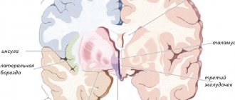
Insula
It is considered responsible for the formation of consciousness in a person. In moments of compassion, empathy, listening to music and the sounds of laughter and crying, active work of the insular lobe is observed. This is also where the processing of feelings of aversion to dirt and unpleasant odors, including imaginary stimuli, takes place.
the impulse is transmitted along the axons, and the impulse is received along the branched neurons. The gray substance, which makes up the cortex, is formed by the cell bodies of neurons. The white substance is located under the cortical region and is formed by axons woven into nervous tissue, through which impulses are transmitted to the subcortical layer.
On the outside, the human brain is protected by the cranial bones, and on the inside, it is surrounded by three membranes, between which the circulation of spinal cerebrospinal fluid occurs, which performs a shock-absorbing function in case of microtrauma. Parts of the brain are interconnected and perform specific tasks. To understand how the functions of the brain are performed, its structure should be studied in detail.
The areas of responsibility are localized in the cortical layer of the white substance. Cerebral structures are hollow: they consist of four ventricles, separated by ducts where cerebral cerebrospinal fluid circulates.
Thanks to the cognitive functions of the brain, a person controls his actions, reproduces and remembers information, thinks figuratively and associatively.
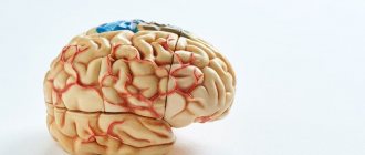
Adult GM weighs approximately 1.5 kg (2% of total weight). For normal functioning, the brain absorbs 20% of the oxygen supplied to the lungs. If a person has lost 50% of their weight, then the GM will lose only 15% of their body weight.
The GM works even at night, and in patients who are in a comatose state, some of its parts do not stop working. Basic brain functions are performed by two hemispheres of different sizes. The right is larger than the left, responsible for visual perception; thanks to the activity of this zone, a person thinks creatively. The left hemisphere is responsible for the ability to think logically and analyze.
There are several main parts of the brain:
- large hemispheric parts;
- middle with medulla oblongata and diencephalon;
- cerebellum;
- cortical region;
- pons.
All these departments perform specific tasks.
GM shells
On the outside, the GM is protected by the cranial bones, under which it is covered with three membranes:
- Solid, having a film structure, one side of which is adjacent to the cranial bones, and the other to the cortex.
- Soft. It has a loose tissue structure; this membrane closely envelops the hemispheres and convolutions of the brain. It supplies blood to the GM.
- Arachnoid, located between the two membranes, which circulates cerebrospinal fluid, protecting cerebral structures from microtrauma.
The structure of the human brain is divided into several sections.
The formation of brain activity occurs during intrauterine development thanks to the rhomboid, midbrain, and forebrain.
The parts of our brain are responsible for various tasks, characterized by the telencephalon and medulla oblongata, intermediate and middle brain, as well as the hindbrain, pons and cerebellum.
| Medulla | Another name for this zone is the bulbus, located in the back of the skull, between the cerebellar region, the pons and the dorsal segment. The bulbus is a continuation of the spinal cord. The white matter of the brain in this area is represented by neurons, and the gray matter by nuclei:
If the functioning of this department is disrupted, heart problems will arise and the transmission of impulses to the brain centers will be disrupted. |
| Diencephalon | This brain region “filters” the impulses of neurons. It will accept all incoming data and decide where and how it will go next. It consists of a lower zone and a posterior zone, consisting of the epithalamus and thalamus. This department is responsible for the functioning of the endocrine system. The hypothalamus is part of the inferior region. This dense neuronal bundle regulates body temperature and the cycle of wakefulness and sleep. It synthesizes hormonal compounds that “tell” a person when to drink or eat. This is the pleasure zone, responsible for interest in the opposite sex. The medullary zone is connected to the pituitary gland, which regulates all glands. Impulses come from the hypothalamic zone to the pituitary gland, and the order to synthesize or stop the release of hormones is “executed.” The thalamus processes impulses from receptors responsible for vision, taste, hearing, and tactile sensitivity. The signals are distributed to the corresponding brain areas. The epithalamus synthesizes the melatonin hormone, which is responsible for the cyclic processes of wakefulness, the emotional sphere, and puberty. |
| Midbrain | The brain section is small in size and consists of two halves: on the roof in the subcortex there are centers of hearing and vision, and conductive pathways are located on the legs. This brain segment includes substantia nigra with red nuclei. There is a temporoparietal node and nuclei of neurons that control the ocular myofibers and temporal zones, which process sound effects that are transformed into recognizable sounds. Reflex activity and reaction to the stimulus are controlled. This organ is responsible for spatial orientation. |
| Finite brain | This is the youngest part of the brain, the main part of the brain, responsible for higher nervous activity, and has numerous grooves with convolutions. The corpus callosum separates the right and left hemisphere zones. Each hemisphere is equipped with a nucleus, mantle, and olfactory brain. |
| Pons | This anatomical formation is part of the hindbrain, which contains the cerebellar region. The functions of the bridge are similar to its name; it consists of nerve fibers. Through it there are impulses passing from the body to the GM and vice versa. It makes up the brain stem, located between the midbrain and medulla oblongata. It contains the nuclei of nerves that control chewing, facial expressions, and some ocular myofibers. It receives signals from receptors responsible for the sensory organs, skin, and inner ear. Thanks to this department, a person feels taste, maintains balance, and hears sounds. |
| Cerebellum | Consists of 2 hemispheric areas and an unpaired formation connecting them. The cerebellar surface is covered by the cortex, which forms 2 nuclei in the thickness of the hemispheric zones. In the deep layers, the lobules consist of a white substance that connects the cerebellar segment with three pairs of legs with the spinal trunk and GM. Responsible for coordinating and regulating the movements of myofibers and muscle memory. Thanks to him, a person maintains a certain body position. |
Content
- 1 Anatomy 1.1 Pathways
- 1.2 Cytoarchitecture
- 1.3 Brodmann fields
- 3.1 Processing of multimodal sensory information
- 4.1 Progressive expressive aphasia
Finite brain
This part has the largest volume (80%) compared to the rest. It consists of two cerebral hemispheres, the corpus callosum connecting them, and the olfactory center.
The large hemispheres of the brain, left and right, are responsible for the formation of all thought processes. Here there is the greatest concentration of neurons and the most complex connections between them are observed. In the depths of the longitudinal groove, which divides the hemispheres, there is a dense concentration of white matter - the corpus callosum. It consists of complex plexuses of nerve fibers that intertwine different parts of the nervous system.
Within the white matter are clusters of neurons called the basal ganglia. The close location to the “transport junction” of the brain allows these formations to regulate muscle tone and carry out instant reflex-motor reactions. In addition, the basal ganglia are responsible for the formation and operation of complex automatic actions, partially repeating the functions of the cerebellum.
Large hemispheres
The right and left hemispheres make up 80% of the central nervous system; they are responsible for mental activity and consist of gray substance (cortex, basal ganglia) and white substance (fibers). In this part of the brain, the density of neurons is highest, they are closely interconnected. A longitudinal groove separates the hemispheric parts of the brain; in its deep layers there is a white substance - the corpus callosum, consisting of bundles of nerve tissue.
The inner layer of the substantia alba contains the basal ganglia, which regulate muscle tone. These structures also control reflexes, motor reactions, and the automaticity of actions, repeating cerebellar tasks.
The lobes of the brain are responsible for specific tasks. The occipital lobe of the brain is responsible for vision, and underneath it is the cerebellum, which controls balance and coordination. The parietal lobe of the brain controls body sensations, the frontal lobe controls movement, and the temporal lobe controls hearing, memorization, and speech.
The thickness of the cortical layer ranges from 3 to 5 mm; the white substance of the right and left hemispheres is covered with the cortex. Nerve cells and woven filamentous processes, afferent, efferent types of neurons, neuroglia form the cerebral cortex.
It is formed by layers:
- granular and internal granular;
- pyramidal external and internal;
- spindle cells, molecular.
The cortex occupies approximately 50% of the volume of the hemispheric regions, the size is about 2200 cm². There are grooves on the surface cortical layer; a third of the total area of the cortex is localized in their depths. The furrows have different sizes and shapes.
The cortex is responsible for higher nervous activity and consists of new, old, ancient and intermediate cortex. The neocortex covers more than 95%, this is the outer part of the cortex. The archicortex covers approximately 2%. This old area controls instincts and the emotional sphere. The paleocortex covers 0.6%; this zone controls vegetative functions and the internal physiological balance of the body. The intermediate layer covers 1.6% of the bark.
Thanks to conditioned reflexes, the relationship of the GM with nerve tissues located in different parts of the body, the body functions fully. The cortex synchronizes mental activity, motor skills of internal organs, and the area that analyzes incoming impulses.
The tasks performed depend on the segment of localization of nerve fibers that receive one of the types of impulses (sensory, motor, associative), responsible for perception. The associative segment, responsible for acquiring and processing data from the sensory area, occupies more than 70% of the cortex. It coordinates the activities of sensory and motor areas.
The grooves of the hemispheric surfaces divide them into 5 segments. In front of the central sulcus, where pyramidal cell structures are localized, there is a frontal segment. This is the main motor area responsible for the precision of movement of the body, arms, and facial muscles.
The premotor cortical area is located above the motor area, which controls complex actions that depend on sensory feedback (grasping an object, moving over an obstacle).
Broca's center is localized in the lower part of the left dominant hemispheric zone, responsible for speech; with the activity of the right hemisphere, the emotional coloring of words is maintained. This segment takes part in short-term memorization of speech; thanks to the activity of this center, a person prefers to work with his left or right hand.
The optic lobe is a motor region that controls rapid movement of the eyes when observing a moving object.
The olfactory segment is located at the base of the frontal regions. Thanks to the activity of the olfactory region, a person perceives odors. This zone of the cortex is connected to the olfactory centers in the lower parts of the brain structures responsible for behavior, motivation, learning, memory, instincts (limbic system).
The prefrontal cortical area is a large area of the frontal lobe, responsible for memory and mental activity. Its activity shows how a person perceives the world, thinks abstractly, and how much control he has over himself.
The parietal lobe is responsible for how a person perceives different sensations through the skin (heat, cold, pain), taste. The correct functioning of this zone helps a person feel the size of his body.
The occipital region is responsible for vision. She perceives the received image from the retina into a meaningful image: a person recognizes words and faces. Below this zone is the cerebellar region responsible for the coordination of movements.
The auditory and vestibular areas are located in the temporal lobe: responsible for the perception and memorization of audio effects. There are centers responsible for speech, sound perception, emotions (fear, anger, joy, pleasure).
The extensive area responsible for speech function is represented by zones of speech formation and understanding.
The two hemispheres coordinate opposite parts of the body. For the majority of humanity (95%), the left hemisphere is responsible for understanding languages, mathematical and logical abilities. The right hemisphere controls visual perception: facial expressions, creative abilities (drawing, music), the emotional sphere.
The activity of the insular lobe appears when a person laughs, cries, listens to music, or empathizes. Here the feelings of aversion to bad smells and dirt are processed.
Basal ganglia
They lie in the deep layers of the white substance, interact with the cortex, and control motor activity. Thanks to this complex nervous structure, movements begin, stop, and their intensity is regulated. The selection of myofibers for movement occurs, and opposing muscle tissues are inhibited.
- caudal and red nucleus;
- shells;
- substantia nigra;
- globus pallidus;
- reticular formation.
Normal functioning of the autonomic nervous system is impossible with dysfunction of the basal ganglia. Thanks to them, a person performs automatic actions, while the central nervous system reserve is not consumed.
Spheres of influence of the cerebral hemispheres
The main functions of the left hemisphere are logical thinking and speech. The left hemisphere is focused on working with numbers and letters.
The left hemisphere determines the following human abilities:
- logical information processing;
- analytical thinking: analysis of facts, recognition of written characters and symbols;
- mathematical abilities: solving mathematical problems, recognizing mathematical symbols and numbers;
- reading ability: recognizing letters, understanding the literal meaning of words;
- writing ability;
speech and control over it;- ability to learn foreign languages.
The main functions of the right hemisphere are imaginative thinking and intuition. The right hemisphere focuses on images and emotions.
The right hemisphere determines the following human abilities:
- figurative perception and processing of all information at once, in its entirety, the ability to perceive the totality of something as a single image;
- processing of non-verbal signals: understanding of non-verbal information coming in the form of images and symbols, body signals both from outside and from inside the human body;
- orientation in space: the ability to determine one’s location and the position of objects in space;
- creative abilities: music, drawing, poetry and others;
- understanding the meaning of words and metaphors;
- imagination: the ability to imagine, dream, fantasize and understand the results of another person’s imagination;
- emotional perception (although another part of the brain is responsible for the emotional sphere of a person, the right hemisphere connects it to the process of perceiving the world);
- the ability to perceive and believe in irrational phenomena.
Diencephalon
The diencephalon serves as a kind of filter for neural signals - it receives all incoming information and decides where it should go. Consists of the lower and posterior parts (thalamus and epithalamus). In this department, the endocrine function is also realized, i.e. hormonal metabolism.
The lower part consists of the hypothalamus. This small, dense bundle of neurons has a tremendous impact on the entire body. In addition to regulating body temperature, the hypothalamus controls sleep and wake cycles. It also releases hormones that are responsible for the sensations of hunger and thirst. As a pleasure center, the hypothalamus regulates sexual behavior.
It is also directly connected to the pituitary gland and converts nervous activity into endocrine activity. The functions of the pituitary gland, in turn, are to regulate the functioning of all glands of the body. Electrical signals go from the hypothalamus to the pituitary gland of the brain, “ordering” the production of which hormones should be started and which ones should be stopped.
The diencephalon also includes:
- Thalamus - it is this part that performs the functions of a “filter”. Here, signals coming from visual, auditory, taste and tactile receptors undergo primary processing and are distributed to the appropriate departments.
- Epithalamus - produces the hormone melatonin, which regulates wakefulness cycles, participates in the process of puberty, and controls emotions.
How to develop the hemispheres of the brain. Hemisphere synchronization
A very important organ responsible for human thinking is the pineal gland. It is located between the hemispheres and is responsible for the functioning of the entire endocrine system and the production of melatonin (the hormone of youth).
In many esoteric teachings, very great importance is attached to its activation. So in Taoism (Chinese traditional teaching) there are such understandings as “operating” with it, which boils down to elementary concepts of concentration. No matter how much we talk about concentration and focusing, if consciousness is not synchronized, a person will not master them fully. He can only have observations related to the work of one or another part of the brain.
To bring the work of both parts into harmony, you should perform certain exercises developed by psychophysiologists. The main instrument here is played by the hands. Acting with two hands, a person develops both hemispheres.
You are ready? Let's get started!
Midbrain
First of all, it regulates auditory and visual reflex activity (constriction of the pupil in bright light, turning the head towards a source of loud sound, etc.). After processing in the thalamus, the information goes to the midbrain.
Here its further processing takes place and the process of perception begins, the formation of a meaningful sound and optical image. In this department, eye movements are synchronized and binocular vision is ensured.
The midbrain includes the peduncles and quadrigeminal region (two auditory and two visual colliculi). Inside there is a cavity of the midbrain that unites the ventricles.
Functions
Processing multimodal sensory information
Functional imaging studies show activation of the insular cortex throughout the performance of audio-visual integration tasks[6][7].
Interoceptive self-awareness
There is evidence that in addition to its basic function, the insula may play a role in some higher mental functions. Functional imaging studies have shown that activity in the right anterior insula correlates with a person's ability to sense their own heartbeat or empathize with the pain of others. It is believed that these functions do not differ from the basic functions of the insula, since they arise as a result of the insula’s perception of homeostatic information from the thalamus [8][9]. Thus, the insula is involved in the perception of heat and cold (without pain) on the skin. This includes a feeling of fullness in the stomach and bladder[10][11][12][13][14][15].
Insula activity has been found to be involved in blood pressure control[16], particularly during and after exercise; in addition, its activity depends on the amount of conscious effort [17][18].
The central lobe stands out as the center for evaluating the sensations that arise[19], which is also expressed in empathy, for example, when a person experiences painful sensations when looking at someone else's pain[20].
One tomography study showed that the sensation of shortness of breath is processed in the insula and amygdala[21].
Cortical processing of the vestibular sensation (balance) also involves the central lobe cortex[22], so with minor damage to the anterior insula the patient may experience dizziness[23].
Other interoceptive perceptions that are processed in the insular cortex are passive listening to music[24], laughter and crying[25], compassion and empathy[26], language[27].
Motor control
The central lobe cortex is involved in hand and eye movements[28][29], swallowing[30][31], gastric motility[32] and language articulation[33][34]. Studies of the insular cortex during conversation have shown its connection with the ability to speak for a long time and complex phrases[35]. The insular cortex is also involved in motor learning[36] and has been identified as playing an essential role in recovery and motor restoration after stroke[37].
Social emotions
In the central lobe, processes of processing sensations of disgust take place, both to odors[38], to the sight of dirt and mutilation[39] - even imaginary ones[40]. In the social aspect, the insular cortex is involved in processing information about violation of generally accepted norms of behavior[41], emotional processes[42], empathy[43] and orgasm[44]. Activity of the lobe has been discovered when making social decisions made as a result of passing various tests[45].
Medulla
This is an ancient formation of the nervous system. The functions of the medulla oblongata are to ensure breathing and heartbeat. If this area is damaged, the person dies - oxygen stops flowing into the blood, which is no longer pumped by the heart. In the neurons of this department, protective reflexes such as sneezing, blinking, coughing and vomiting begin.
The structure of the medulla oblongata resembles an elongated onion. It contains the nuclei of gray matter: the reticular formation, the nuclei of several cranial nerves, as well as neural ganglia. The pyramid of the medulla oblongata, consisting of pyramidal nerve cells, performs a conducting function, uniting the cerebral cortex and the spinal region.
The most important centers of the medulla oblongata:
- breathing regulation
- regulation of blood circulation
- regulation of a number of functions of the digestive system
Clinical significance
The central lobe is believed to be involved in the functioning of consciousness and plays an important role in various functions, usually related to the regulation of homeostasis and emotions. Functions of the insula include, but are not limited to: perception, motor control, self-awareness, cognition, and interpersonal experience. This gives rise to a role in the corresponding psychopathological processes.
Progressive expressive aphasia
Type of semantic aphasia. In progressive expressive aphasia, there is a deterioration of normal language function, resulting in loss of fluent speech with preserved ability to understand single words and unaffected nonlinguistic cognitive functions. Occurs in a variety of degenerative neurological diseases, including Pick's disease, motor neuron disease, corticobasal degeneration, frontotemporal dementia, and Alzheimer's disease. This is associated with hypometabolism[46] and atrophy of the left anterior central lobe[47].
Addiction
A number of functional brain imaging studies have shown that the central lobe cortex is activated when drug abusers are exposed to the environment and cues that trigger cravings to use. This has been shown for a variety of drugs, including cocaine, alcohol, opiates and nicotine. Despite these findings, the role of lobe has been ignored in the addiction literature [ source unspecified 525 days
].
A 2007 study showed that smokers who have central lobe damage after a stroke are able to quit tobacco addiction[48]. This has been confirmed by newer studies[49][50][51], making the central lobe a promising site for new research and a target for new anti-drug drugs[52][53].
Other clinical conditions
The central lobe is believed to play a role in the onset and progression of disease states such as anxiety disorders[54], emotional dysfunction[55], and anorexia[56].
Hindbrain: pons and cerebellum
The structure of the hindbrain includes the pons and the cerebellum. The function of the bridge is very similar to its name, since it consists mainly of nerve fibers. The cerebral pons is essentially a “highway” through which signals travel from the body to the brain and impulses travel from the nerve center to the body. Along the ascending pathways, the brain bridge passes into the midbrain.
The cerebellum has a much wider range of capabilities. The functions of the cerebellum are to coordinate body movements and maintain balance. Moreover, the cerebellum not only regulates complex movements, but also contributes to the adaptation of the motor system to various disorders.
For example, experiments using an invertoscope (special glasses that invert the image of the surrounding world) have shown that it is the functions of the cerebellum that are responsible for the fact that when wearing the device for a long time, a person not only begins to navigate in space, but also sees the world correctly.
Anatomically, the cerebellum follows the structure of the cerebral hemispheres. The outside is covered with a layer of gray matter, under which there is an accumulation of white matter.
Artist or thinker
Depending on which hemisphere is predominantly dominant, two personality types :
- A thinker , a person with a technical mindset is a left-hemisphere type.
- An artist , a person with a humanitarian mindset is a right-hemisphere type.
How to determine the dominant hemisphere? There are several methods, the simplest and most popular is the “Leading Hemisphere of the Brain” test.
To determine the dominant hemisphere you need:
- Bring your hands together, interlocking your fingers. Look and write down on paper the thumb of which hand is on top.
- Make a small hole in a sheet of paper (you can use a pencil), and look through it with both eyes at any object. Then alternately close your left and right eyes. Please note that the object moves when the right or left eye is closed. Record your answer on a piece of paper.
- Cross your arms over your chest so that your palms are on your forearms. Mark which hand is on top.
- Clap your hands. Write down which palm is on top.
If there are more answers “right hand” - a right-hemisphere type, “left hand” - a left-hemisphere type.
An interesting fact is that the right hemisphere of the brain controls the left half of the body, sending signals there in the form of electrical impulses, and the left hemisphere controls the right side of the human body.