| Lateral ventricles of the brain | |
| Latin name | ventriculi lateralis |
| Catalogs |
|
| Lateral ventricles of the brain at Wikimedia Commons | |
Lateral ventricles of the brain
(lat. ventriculi laterales) - cavities in the brain containing cerebrospinal fluid, the largest in the ventricular system of the brain. The left lateral ventricle is considered the first, the right - the second. The lateral ventricles communicate with the third ventricle through the interventricular (Monroy) foramina. They are located below the corpus callosum, symmetrically on the sides of the midline. In each lateral ventricle, there is an anterior (frontal) horn, a body (central part), a posterior (occipital) and an inferior (temporal) horn.
Choroid plexuses
The lateral ventricle(s) are only partially involved in such a concept as the choroid plexus. The bulk of these structures are located in the roofs of the third and fourth ventricles. They are responsible for most of the production of cerebrospinal fluid. In addition to them, this function is performed directly by the nervous tissue, as well as by the ependyma, which covers the ventricles of the brain from the inside.
Morphologically, the choroid plexuses are outgrowths of the pia mater, immersed in the ventricles. On the outside, these protrusions are covered with cuboidal specific choroid epithelium.
Ependymocytes
The lateral ventricles of the brain are lined from the inside with a special tissue that can both produce cerebrospinal fluid and absorb it. This helps maintain the optimal amount of fluid in the cavity and prevent increased intracranial pressure.
The cells of this epithelium have many organelles and a large nucleus. Their outer surface is covered with a large number of microvilli; they help promote the cerebrospinal fluid, as well as its absorption. Outside the ependyma are Kolmer cells, which are considered a special type of macrophages capable of moving throughout the body.
Through multiple small gaps in the basement membrane of epindemocytes, blood plasma sweats into the ventricular cavity. Proteins produced directly by the cells of the internal epithelium of the brain cavities are added to it, and this is how cerebrospinal fluid is obtained.
Lateral divisions
Conventionally, they are called the first and second. Each lateral ventricle of the brain includes three horns and a central section. The latter is located in the parietal lobe. The anterior horn is located in the frontal, the lower - in the temporal, and the posterior - in the occipital zone. In their perimeter there is a choroid plexus, which is distributed quite unevenly.
So, for example, it is absent in the posterior and anterior horns. The choroid plexus begins directly in the central zone, gradually descending into the lower horn. It is in this area that the size of the plexus reaches its maximum value. For this reason, this area is called a tangle. Asymmetry of the lateral ventricles of the brain is caused by a disturbance in the stroma of the tangles.
Structural features
The ventricles of the human brain are important elements on the condition of which the patient’s well-being depends.
An adult has the following structure:
Between the first and second there is a subarachnoid space filled with cerebrospinal fluid, or cerebrospinal fluid. It washes various parts of the vital organ, provides them with nutrition and protection, and removes waste products using capillaries running in the soft (vascular) membrane.
Another important role of CSF is protection against trauma, concussions, and impacts of the brain on the hard membranes and vaults of the skull.
To allow fluid to move freely, the surfaces are covered with special ependymal cells with ciliated processes. They have another important role - they produce myelin, the substance that covers the sheaths of nerve fibers. It protects them during the transmission of electrical impulses between neurons.
In total, humans have 4 ventricles:
- The first two are the lateral ventricles of the brain, cavities with cerebrospinal fluid, located symmetrically in the right and left hemispheres.
- The third ventricle of the human brain resembles a donut, located between the visual thalamus.
- The fourth ventricle of the brain communicates with the third and is located below it between the cerebellum and the medulla oblongata. It is the smallest of all four ventricles and is the cavity connecting the third ventricle to the central canal of the spinal cord.
The ventricles are not separate structures with clear boundaries such as walls or membranes. These are cavities in the gray matter, filled with a special fluid, communicating with each other and with the spinal canal.
Third cavity of the system
This ventricle is located in the diencephalon. It connects the lateral sections with the fourth. As in other ventricles, the third contains choroid plexuses. They are distributed along its roof. The ventricle is filled with cerebrospinal fluid. In this department, the hypothalamic groove is of particular importance.
The third ventricle is special, although they all form a single system. If any malfunctions are detected, you should immediately contact a specialist, as serious consequences may occur. The size of this cavity is 6mm in adults, 5mm in children. It plays a huge role in the processes that provide inhibition of the ANS (autonomic nervous system), and is closely related to visual function.
Disorders of this ventricle can be fatal, unlike dysfunctions of others.
Its role is important for the central nervous system. Certain disorders can lead to major problems in the body and, as a result, to disability.
Important Features:
- protects the central nervous system;
- monitors metabolism;
- regulates the production of cerebrospinal fluid;
- monitors the normal functioning of the central nervous system.
Correct, coordinated operation of the liquor system is an important, refined process. If failures occur, this affects the health of adults and children.
The cerebrospinal fluid is produced with some disturbances, something goes wrong, you need to look at the norm:
- infants – 5 mm;
- up to three months – no more than 5mm;
- child under six years old – 6mm;
- adult – no more than 6 mm.
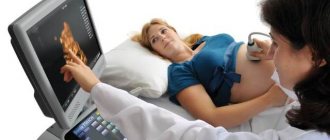
This problem (fluid outflow dysfunction) is more common in children under 12 months. The most common complication is hydrocephalus. This can be avoided by having an ultrasound done during pregnancy, which makes it possible to identify certain abnormalities at an early stage. If the doctor discovers that the 3rd cavity is enlarged, you need to undergo further examination and then be observed by a doctor.
There is a mandatory examination of babies at the age of two months by a doctor to exclude problems with the third cavity.
Violations can be tracked by the following symptoms:
- constant strong crying;
- divergence of cranial sutures;
- head enlargement;
- the baby does not latch on well;
- enlarged veins on the head.
If you track the disease in time and resort to treatment: massage, medications, you can avoid radical interventions.
In adults, diseases associated with the third ventricle are also diagnosed. A colloid cyst may occur; it is a benign tumor that grows slowly and practically does not metastasize. It affects people mainly after 20 years of age.
The cyst itself is not life-threatening, but if it begins to grow and interferes with the outflow of cerebrospinal fluid, the following symptoms may occur: vomiting, severe headache, seizure disorders, and vision problems. If the cyst reaches a large size, surgical intervention is indicated to restore normal circulation of spinal cord fluid. After this, all functions are restored, unpleasant symptoms disappear.
Cerebrospinal fluid
The norm for the lateral ventricles is to produce half a liter of cerebrospinal fluid per day, but only one hundred and forty milliliters of this amount constantly circulates in the subarachnoid space. Despite the fact that the basis for cerebrospinal fluid is blood plasma, they have significant differences in the amount of electrolytes and protein.
– detoxification (transportation of metabolic products);
– depreciation (when walking, falling, sharp turns);
– formation of a hydrostatic membrane around the elements of the nervous system;
– maintaining a constant composition of fluids in the central nervous system;
– transport (transfer of hormones and some drugs).
Fourth cavity
This section is located between the pons, cerebellum and medulla oblongata. The shape of the cavity is similar to a pyramid. The floor of the ventricle is called the rhomboid fossa. This is due to the fact that anatomically it is a depression that looks like a diamond. It is lined with gray matter with a large number of tubercles and depressions.
The roof of the cavity is formed by the lower and upper brain sails. It seems to hang over the hole. The choroid plexus is relatively autonomous. It includes two lateral and medial sections. The choroid plexus attaches to the lower lateral surfaces of the cavity, extending to its lateral inversions.
Bark
It is difficult to imagine that the cortex, which provides the main characteristics of consciousness and intelligence, is only 3 mm thick. This thinnest layer reliably covers both hemispheres. It is composed of the same nerve cells and their processes, which are located vertically.
The layering of the crust is horizontal. It consists of 6 layers. The cortex contains many vertical nerve bundles with long processes. There are more than 10 billion nerve cells here.
The cortex is assigned various functions that are differentiated between its different sections:
- temporal – smell, hearing;
- occipital – vision;
- parietal – taste, touch;
- frontal – complex thinking, movement, speech.
In the hemispheres themselves there are basal ganglia - these are large clusters that consist of gray matter. It is the basal ganglia that transmit information. Between the cortex and the basal ganglia are the processes of neurons - the white matter.
It is the nerve fibers that form the white matter; they connect the cortex and those formations that are under it. The subcortex contains the subcortical nuclei.
Diseases associated with the ventricles of the brain
Several diseases can affect the ventricles of the brain. The most common are: hydrocephalus, meningitis and ventriculitis.
It is important that the production of cerebrospinal fluid is balanced with its resorption so that it does not accumulate more than necessary. Many pathologies that affect the ventricles of the brain are associated with obstruction of these.
hydrocephalus
Hydrocephalus is the accumulation of cerebrospinal fluid in the ventricles of the brain when it is not absorbed properly. If left untreated, it causes increased intracranial pressure and brain atrophy.
– Communicating hydrocephalus: occurs when fluid accumulates without obstruction in the circulation. This usually occurs due to deterioration of the arachnoid granulations, which reabsorb the cerebrospinal fluid.
– Non-infectious or obstructive hydrocephalus: This is due to obstruction in the ventricular system. They are usually found in the cerebral aqueduct, which connects the third and fourth ventricles.
Symptoms of hydrocephalus include headaches, drowsiness, loss of coordination, blurred vision, seizures, nausea, and cognitive changes such as difficulty maintaining attention or psychomotor retardation.
If this process occurs before the fontanelles merge, that is, before joining different areas of the skull, macrocephaly can be observed. In this case, the size of the skull grows abnormally.
Whereas, if the fontanelles have fused, it is more likely to compress and damage adjacent tissue.
It has also been noted that the ventricles dilate in neurodegenerative diseases in parallel with brain atrophy. This is what happens, for example, in Alzheimer's disease.
meningitis
Meningitis is a disease in which the meninges and spinal cord, that is, the layers that cover it and which contain cerebrospinal fluid, become inflamed. Usually caused by viruses, fungi or bacteria, which leads to increased intracranial pressure and obstruction of the circulation of cerebrospinal fluid.
It is accompanied by headache, cognitive impairment, nausea, sensitivity to light, sudden fever, muscle weakness, etc.
ventriculitis
Ventriculitis, as its name suggests, is an inflammation of the ventricles of the brain that involves four cavities.
Ventriculitis is a serious complication of any meningitis. This is due to lack of antibiotic treatment. It is accompanied by hydrocephalus and is associated with arachnoiditis, encephalitis, cerebritis and encephalomyelitis.
schizophrenia
Some scientists have discovered a connection between schizophrenia and the size of the ventricles of the brain. In particular, schizophrenics seem to have larger ventricles than healthy people.
However, it is unclear whether psychiatric disorders cause ventricular dilatation or whether ventricular dilatation is responsible for psychiatric disorders.
On the other hand, obstructions in the ventricular system due to tumors, cysts, trauma, developmental abnormalities, vascular malformations (aneurysms), etc. can also occur.
On the other hand, asymmetry in the lateral ventricles is often observed on brain scans. A paper that examined ventricular asymmetry in the human fetal brain found that it is associated with a normal variant that does not suggest any pathology.
According to Orellana (2003), what helps determine that asymmetry is an anatomical variant and not a pathology is that, typically, in this variant the temporal horns are the same size, and even sometimes the contralateral one is more widened.
When one lateral ventricle (or both) produces more fluid than it can absorb, a pathological condition called hydrocephalus develops. The internal volume of the ventricles of the brain gradually increases, compressing the brain tissue. Sometimes this leads to irreversible ischemia and necrosis.
In newborns and young children, the symptoms of this disease are disproportionate sizes of the skull compared to the facial skull, bulging of the fontanelles, and causeless restlessness of the child, turning into apathy. Adults complain of headache, pain in the eye area, nausea and vomiting.
For diagnosis, neuroimaging methods are used: magnetic resonance therapy or computed tomography. Timely detection and treatment of this disease allows you to avoid a significant number of complications and maintain the possibility of normal life.
Treatment
Treatment for tumors, hematomas and parasites of g. g. m. is only surgical. To remove the intraventricular focus, ventriculotomy is performed (see) - dissection of the wall of the ventricle with opening of its lumen.
In inflammatory processes, surgical intervention is resorted to in cases of development of occlusive phenomena (see Hydrocephalus). Ventriculopuncture is used as a temporary measure for acute occlusions of the cerebrospinal fluid outflow tract to reduce intraventricular pressure (see).
In cases where the occlusion cannot be surgically eliminated, palliative operations are performed aimed at creating a roundabout path for the outflow of cerebrospinal fluid from the ventricles (ventriculostomy operations, perforation of the terminal plate, ventriculosubdural anastomosis, ventriculocisternostomy).
Among the conservative methods of treating ventriculitis, dehydration is used to reduce intracranial pressure and reduce hypertensive syndrome (see Dehydration therapy). For acute and chronic infectious ventriculitis, anti-inflammatory treatment is carried out.
The prognosis for tumors of the gastrointestinal tract is always serious and is determined by their good quality, localization within the ventricular system, and relationship to adjacent brain formations. With hemorrhages in the gastrointestinal tract, in most cases the prognosis is unfavorable. In inflammatory processes, the prognosis largely depends on concomitant disturbances in the circulation of cerebrospinal fluid.
See also Intracranial pressure, Hypertensive syndrome.
Bibliography: Multi-volume guide to neurology, ed. S. N. Davidenkova, vol. 5, M., 1961; Multi-volume guide to surgery, ed. B.V. Petrovsky, vol. 3, book. 2, M., 1968; Patten B. M. Human embryology, trans. from English, M., 1959; Shelia R. N. Tumors of the ventricular system of the brain, L., 1973; G 1 a g a M.
Das Nerven-system des Menschen, Lpz., 1953; G o r-rales M. a. T orre a 1 ba G. The third ventricle, Normal anatomy and changes in some pathological conditions, Neuroradiology, v. 11, p. 271, 1976, bibliogr.; Messert B., Wanna-maker BB a. Dudley AW Reevaluation of the size of the laterol ventricles of the brain, Postmortem study of an adult population, Neurology (Min-neap.), v. 22, p. 941, 1972.
E. P. Kononova, S. S. Mikhailov; N. Ya. Vasin (neurosurgeon).
General system structure and some important terms
The expansion of the ventricles of the brain negatively affects the activity of the nervous system. Their condition can be assessed using diagnostic methods. For example, a computed tomography scan reveals whether the ventricles of the brain are enlarged or not. MRI is also used for diagnostic purposes.
Asymmetry of the lateral ventricles of the brain or other disorders can be caused by various reasons. Among the most popular provoking factors, experts call increased formation of cerebrospinal fluid. This phenomenon accompanies inflammation in the choroid plexus or papilloma.
Asymmetry of the ventricles of the brain or changes in the size of the cavities may be a consequence of impaired outflow of cerebrospinal fluid. This happens when the holes of Luschka and Magendie become impassable due to inflammation in the membranes - meningitis. The cause of obstruction may also be metabolic reactions due to venous thrombosis or subarachnoid hemorrhage.
Cavities containing cerebrospinal fluid communicate with a number of organs. In particular, with the spinal cord canal, subarachnoid space. The structure of the system is as follows:
- 2 lateral ventricles;
- third and fourth ventricles;
- choroid plexuses;
- choroid ependymocytes;
- tanycytes;
- blood-cerebrospinal fluid barrier;
- liquor fluid.
Contrary to their name, the ventricles are not bags filled with cerebrospinal fluid, but hollow spaces, or cavities, located in the brain. The produced liquor performs a huge number of functions. The common cavity, formed from the ventricles of the brain with canals, overlaps with the subarachnoid space and the median canal of the spinal part of the central nervous system.
Most of the total cerebrospinal fluid is produced in the area of the choroid plexuses located above the 3rd and 4th ventricular cavities. A little substance is located in the wall areas. Soft membranes emerge into the lumen of the cavities, from which plexuses of blood vessels are created. Ependymal cells (choroid ependymocytes) play a huge role and are quite functional in stimulating nerve impulses.
An important criterion is the promotion of cerebrospinal fluid with the help of special cilia. Tanycytes provide connections between blood cells and spinal cord fluid in the ventricular lumens; they have become a specialized variety of ependymal cells. The blood-cerebrospinal fluid barrier is a highly selective filter. It performs the function of selectivity in the flow of nutrients into the brain. It also displays metabolic products. Its main purpose is to maintain the homeostasis of the human brain and the multifunctionality of its activities.
Pathology
The pathology may be due to the development of inflammatory processes, hemorrhages, localization of parasites, and tumors in the gastrointestinal tract.
Inflammatory processes in the gastrointestinal tract (ventriculitis) can be observed with various infectious lesions and intoxications of c. n. With. (for example, with meningoencephalitis, etc.). In acute ventriculitis, a picture of serous or purulent ependymatitis may develop (see Chorioependimitis). With hron, productive periventricular encephalitis, the ventricular ependyma thickens, sometimes taking on a granular appearance, which is caused by warty reactive growths of the subependymal layer.
Clinically, disturbances in the circulation of cerebrospinal fluid in ventriculitis are manifested by paroxysms of headaches, during which patients, depending on the level of difficulty in the outflow of cerebrospinal fluid, take characteristic forced positions with the head tilted forward, tilting it back, etc. (see.
Occlusion syndrome). Neurol, symptoms of ventriculitis are polymorphic; it manifests itself with a wide range of symptoms from the periventricular (periventricular) structures of the diencephalic parts of the brain (arterial hypertension, hyperthermia, diabetes insipidus, narcolepsy, cataplexy), midbrain (oculomotor disorders), hindbrain and medulla oblongata - the bottom of the fourth ventricle (vestibular disorders, symptoms of damage nuclei of VI,VII cranial nerves, etc.).
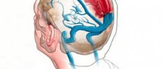
Primary hemorrhages in the gastrointestinal tract are rare and in the vast majority of cases are of traumatic origin. Secondary hemorrhages are more often observed, resulting from the breakthrough of intracerebral hematomas (traumatic, after a stroke) into the ventricular cavity. These hemorrhages are manifested by the acute development of a coma with pronounced reactions from the cardiovascular system, respiratory disorders, hyperthermia, dissociated meningeal symptoms, and often hormetonic syndrome (see Hormetonia). An admixture of blood is detected in the cerebrospinal fluid.
Of the parasitic lesions of the stomach, the most common are cysticercosis, echinococcosis, and coenurosis. The main wedge, their manifestation is the symptoms of aseptic ependymatitis with disturbances in the circulation of cerebrospinal fluid. The latter may also be due to obstruction of the cerebrospinal fluid outflow tract by a parasite freely floating in the ventricular fluid.
Headaches that appear with a certain position of the head, forced position of the head, and hypertensive-hydrocephalic syndrome are also noted. Analysis of cerebrospinal fluid reveals a picture of aseptic meningitis. When parasites are localized in the fourth ventricle, Bruns syndrome may develop (see Occlusion syndrome).
Rice. 5. Frontal section of the brain at the level of the pons. Ependymoma of the fourth ventricle, filling its cavity (indicated by an arrow).
Rice. 6. Frontal section of the brain: choroidpapilloma of the lateral ventricle (indicated by an arrow), obstructing the interventricular foramen.
Rice. 7. Sagittal section of the brain: astrocytoma of the third ventricle (indicated by an arrow), obstructing the cerebral aqueduct (1) and the interventricular foramen (2).
Rice. 8. X-ray of the skull in the lateral projection with pneumoventriculography: ependymoma in the area of the triangle of the lateral ventricle (indicated by arrows) obstructs the interventricular foramen, causing hydrocephalic expansion of the anterior horn, body and lower horn of the lateral ventricle (on the right - a similar X-ray of the skull of a healthy person): 1 - anterior horn lateral ventricle; 2 - body of the lateral ventricle; 3 - lower horn of the lateral ventricle.
Rice. 9. X-ray of the skull in a direct projection during ventriculography with the introduction of mayodil emulsion: astrocytoma of the third ventricle obstructs the interventricular foramen, causing severe hydrocephalus of the lateral ventricles and displacement to the right side of the third ventricle (on the right - a similar radiograph of a healthy person); 1 - lateral ventricles; 2 - third ventricle.
Rice. 10. X-ray of the skull in a lateral projection during ventgiculography with the introduction of mayodil emulsion. Inflammatory occlusion (indicated by an arrow) of the fourth ventricle causes hydrocephalus of the lateral ventricles (1).
Tumors of the gastrointestinal tract are divided into primary, which develop from the choroid plexuses, ependyma, or subependymal glia, and secondary, which grow into the ventricles from neighboring brain formations. Among primary tumors, the most common are ependymomas (Fig. 5), choroid papillomas (Fig. 6), meningiomas, and less commonly astrocytomas (Fig.
7) and spongioblastoma (see Brain, tumors). Tumors of the lateral ventricles are clinically manifested by a remitting course with occlusive-hydrocephalic paroxysms due to obstruction of the interventricular foramina. During paroxysms, there is a forced position of the head and symptoms of infringement of the brainstem (upward gaze paralysis, bilateral patol.
reflexes on the legs, disorders of cardiovascular activity and breathing). Dissociated meningeal symptoms are often observed as a manifestation of tonic reflexes due to irritation of brain stem structures. In addition, periventricular symptoms may be detected as a result of the impact of the tumor on the adjacent parts of the brain (motor and sensory disturbances varying in severity over time, hemianopia, unilateral symptoms of subcortical lesions, general epileptic seizures with a tonic convulsive component, etc.).
Tumors of the third ventricle are characterized by a combination of hypertensive-hydrocephalic symptoms due to occlusion of the cerebrospinal fluid circulation pathways - the cerebral aqueduct and interventricular (Monroy's) foramina with various metabolic-endocrine and autonomic-vascular disorders, which often serve as the first manifestations of the disease.
Cataplectoid type seizures, sleep rhythm disturbances, sometimes patol, drowsiness are observed. In the later stages of the disease there are attacks of decerebrate rigidity with respiratory and cardiovascular disorders. There is usually a significant increase in protein in the cerebrospinal fluid, sometimes with an increase in the number of cells and xanthochromia.
Wedge, the picture of a tumor of the fourth ventricle consists of symptoms of damage to the nuclear formations of the periventricular structures of its bottom and hypertensive-hydrocephalic symptoms due to obstruction of the outflow tract of cerebrospinal fluid. Paroxysms of headaches with vomiting, dizziness and impaired cardiovascular activity and breathing (Bruns' attacks) are characteristic. A constant symptom is pronounced brainstem nystagmus.
When diagnosing the pathology of gastrointestinal tract, in addition to analyzing the characteristics of the wedge, manifestations, ventriculography (see), ventriculoscopy (see) and encephalography (see) are used using water-soluble emulsion and gas radiopaque substances and radioisotopes (Fig. 8— 10).