Our brain is a unique multi-complex system that simultaneously controls the sensory and vestibular apparatus, movement, thinking, speech, vision and much more.
In this article we will talk about how the brain controls voluntary and involuntary movement. And about what neurological abnormalities are associated with damage to the pyramidal system of the brain.
Pyramidal and extrapyramidal tract
The pyramidal system consists of pyramidal and extrapyramidal tracts. What is their difference? The pyramidal tract, or tractus pyramidalis, is a pathway that connects cortical neurons responsible for motor activity with the nuclei of the spinal cord and cranial nerves. Its job is to control voluntary muscle movements by transmitting signals from the central nervous system to the body. But the extrapyramidal one, it controls the unconscious conditioned reflexes of our body. This is an older and deeper structure of the brain, and its signals are not reflected in consciousness.
Extrapyramidal and pyramidal are descending pathways. And the ascending main pathways are responsible for transmitting information from the senses to the brain. These include: the lateral spinothalamic tract, the anterior spinocerebellar tract and the posterior spinocerebellar tract.
Extrapyramidal system: structure and functions
Why are we moving? The answer would seem obvious. Thanks to the movement of arms and legs, muscle contraction. However, this is only the last stage in the implementation of the motor act.
Our limbs are controlled by the central nervous system. First of all, the brain.
Namely, two structures located in parallel, but at the same time being in constant interaction: the pyramidal and extrapyramidal systems.
Pyramid system - what is it?
It is a collection of nerve cells (motoneurons) grouped into a complex of pathways that stretch from the cerebral cortex (the center of higher nervous activity) through the anterior horns of the spinal cord to the receptors located in the muscles. Thanks to this transmission of nerve impulses, movement occurs.
The peculiarity is that the transmission of these nerve impulses is voluntary, that is, controlled by consciousness.
What is the extrapyramidal system?
This is a system of neurons and pathways in the brain that makes clear, fast, highly precise movements possible. Although these paths do not intersect with the paths of the pyramidal system, they constantly interact.
The main difference between the pyramidal and extrapyramidal systems is the involuntary functioning of the extrapyramidal pathways.
For example, when a desire arises to take a pen, a person voluntarily, consciously reaches for a specific pen (pyramid paths).
However, in order to reach it, he does not need to aim, calculate the distance and force, all this happens automatically (extrapyramidal pathways).
Structure
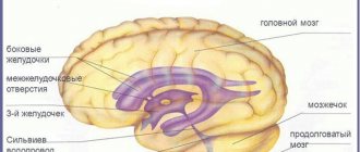
The extrapyramidal system consists of the gray matter of the brain, that is, a collection of nerve cell bodies. The same gray matter is present in the cerebral cortex. The main structural unit is the basal nucleus or basal ganglion. These are kernels such as:
- The striopalidal system, consisting of the lentiform and caudate nuclei. The lenticular ganglion in turn consists of the putamen and the globus pallidus.
- The striatum is a more phylogenetically ancient component of this part of the basal ganglia; it includes the caudate nucleus and putamen.
- The pallidum is a younger component, represented by the globus pallidus.
- Subthalamic nucleus.
- Red core.
- Substantia nigra located in the midbrain.
In addition to the basal ganglia, the extrapyramidal system of the brain also includes:
- thalamus;
- cerebellum;
- olive and vestibule nuclei in the medulla oblongata;
- reticular formation;
- association centers of the cerebral cortex.
In addition, there is a division of the above structures into two departments:
- Neostriatum - includes the cortex, putamen and caudate nucleus. This system is evolutionarily younger;
- Paleostriatum - consists of the globus pallidus, vestibular nuclei, subthalamic nucleus, midbrain structures (tegmentum and substantia nigra).
Striopallidar system
Perhaps the most significant part of the basal ganglia, which has the largest number of connections with the rest of the central nervous system, is the striopallidal system.
The putamen and caudate nucleus receive information via nerve fibers from the cerebral cortex, thalamus, and midbrain (substantia nigra).
From the striatum, fibers are first sent to the pallidum, and then to the rest of the nervous system: the thalamus, hypothalamus, subthalamic nucleus, brainstem (consists of the midbrain, pons and medulla oblongata).
Through the thalamus, the striopallidal system also interacts with the cerebellum, the main coordinator of movements and balance.
Pathways
The structure of the brain, including the extrapyramidal pathways, is truly complex. And to remember it better, it’s worth imagining all the structures through which the path passes. And the best method is to sketch them.
The following pathways of the extrapyramidal system are distinguished:
- reticular-spinal;
- red nucleus spinal;
- vestibulospinal;
- roof-spinal;
- olivospinal.
The reticular-spinal tract originates in the reticular formation in the region of the brain stem. The first neuron is here. The impulse along the nerve fibers spreads down to the second neuron. Its localization is the anterior columns of the spinal cord. Then the impulse travels along the spinal nerves to the striated muscles, where this path ends.
The red nucleus spinal tract begins with the first neuron in the red nuclei located in the midbrain.
The processes of this neuron move to the opposite side and then along this side continue their course to the segments of the spinal cord, where they end with an interneuron (interneuron) - in its gray matter.
This pathway influences the motor nuclei of the spinal cord, from which the impulse goes further to the skeletal muscles, indirectly - through the interneuron.
The vestibular spinal tract consists of the first neuron located in the vestibular nuclei (lateral - Deiters' nucleus, inferior - Roller's nucleus). Second neurons are also located in the anterior columns of the spinal cord, to which the impulse reaches through the medulla oblongata and the anterior funiculus of the spinal cord. The axons of the second neurons then reach the skeletal muscles.
The roof-spinal tract is considered the youngest of all the tracts that make up the anatomy of the extrapyramidal system. Starting in the superior colliculi of the midbrain, where visual information arrives, it then moves to the opposite side, heading similarly to other pathways to the corresponding segments of the spinal cord.
The olivospinal tract is essential for maintaining muscle tone in the neck and maintaining balance. It begins with the formation of the olive nucleus in the medulla oblongata and reaches the sixth segment of the cervical spinal cord. From there, the processes of motor neurons conduct impulses to the muscles of the neck.
Main functions
As noted above, the extrapyramidal system is an important component of the central nervous system, which makes it possible to perform everyday activities. But how does it regulate our movements, making them so precise and accurate?
The main functions of the extrapyramidal system are listed below:
- the orderliness of voluntary movements, initially regulated by the pyramidal system;
- regulation of automatic motor acts of both congenital and acquired nature;
- maintaining balance;
- regulation of muscle tone;
- involuntary contraction of facial muscles;
- regulation of movements that serve as accompanying movements (for example, rapid movement of the arms when running).
Pathological changes
Violation of the function and structure of the extrapyramidal system is called dyskinesia, literally a movement disorder.
It can change both in the direction of increased motor activity - hyperkinesis, and in the direction of a decrease in the amount of movement, the appearance of their stinginess - hypokinesis.
It is characteristic that such a violation cannot be changed in any way with the help of consciousness. Hyperkinesis is involuntary, appears suddenly and also suddenly stops.
Hyperkinesis
The following types of hyperkinetic disorders of the physiology of the extrapyramidal system are distinguished:
- Chorea is rapid, sudden, erratic, involuntary movements of the arms, legs, and facial muscles. This is manifested by the appearance of grimaces on the face and strange gestures.
- Athetosis - movements of the fingers, can also occur in the muscles of the tongue and face. Manifested by arching, worm-like movements of the fingers, twisting of the tongue.
- Torsion dystonia – sudden turns of the body in different directions, arching of the whole body. They often have a corkscrew appearance. First of all, the neck muscles are damaged.
- Hemibalism is one-sided, sweeping movements, most often of the hands, reminiscent of the flapping of a bird's wing.
- Tics are fast, simple, stereotypical movements of small muscle groups.
- Myoclonus is short twitching of individual muscle fibers at a very fast pace. Often, movements of the limbs are not observed.
Parkinson's disease
The classic manifestation of hypokinetic syndrome is Parkinson's disease or parkinsonism syndrome. Their difference lies in the fact that Parkinson's disease occurs with direct damage to the structure of the extrapyramidal system, and parkinsonism syndrome is one of the manifestations of any other diseases not associated with damage to the basal ganglia.
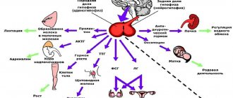
Parkinson's disease develops when the substantia nigra of the midbrain is damaged, which begins to produce less dopamine. Its main function is to reduce the inhibitory effect of the caudate nucleus on motor activity. When this function decreases, the caudate nucleus inhibits motor skills to a greater extent, which leads to the development of hypokinesis.
Parkinson's disease: symptoms
In addition to stiffness and decreased movement, Parkinson's disease also manifests itself:
- increased muscle tone - in neurology the term “increased tone of the hyperplastic type” is used;
- stiffness of facial expressions;
- stooped posture;
- The supplicant's pose is characteristic - the head is tilted down, the arms are bent at the elbows, the torso is tilted;
- tremors of the limbs (tremor);
- Difficulties at the beginning of the movement and at its completion (braking).
The structure of the brain is truly complex and multifaceted. This is due to the many functions it performs. What is the value of just one extrapyramidal system! In order for us to make a basic movement, we need to simultaneously use many brain structures. It is also interesting that a lot of processes happen in a fraction of a second.
Yes, medicine has come a long way in recent decades. However, neurosurgeons have no idea how many more secrets the brain hides.
Pyramidal tracts of the brain. Structure
They are divided into 2 types: corticospinal and corticonuclear. The corticospinal cord is responsible for the movements of the torso, the cortico-nuclear cord controls the facial and swallowing muscles.
How does the corticospinal pyramidal tract work? This electrical path begins from the cerebral cortex - the area that is responsible for higher mental activity, for consciousness. The entire cortex is made up of interconnected neural networks. More than 14 billion neurons are concentrated in the cortex.
In the hemispheres, information is redistributed in this way: everything that concerns the work of the lower extremities is located in the upper sections, and everything that concerns the upper, on the contrary, is in the lower structures.
All signals from the upper and lower parts of the cortex are collected and transmitted to the internal capsule. Then, through the midbrain and through the middle part of the pons, a bundle of nerve fibers enters the pyramids of the medulla oblongata.
Here branching occurs: most of the fibers (80%) pass to the other side of the body and form the lateral spinal tract. These branches “launch” motor neurons, which then transmit signals to contract or relax directly to the muscles. A smaller part of the fiber bundle (20%) innervates the motor neurons of “their” side.
The corticonuclear pyramidal tract initially passes through the same brain structures as its “partner”, but crosses over in the midbrain and goes to the facial neurons.
Structure
The extrapyramidal system consists of the gray matter of the brain, that is, a collection of nerve cell bodies. The same gray matter is present in the cerebral cortex. The main structural unit is the basal nucleus or basal ganglion. These are kernels such as:
- The striopalidal system, consisting of the lentiform and caudate nuclei. The lenticular ganglion in turn consists of the putamen and the globus pallidus.
- The striatum is a more phylogenetically ancient component of this part of the basal ganglia; it includes the caudate nucleus and putamen.
- The pallidum is a younger component, represented by the globus pallidus.
- Subthalamic nucleus.
- Red core.
- Substantia nigra located in the midbrain.
In addition to the basal ganglia, the extrapyramidal system of the brain also includes:
- thalamus;
- cerebellum;
- olive and vestibule nuclei in the medulla oblongata;
- reticular formation;
- association centers of the cerebral cortex.
In addition, there is a division of the above structures into two departments:
- Neostriatum - includes the cortex, putamen and caudate nucleus. This system is evolutionarily younger;
- Paleostriatum - consists of the globus pallidus, vestibular nuclei, subthalamic nucleus, midbrain structures (tegmentum and substantia nigra).
Anatomical features important for diagnosis
The pyramidal pathway has some structural features that should not be overlooked when it is necessary to determine the localization of the pathology. What specific features do you need to know?
- Some of the nerve fibers of the corticospinal tract, in addition to the lateral decussation, also intersect in the area of the white commissure of the spinal cord segment, where they end.
- Most of the muscles in the trunk are controlled by both hemispheres of the brain. This is an important protection. In the event of a stroke or stroke, those patients diagnosed with hemiplegia can support the body upright.
- In the area of the pons, the fibers of the corticospinal tract are separated by other fibers - the cerebellar tract. Divided bundles emerge from the bridge. In this regard, movement disorders are often dispersed. Whereas the pathological focus may be single.
Symptoms of damage to the pyramidal tract are sometimes quite obvious, as in the case of paraplegia, for example. But sometimes it is difficult to determine the cause. It is important to notice minor disturbances in motor skills in time and see a doctor.
Extrapyramidal system (pathways) - structure, functions and significance
The relevant articles described in sufficient detail the pyramidal system, which provides voluntary or conscious movements in humans. But, obviously, there must also be a system that is responsible for providing involuntary or automatic movements? Yes, such a special, more ancient motor nervous system exists, and separately from our consciousness.
"Fossils" of the brain
Its name is the extrapyramidal system. It has taken root in neurology, but in fact it is not entirely successful. Anatomically, the extrapyramidal system does not lie outside the pyramidal fascicles, that is, it is not located “extra”.
It is located “in the depths” of the brain. This structure evolved phylogenetically.
After all, automated, reflex movements were almost the only type of motor activity until a large cerebral cortex developed.
The telencephalon, like a cloak, wrapped and covered these ancient paths and centers, subordinating them to its will. It is no coincidence that another name for the bark is a cloak, pallium. Therefore, the name would be more correct not “extrapyramidal system”, but “intrapyramidal”, “internal”. This term would correctly reflect the phylogenetic development of this structure.
In fact, extrapyramidal tracts appear when the pyramidal tract does not yet exist at all, for example, in ancient fish.
In amphibians (amphibians), the paths of this system become more complex, and additional formations (subcortical centers) appear.
This name is arbitrary, because there is no full-fledged cortex yet and these centers are the highest organs of regulation of motor skills and tone, especially since amphibians can hunt and move without the participation of thinking, but on reflex acts.
A more advanced system gradually developed in humans, the functions of the extrapyramidal system were reduced to servicing skeletal muscles and maintaining higher unconditioned reflexes, and the role of “leader” passed to the cortex. Let's get acquainted with the movements that this system provides, and then turn to its anatomy.
Functions
If you think carefully, you can understand which motor functions are unconscious in a person and do not require the participation of the cortex. These are those movements that have only an unambiguous interpretation. What does this mean and how to understand it?
For example, the pyramidal tract works with subtle, conscious movements. Even in order to bring a spoon to your mouth, you need to think. It can be brought sideways, butt-wise, quickly, slowly. The spoon can be held at the mouth or quickly pulled away. All this can be done according to your will. And the extrapyramidal system “manages” the following functions :
- Regulation of muscle tone. It is known that we have some tone of the resting muscles; they are never completely relaxed, even in sleep. However, we never consciously think about somehow changing or maintaining tone; this ensures “readiness” for movement and includes the extrapyramidal system;
- Security reflexes. These include blinking and flinching of the whole body when there is a sudden gunshot or loud sound. It is absolutely clear that these movements are reflexive and are produced without the participation of thinking and consciousness;
- Maintaining balance. An excellent example of such a “set of higher reflexes” is the behavior of a person who slipped on ice in an attempt to maintain his balance. He can sharply tilt his body, unconsciously wave his hand, and only then it turns out that in his hand there was a string bag with eggs, in the path of which he suddenly met a wall. The eggs were broken, but the person was unable to stop this movement: the activation of a sharp motor act, which returned the center of gravity of the body to its place, was automatic;
In the last example, not only the structures of the extrapyramidal system were involved, but also the “whole color” of autonomous formations, for example, the vestibular-cochlear organ of balance (statokinetic analyzer), the cerebellum and the red nuclei, which will be discussed separately.
- Acquiring and consolidating skills.
This is a most interesting phenomenon. We see how deftly a jeweler or woodcarver can work, a musician can play an instrument by reading notes directly from a sheet, and a grinder can sharpen a knife in one motion, obtaining a perfectly smooth edge. At the same time, they talk to us, get distracted, and do their work, as if joking.
This extrapyramidal system, after much repetition and training, was able to take over the stereotypical motor act. Repeated too often, it “taught” the extrapyramidal nervous system. The cerebral cortex “understood” that a movement repeated so many times can then be transferred from “higher control” to the “periphery”, in connection with the “complete mastery” of the technique into reflex acts.
In addition, rhythm, plasticity and tempo of movements, which we do not think about at all, also belong to the extrapyramidal system.
If this department is damaged, neurological movement disorders occur. These include impaired muscle tone, immobility, or, conversely, the appearance of unconscious hyperkinesis. But we will talk about the neurology of these lesions in other articles, and now let’s return to the anatomy.
Features of functioning and relationships
The subtle relationships between all the structures of the extrapyramidal system and other formations are truly inexhaustible and still remain largely unexplored. So, in addition to the named structures, there are connections with the thalamus and the nuclei of the reticular formation, with the olives, the quadrigeminal nuclei, and the Darkshevich nucleus.
The pathways penetrate the pons and cerebellum. Descending fibers go from different parts of the cerebral cortex (frontal lobes and hippocampus) to the extrapyramidal system.
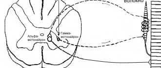
Gamma motor neurons of the spinal cord and ascending pathways of proprioceptive sensitivity (joint-muscle sense analyzer) also take part in the operation of the system..
The pathways of the extrapyramidal system also include the efferent, motor descending pathways from the cerebellum, which is of great importance in maintaining unconscious postural balance, and the reticulospinal pathway, which regulates the motor activity of the spinal cord.
The activity and activity of the extrapyramidal system may exceed the faster pyramidal system in terms of the number of neural sequences. Thus, there is a six-neuron pathway, the first neuron of which is located in the premotor zone of the cortex, the second in the pons, the third in the cerebellar cortex, the fourth in the dentate or cortical nuclei, the fifth in the red nuclei, the sixth in the anterior horns of the spinal cord.
Thus, part of the conscious, cortical impulses, after processing in the cerebellum and “correction of motor coordination,” enters the unconscious rubrospinal tract, which is called the “Monaco bundle.”
Of course, we can make a small number of targeted movements, while the regulation of muscle tone and posture involves all muscles during wakefulness.
It is important to remember that the deep integration of the extrapyramidal and limbic systems leads to the fact that emotional disorders change a person’s involuntary gestures and facial expressions, and vice versa - lesions of the extrapyramidal system cause violent crying or laughter, and lead to speech disorders.
It was said above that unconscious blinking and flinching at the sound of a shot indicates that the primary analyzers of vision and hearing - the lateral and medial geniculate bodies, receiving an excessive stimulus that indicates danger, first switch it to extrapyramidal fibers, and only then, after closing the eyes , comes the realization of what happened.
In this case, the person may turn pale, have palpitations and other autonomic reactions to the stimulus.
This suggests that the pathways of hearing and vision are associated not only with the system of unconscious movements, but also with the thalamus and centers of autonomic regulation.
Despite all the discoveries, such integral, holistic brain activity still poses a great mystery to researchers.
Symptoms of defeat. Levels
Clinical manifestations of pyramidal tract disorders depend on the specific part of the nerve fibers damaged. There are several levels of damage to motor activity: from complete paralysis to relatively favorable impairments.
So, neurology identifies the following levels of damage to the pyramidal tract:
- Central monoparesis (paralysis). The disorders are localized in the cerebral cortex (left or right).
- Central hemiparesis. The internal capsule is damaged.
- Various alternating syndromes - the brain stem area is affected.
- Paralysis of limbs. One of the lateral cords in the spinal cord.
Central paralysis with damage to the brain capsule and cerebral hemispheres is characterized by the fact that muscle function is impaired on the opposite side of the body relative to the affected area. After all, the intersection of the pyramidal tract works in the nervous system. That is, the fibers move to the lateral or lateral spinal tract. The simplified diagram shows how the pyramidal path, the anatomy of which was discussed above, crosses and moves on.
If the lateral cord in the spinal cord is damaged, the work of the muscles on the same side as the injury is disrupted.
Pyramid system - what is it?
It is a collection of nerve cells (motoneurons) grouped into a complex of pathways that stretch from the cerebral cortex (the center of higher nervous activity) through the anterior horns of the spinal cord to the receptors located in the muscles. Thanks to this transmission of nerve impulses, movement occurs.
The peculiarity is that the transmission of these nerve impulses is voluntary, that is, controlled by consciousness.
Pyramidal insufficiency in newborns. Causes
Symptoms of motor impairment in a child include strange jerking movements, or he may walk differently than other children - on tiptoe; or the placement of the feet is incorrect. The reasons for this condition in a child may be:
- underdevelopment of the brain (spinal or brain);
- birth trauma, if the parietal lobe of the brain or the brain stem itself is damaged, there will definitely be disturbances in the pyramidal tract;
- hereditary diseases of the nervous system.
- hypoxia;
- cerebral hemorrhage after childbirth;
- infection such as meningitis or arachnoiditis.
Treatment for adults is often medicinal. But for children it is much better to use methods such as exercise therapy, massage and taking vitamins. If there are no brain abscesses or other serious injuries, the condition improves by the first year of life.
Pallidostrial system in evolution
The pale body is considered more ancient than the striped one. The system itself, at that stage of evolution when the cerebral cortex of living beings was not fully developed, completely controlled the animal’s behavior and was its motor center.
The striopallidal locomotor system made it possible to perform massive diffuse movements of the body - swimming, locomotion, etc. After the “reign” of the cerebral cortex, the striopallidal system came under its control and began to provide preparation for performing a particular movement. At the present stage, it is responsible for the redistribution of muscle tone - coordinated contraction and relaxation of muscle groups.
It is the striopallidal system that helps save muscle energy during movement, and also allows you to bring some actions to “automatic” - driving a car, swinging a mower’s hand, running a musician’s fingers, etc. People inherited it from birds and reptiles. In young children, at certain stages of development, you can very clearly notice its work:
- Pallidum (premature infants, newborns): crawling, axial movements of the body.
- Striatum (second half of life): excessive fussy movements, hand support reaction.
Paresthesia and myoclonus
Disturbance in the cervical spine leads to paresthesia. This is a neuropathy that is characterized by impaired sensitivity. A person may either lose touch sensation in the skin altogether or experience tingling sensations throughout the body. Paresthesia is treated with reflexology, manual therapy or physiotherapy. And, of course, you need to remove the main cause of neuropathy.
Another lesion of the pyramidal tracts and, consequently, motor activity is myoclonus - involuntary twitching.
There are several types of myoclonus:
- rhythmic myoclonic contractions of a separate muscle group;
- velopalatine contractions - sudden non-rhythmic contractions of the tongue or pharynx;
- postural myoclonus;
- cortical;
- myoclonus in response to physical activity (in athletes).
Myoclonus or cortical myoclonus is a disease of the nerve pathway caused by a disorder in the motor centers of the brain. That is, at the very beginning of the pyramid path. If there is a “failure” in the cortex, the signals reach the muscles already distorted.
However, the causes of disturbances in the motor pyramidal pathway can be a lack of magnesium, psycho-emotional or physical fatigue, and many other reasons. Therefore, the diagnosis must be made by a doctor after checking with an MRI.
The main reasons for the development of central paralysis
First of all, it should be noted that it is necessary to distinguish between phenomena such as paralysis and paresis. Paresis is a condition in which muscle strength and reflexes are significantly weakened, but are preserved to a certain extent. With paralysis, a person loses the ability to control certain parts of the body. These 2 conditions have something in common, that is, etiology. The reasons for their appearance are rooted in various types of damage to the human nervous system. As a rule, the development of central paralysis is associated with damage to the motor centers or the central pathways of their departments. Such damage to the human nervous system can be caused by:
- injuries;
- infectious lesion;
- metabolic disorders of various etiologies;
- hereditary predisposition;
- congenital malformations;
- intoxication;
- malignant neoplasms;
- unbalanced diet.
Central paralysis occurs predominantly in people over 45 years of age, but currently there is a trend towards “rejuvenation” of this pathological condition. Statistically, more than 60% of cases of central paralysis are the result of a previous stroke.
Instability of the circulatory system of the brain can provoke damage to neurons not only due to hemorrhages, but also when a blood clot forms and blocks blood flow to a certain part of the motor center or pathways. Children, as a rule, have hereditary spastic paraplegia, which manifests itself at an early age.
Diagnosis of disorders
The descending pyramidal tract is a projection one, while the ascending tract is considered to be the one that transmits body signals through the spinal cord to the central nervous system. Descending, on the contrary, transmits brain signals to neurons.
To determine which system has been damaged and to what extent, during the examination, the neurologist examines many parameters relating to muscles, joints, and nerve reflexes.
A neurologist performs the following diagnostic procedures:
- examines the range of motion of all joints;
- checks deep reflexes, looks for pathological reflexes;
- checks the functioning of all facial nerves;
- measures electrical conductivity of muscles, their biopotentials;
- examines muscle strength;
- and is also required to check whether pathological clonic contractions are present.
When a neurologist checks range of motion, he begins by examining the larger joints first, and then examines the smaller ones. That is, first examines the shoulder joint, then the elbow and wrist.
Symptoms of central paralysis
There are many characteristic signs that distinguish central paralysis from other disorders of the motor function of individual muscle groups associated with damage to the brain or spinal cord. The most characteristic features of central paralysis include:
- muscle hypertension;
- expansion of the distribution area of reflexes;
- hyperreflexia;
- clonus of the kneecaps or feet.
Muscular hypertension is a phenomenon characterized by pathological muscle tension. With this pathology, the muscles have a dense consistency upon palpation. In addition, a distinctive feature of this condition is increased muscle resistance both during passive movement and under directed influence. In the presence of severe muscle hypertension, the appearance of contractures is observed, which leads to a significant or complete limitation of possible passive and active movements. When contracture occurs, the limb usually freezes in an unnatural position.
Hyperreflexia, accompanied by an increase in the area of influence of reflexes, can provoke many visible manifestations of central paralysis. Clonus of the kneecaps and feet are characterized by the appearance of rhythmic contractile movements of individual muscles as a reaction to tendon stretching. As a rule, clonus is a consequence of a significant increase in tendon reflexes. Typically, foot clonus is a consequence of rapid dorsiflexion. In response to such an impact, a reflex rhythmic twitching of the foot is observed. Patella clonus usually develops as a result of a sharp abduction of the leg downwards.
The appearance of pathological reflexes is an indicative sign of damage to any level of the pyramidal tract. Pathological reflexes of the hand and foot can be distinguished. The most indicative pathological reflex movements include the reflexes of Zhukovsky, Babinsky, Gordon, Rossolimo, Oppenheim and Schaeffer.
Among other things, protective reflexes are indicative, which provoke twitching of a paralyzed limb in response to mechanical or thermal influence.
Another characteristic sign of the development of central paralysis is synkinesis. Synkinesis is involuntary cooperative movements in the damaged limb against the background of active directed actions. An example of synkinesis is the swinging of arms while walking, bending or straightening of arms or legs when performing active movements on the half of the body that is not subject to central paralysis. There are many types of synkinesis that may indicate spastic paralysis.
Muscle spasticity, which is a consequence of increased reflex tone, is often unevenly distributed. In most cases, with central paralysis, half the body is affected at once, and the arm in this case, as a rule, is close to the body, the hand and all fingers are bent, while the leg is fully extended at both the hip and knee joints, and the foot is bent and turned inward. This position of the limbs is a very common manifestation of central hemiplegia. The characteristic posture resulting from this arrangement of the limbs in central palsy is known as the Wernicke-Mann manifestation. The gait of people with a similar manifestation of central paralysis is very peculiar, since in order not to cling to the floor with the toe of the affected leg, the patient is forced to move it far away. Central facial paralysis is usually accompanied by numbness of the tongue and palate, facial tics, involuntary eye movements, etc.
Despite a significant increase in tendon reflexes, with central paralysis there is a significant decrease or complete loss of abdominal, cremasteric and plantar reflexes. Among other things, central paralysis is characterized by the absence of pronounced muscle atrophy. The most noticeable manifestations of central paralysis include:
- unnatural posture of the patient;
- low or increased mobility;
- paresis of facial muscles;
- phonation disturbances during speech;
- speech disorders;
- convulsive muscle twitching;
- muscle tremors;
- incorrect gait;
- upward wrinkling of muscles;
- involuntary opening of the mouth;
- closing of eyelids;
- involuntary shrugging of shoulders;
- involuntary flexion and extension of the hands, elbows and other joints;
- increased muscle tone when palpated.
All the symptoms accompanying spastic paralysis make it possible not only to distinguish it from the peripheral form of motor impairment, but also to identify the main area of damage to the pyramidal tract.
Damage to the corticonuclear tract
The pyramidal path is the basis of all movements not only of the muscles of the body, but also of the face. The axons of various facial motor neurons transmit signals to the muscles. Let's take a closer look. The motor neurons of the nucleus ambiguus innervate the muscles of the pharynx, larynx, soft palate, and even the muscles of the upper esophagus. Motor neurons of the trigeminal nerve are responsible for the work of some muscles of mastication and those that give the signal to the eardrum to contract. Individual motor neurons contract the facial muscles when we smile or frown. These are facial neurons. Another group of muscles is responsible for eye and eyelid movements.
The defeat of the leading neuron affects the work of the “subordinate” muscles. The entire pyramidal path is based on this principle. Neurology of the facial nerve leads to very unpleasant consequences. However, eye movements and swallowing are usually preserved.
It is worth noting that complete disconnection of the facial muscles from the controlling segment of the brain occurs only if both the right and left hemispheres are affected. Most facial neurons are controlled bilaterally, as are the muscles of the trunk. One-way crossed fibers go only to the lower part of the face, namely to the muscles of the tongue and lower jaw.
Pyramidal pathways (system) - description and functions
Neurology is the most precise science in medicine. This is what topical diagnostics does.
Topical diagnostics allows a neurologist, with the help of a hammer, questioning and examination, as well as when performing samples and tests, in some cases to localize the lesion located in the head or spinal cord with an accuracy of a millimeter. But earlier this science was applied, and even earlier it was descriptive, like all anatomy.
As a result, such names as “cerebellar peduncles”, “putamen”, “fence”, “tubercles of the quadrigeminal tuberosity”, “aqueduct” passing deep into the brain, and many other formations appeared.
Their functions were completely unknown for a long time. Scientists understood that the brain and spinal cord consisted of gray and white matter, but this was perhaps the only difference.
The internal structure was not amenable to analysis.
Dyes have not yet been invented that would make it possible to accurately show neurons and prove that the central nervous system consists of cells with the longest processes (sometimes up to a meter in length). The science of neuroanatomy was not invented.
And only after the advent of Virchow’s cellular theory, which placed the function of an organ in direct dependence on its cellular composition, after the advent of physiology, which studied the functions of neurons and how they differ, did an understanding of the integral work of a nerve cell arise.
The subsequent steps were taken by Sechenov and Pavlov.
An excellent illustration of this “internal formation” is the pyramid system. It is the main effector: with its help all motor conscious acts are performed.
If the pyramidal system were absent, then we would be immobile animals, like sponges or bryozoans, and all civilization would be impossible.
They usually say that civilization was created by the human brain and his hands, but they forget to say that the pyramidal path is precisely the intermediary that brings brain impulses for movement to the muscles.
What are the functions of a pyramid system? What is the diagram of the pyramid paths? We'll tell you about this .
What it is?
Pyramidal tracts (or system) are the second name for the corticospinal, efferent, or descending tracts. These pathways begin in the precentral gyrus, in its gray matter.
It is there that the bodies of neurons are located, which generate impulses for commands to the striated, or skeletal muscles.
These impulses are conscious, the pyramidal system obeys our will.
The pyramidal and extrapyramidal systems (unconscious) are combined into a single system of movement, coordination of balance and muscle tone.
The pyramidal tracts end in the anterior horns of the spinal cord, at its various levels - from the neck to the sacrum.
It is there that the switch occurs to large motor neurons, which directly terminate at the neuromuscular junction, in which the neurotransmitter acetylcholine gives the signal for muscle contraction.
This definition is a little vague, but it explains the essence. Let us consider in detail the anatomy and organization of the corticospinal tract and its structures at different levels.
Bark
Pyramidal tracts begin in the precentral gyrus, or more precisely, in its special field, which is projected along it in a narrow strip, from bottom to top. This band is called cytoarchitectonic field No. 4 according to Brodmann (yes, the cerebral cortex, like the earth’s crust, has its own architecture and architectonics).
In this field there are giant (they are indeed very large) pyramidal Betz cells (in memory of the Russian histologist and anatomist Vladimir Betz, who discovered these cells in 1874). Their task is to generate impulses for precise and targeted movements.
In the lower sections there are neurons responsible for the movement of the pharynx and phonation, then, higher, there are cells that innervate the facial muscles, arms, then the torso and legs.
If you type the name “motor homunculus” into a search engine, you will see a picture of how the relative “powers” of this cortical zone are distributed among themselves.
A huge part of the nerve cells are responsible for fine movements of the hands and fingers, as well as for facial and vocal muscles.
But the innervation of the legs, which produce a small number of stereotypical movements, requires a small number of cells.
Cortical impulses generated by large Betz cells (for example, you are planning to move your finger) must reach the muscle as quickly as possible. After all, this is not the autonomic nervous system, which “slowly manages” inside the body. For example, the extraction of food depends on the quality and speed of voluntary movements.
Therefore, the axons of these neurons are well isolated, “of the highest class.” Their fibers have a thick, myelin sheath. This is the “elite” among all pathways; they include only 3-4% of axons from the total volume of the pyramidal system.
The remaining sources of impulses are smaller neurons located in other layers of the cortex.
In addition to the Brodmann area, there are also premotor areas that give off their impulses; they are also classified as the corticospinal tract.
It must be remembered that all the cortical structures we have mentioned perform movements on the opposite side of the body. Left neurons generate movements on the right, and vice versa.
The fact is that almost 90% of the total volume of fibers crosses below and moves to the other half of the body. What happens next?
The first “fork” and the corticonuclear pathway
Everyone knows that in addition to the muscles on the arms, legs and torso, there are also muscles on the face and head. Even before passing through the inner capsule (see below), the bundles are separated.
Therefore, the first, larger, bundle leaves the cortex to innervate the limbs and torso, and the second, smaller one, goes to the motor nuclei of the cranial nerves, which are also parts of voluntary and conscious movement.
The first remains the pyramidal fasciculus, and the second is called the corticonuclear, or corticonuclear tract . These nerves that “receive pyramidal nutrition” include:
- Oculomotor nerve (3 pairs) – movements of the eyes and eyelids;
- Trochlear nerve (4 pairs) – eye movements;
- Trigeminal nerve (5th pair) – chewing muscles;
- Abducens nerve (6th pair) – eye movement;
- Facial nerve (7th pair) – facial expressions;
- Glossopharyngeal nerve (9th pair) – stylopharyngeal muscle, pharyngeal constrictors;
- Vagus nerve (10th pair) – muscles of the pharynx, larynx;
- Accessory nerve (11th pair) – trapezius and sternocleidomastoid muscles;
- Hypoglossal nerve (12th pair) – muscles of the tongue.
Practically, all nerves, except for purely sensory nerves - 1 pair (olfactory) and 2 pairs (visual), receive innervation through that part of the pyramidal tract, which is called the corticonuclear, or corticonuclear. Then both already separated bundles pass past the inner capsule, where the conductors lie especially densely. This is the place of the highest con of the brain.
The internal capsule appears as a small band in the white matter that lies between the basal ganglia. In it, on a horizontal section, two “hips” are visible, tilted in different directions, and a “knee” connecting them. The corticonuclear tract is located in the “knee” region, and the corticospinal, pyramidal fascicle, which splits off above, occupies the anterior 2/3 of the “posterior thigh”.
After switching to the nuclei of the cranial nerves, the impulse goes further and, along individual nerves, approaches the corresponding muscles . Some of the beams also intersect and carry out movements on the opposite side, but some do not. In other words, part goes contralaterally, and part goes ipsilaterally.
The pyramid path itself
What about the main bundle, which should carry out movements to the arms and legs? It is still traveling in the depths of the brain, and as it moves towards the exit through the foramen magnum, it becomes denser and thicker.
After the axons leave the internal capsule, on the right and left these bundles pass through the middle of the cerebral peduncles and descend into the pons.
There, pyramidal tracts form each of the halves of the bridge, and go down, surrounded by a “retinue” of masses of bridge nuclei, fibers of the reticular formation and other formations.
Finally, leaving the pons and entering the medulla oblongata, the pyramidal tracts begin to become visible to the eye. Elongated and seemingly inverted pyramids appear, which are located symmetrically from the center. That is why the corresponding paths conducting movements are named this way.
Transition to the spinal cord
Having reached the bottom of the medulla oblongata, some of the axons cross over to the opposite side, as discussed above.
This crossed part forms the so-called lateral fasciculus, and the remaining uncrossed part is called the anterior corticospinal tract.
Surprisingly, the axons of the neurons of this bundle still move to the mirror side, but in the segment of the spinal cord that they should innervate. The transition occurs in the area of the white commissure anteriorly, which connects the right and left halves of the spinal cord.
The crossed, massive parts descend down, of course, not on their own, but as part of the same-named (tractus corticospinalis lateralis) path in the spinal cord.
As fibers are given off at the level of each segment, this bundle becomes thinner, like icicles “frozen” into the spinal cord on both sides.
Finally, in the region of the sacral region this bundle becomes very thin and ends.
Almost 90% of all fibers switch not directly to motor neurons in the greater horn of the spinal cord, but to interneurons . These neurons directly form synapses with large motor neurons, and differ from them in function.
We can say that the mass of interneurons, which are closed within one horizontal (segmental) level, are in contact with sensory and motor nerve cells, and have some autonomy. As a result, at the level of each segment there are polysynaptic “relay substations”.
This is what distinguishes the pyramidal and extrapyramidal pathways for regulating movements.
https://www.youtube.com/watch?v=YVIcFGF5AWw
The extrapyramidal system, operating in a completely autonomous mode, does not require such a large number of two-way connections, since it is not accessible to voluntary control.
Instead of a conclusion
There are a few very important words left to say.
What happens if there is an obstacle in the way of the pyramidal beam? If axonal interruption occurs due to injury, will it cause their death or dysfunction (tumor, hemorrhage)? The result is paralysis of the muscle, which is left without a command to move.
Incomplete paralysis, in which some of the axons still “get” to the muscle, is called paresis, and is manifested by weakness and muscle wasting.
The most interesting thing is that when the central neuron dies or the path is interrupted, the second neuron remains unharmed - which lies in the anterior horns of the spinal cord and directly approaches the muscle. He just loses his “boss from above.” This type of paralysis is called central. And what happens as a result of central paralysis, why it occurs and how it manifests itself will definitely be discussed in the following articles
Pogrebnoy Stanislav Leonidovich, neurologist
Rate this article:
Total: 160
4.36 160
Source: https://mozgius.ru/stroenie/piramidnaya-sistema.html
Symptoms of damage to the pyramidal tract at the level of the brain stem
Since fiber crossover occurs at the level of the brain stem (the medulla oblongata and the pons), when these structures are damaged, gamiplasia occurs on the other half of the body. This symptom is called alternating paralysis.
The pyramidal tract is the basis of fine motor skills. If the brain stem is even slightly damaged, fine movements of the fingers are greatly affected.
There are many different syndromes that clearly and in detail characterize disorders that affect the work performed by the pyramidal tract: Avellis, Schmidt, Wallenberg-Zakharchenko syndromes and others. Based on the symptoms of these syndromes, the doctor can often determine the exact location of the pathway disorder before testing.
Pyramidal neurons: functions and location in the brain
Neurons are the main type of cells that make up every element of the nervous system. These structures are well known to most people today.
But although in the collective imagination we have a typical idea or idea of what a neuron is, and many people imagine that all or almost all have the same structure and shape, the truth is that not all neurons are the same: there are very different types in depending on their shape, where they send information from, or even their relationship with other neurons. In this article we will focus on pyramidal neurons and their functions .
- Article on the topic: “Types of neurons: characteristics and functions
Pyramidal neurons
Pyramidal neurons are one of the different types of neurons present in our nervous system . They are one of the most common types of multipolar neurons, accounting for about 80% of the neurons in the cortex (not for nothing, the two layers of the cortex are called the inner and outer pyramids) and are found between some of the the most relevant from the body. They are generally considered to be projection neurons. That is, they operate by sending messages to cells that are far away and separated from the place where they were born.
Discovered by Santiago Ramon y Cajal The name of this type of neuron refers to the shape of its soma, with a triangular or pyramidal appearance.
These are mainly glutamatergic neurons, with glutamate being the neurotransmitter that activates them, and they typically act as excitatory type neurons.
They can have different sizes, the largest of which are giant pyramidal cells or Betz cells.
Like other neurons, the structure of this type of neuron consists of a soma, which, as we have already said, has a pyramidal shape, an axon and dendrites. However, they have their own peculiarity: as for the dendrites, they are quite long compared to the rest, called the apical dendrite and numerous basal and shorter dendrites that branch.
- You may be interested in: “Parts of the Human Brain (and Functions)”
The location of these nerve cells
Pyramidal neurons can be found at various points in the nervous system, but they are much more common in some specific areas. Among them the following stand out.
Cortex
Pyramidal neurons are found primarily in the cerebral cortex, forming part of most of it, and are found in five of the six layers that make up this area of the brain. In particular, they can be observed in the granular and pyramidal layers, both external and internal.
They are especially prominent in the third and fifth layers (which are actually called the outer pyramidal and inner pyramidal), and they are larger the deeper into the cortex. There are also areas within the cortex where its existence has been discovered more frequently.
Motor cortex
In the motor cortex we can find a large number of pyramidal neurons, especially those associated with motor control. In this area of the cortex there are many giant pyramidal neurons known as Betz cells that transmit motor information from the brain to areas of the spinal cord, where they synapse with motor neurons that activate movement.
Prefrontal cortex
Pyramidal neurons can also be found in the prefrontal cortex, influencing higher mental processes. It is believed that these cells are the main primary excitations of prefrontal neurons, participating in numerous functions and considering themselves primordial for the existence of behavioral control.
Corticospinal tract
Pyramidal neurons are especially visible along the corticospinal tract, which sends motor information from different brain nuclei responsible for motor control to motor neurons that will cause muscle contraction as they pass through the spinal cord.
4. Hippocampus
Not only in the cortex we can find pyramidal neurons, but we can also find in subcortical structures . One of them is the hippocampus, associated with aspects such as memory and orientation.
- Article on the topic: “Hippocampus: functions and structure of the memory organ”
5. Amygdala
Another structure in which these neurons are found is in the amygdala, an area of the limbic system associated with emotional memory.
Functions of pyramidal neurons
Like other neurons, pyramidal neurons are structures that transmit information in the form of electrochemical impulses that will be captured by other neurons until they reach their final destination. Being a type of neuron widely distributed in the cerebral cortex, pyramidal neurons are activated and connected to a large part functions and processes performed by humans. Examples of such functions are as follows.
1. Movement
Motor control is one of the functions that is traditionally associated with pyramidal neurons. In particular, these neurons are closely associated with voluntary motor control of muscles.
Cognition and executive functions
The excitatory role of pyramidal neurons in the prefrontal cortex means that their activation may be associated with cognitive processes of high importance, such as executive functions or cognition .
3. Emotions
The activity of pyramidal neurons in the prefrontal cortex is associated with the connection of these areas with various subcortical areas, among them the limbic . In this sense, the amygdala and hippocampus play a fundamental role.
Memory and orientation
Memory and specific orientation are other functions in which there is greater activation in pyramidal neurons, in this case in the hippocampus.
Bibliographical references
- Kandel E.R.; Schwartz, JH & Jessell, T.M. (2001). Principles of neurobiology. Fourth edition. McGraw-Hill Interamericana. Madrid.
- McDonald, A.J. (1992). Cell types and internal connections of the amygdala. Prog. Neurobiol. 55: 257-332.