The functions of cells in the nervous system are very diverse. One type is a motor neuron (motoneuron). Its name translated from Latin means “sets in motion.” It is through this that muscle contraction occurs.
The peculiarity of motor nerve cells is that their cytoplasm does not evenly surround the nucleus, but forms two processes. One of them is shorter (dendrite) receives a nerve impulse, the second (axon) transmits it further.
Thus, the peripheral motor neuron conducts nerve impulses from the central nervous system to the muscle. In muscle tissue, its long process branches and connects with dozens of muscle fibers.
Types of motor neurons
Based on localization, motor neurons are divided into central and peripheral.
The central ones are located in the brain tissue. They are responsible for conscious, controlled muscle contractions. Motor neurons that go directly to muscle fibers are called somatic neurons.
The cell bodies of motor neurons of the somatic nervous system are located in the region of the anterior horns of the spinal cord and are arranged in groups, each of which is responsible for the contraction of strictly defined muscles. For example, motor neurons in the cervical region control the muscles of the arms, while those in the lumbar region are responsible for the innervation of the legs.
Peripheral nerve cells responsible for movement are classified as follows:
- large alpha motor neurons;
- small alpha motor neurons;
- gamma motor neurons;
- Renshaw cells.
Large alpha cells form large conducting trunks. Small alpha and gamma neurons have thinner axons. Renshaw cells are part of large trunks and serve for switching signals.
Gamma motor neuron loop
Motor neuron functions
Central and peripheral motor nerve cells work in concert. Together they provide contraction of certain muscle groups and allow a person to perform any action.
For coordinated movements of the limbs, simultaneous contraction of the flexors and extensors is necessary. When the flexors work, the initial excitation signal arises in the area of the precentral gyrus of the corresponding hemisphere.
Cells called pyramidal cells are responsible for this action. Their processes collected together form the so-called pyramidal motor pathway. Next, the signal goes to the anterior horns of the spinal cord, from where it is transmitted directly to the myofibrils.
Special centers of the posterior sections of the cerebral hemispheres have an activating effect on the motor neurons of the extensor muscles. They form the dorsal and ventral pathways. Thus, two areas of the brain are involved in the formation of coordinated movement.
Based on the nature of their function, the nerve cells involved in the process of muscle contraction are divided into motor neurons and interneurons. The former are responsible for the executive function, while the intercalary ones serve to coordinate nerve impulses. This particular variety is smaller in size and more numerous.
For comparison, in the area of the anterior horns there are 30 times more of them than motor horns. When excitation is carried out along the axon of the motor nerve, it initially passes to the interneuron. Depending on the nature of the signal, it can be amplified or weakened, after which it is transmitted further.
Intercalated cells have more processes and are more sensitive. They have a large number of processes and are also called multipolar.
To optimize the signals emanating from the axons and going to the muscle fibers, special Renshaw cells are used, which transmit excitation from one process to another. This mechanism serves to equalize the intensity of the nerve signal.
Along the process of the motor neuron, the impulse reaches the muscle fiber, which contracts. Each group of motor neurons and the muscle fibers they innervate are responsible for specific movements.
Nerve cells providing motor function:
| Types of neurons | Localization | Function |
| central innervating flexors | precentral gyrus area | contraction of skeletal flexor muscles by transmitting impulse to the anterior horns |
| central innervating extensors | hindbrain region | contraction of skeletal extensor muscles by transmitting impulses to the anterior horns |
| peripheral alpha | anterior horn of the spinal cord | direct contraction of skeletal muscles |
| peripheral gamma | anterior horn of the spinal cord | regulation of tone |
| insertion | all parts of the central nervous system | communication of signals within the central nervous system |
Large alpha neurons conducting a strong impulse cause myofibril contraction. Small ones conduct weak signals and serve to maintain muscle tone.
In addition to the fibers responsible for contraction, muscle tissue also contains special spiral fibrils that regulate the force of muscle tension.
These extrafusal muscle fibers are innervated by gamma neurons.
Excitation of the gamma motor neuron leads to an increase in the stretch of myofibrils and facilitates the passage of the tendon reflex impulse. An example would be the passage of a nerve signal along the arc of the knee reflex.
The coordinated work of peripheral motor neurons achieves fine tuning of muscle tone, which allows for precise coordinated movements. When peripheral motor neurons are damaged, muscle tone disappears and movement is impossible.
Peripheral nervous system
The main components of the peripheral nervous system are nerves, which connect the central nervous system to other parts of the body, and ganglia, groups of nerve cells located at different points in the nervous system.
A nerve is a bundle of motor and sensory fibers along with connective tissue and blood vessels. The large nerves (43 of them) actually come from the central nervous system: 12 pairs come from the lower part of the brain (cranial nerves) and 31 pairs from the spinal cord (spinal nerves).
The cranial nerves serve primarily the sensory organs and muscles of the head, although a very important cranial nerve, the vagus nerve, serves the digestive organs, the heart, and the air passages in the lungs. Some cranial nerves, such as the optic nerve to the eye, contain only sensory fibers. Spinal nerves arise from the spinal cord at regular intervals and always contain both motor and sensory fibers. They serve all parts of the body below the neck. Each spinal nerve is connected to the spinal cord by two roots, one of which contains motor fibers and the other contains sensory fibers. Immediately after leaving the spinal cord, sensory and motor fibers join together to form a nerve, but each group of fibers acts independently of the other, like two wires in an electrical wire. (Although the cranial nerves are also connected to the lower part of the brain by roots, sensory and motor fibers form separate nerves.)
At a short distance from the spinal cord, each spinal nerve branches, and each branch is divided, in turn, into many smaller ones, forming a network covering the entire body.
Both sensory and motor fibers are part of sensory and motor neurons. The sensory and motor fibers of the peripheral nervous system are just the longest fibers of the corresponding neurons. For example, a motor fiber from a neuron in the spinal cord can stretch uninterrupted to a muscle in the foot.
Somatic and autonomic systems
The peripheral nervous system has two main divisions: the somatic nervous system, which is under the constant control of a person, and the autonomic system, which is under his unconscious control.
The somatic system performs a dual task. First, it collects information about the world around us from sensory organs such as the eyes, which contain special receptor cells. Signals from these receptors are carried to the central nervous system via sensory fibers. Secondly, the somatic system transmits signals along motor fibers from the central nervous system to the skeletal muscles, thereby causing movement.
The autonomic system is primarily responsible for maintaining the automatic ( occurring without The autonomic system consists solely of motor nerves that act as a relay between the spinal cord and various muscles.
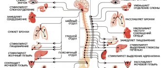
The autonomic nervous system is divided into two parts—sympathetic and parasympathetic. Each of them uses its own mediator, each is designed in its own way, and each has its own special effect on the organ it serves. For example, the parasympathetic nerves that supply the bronchial passages to and from the lungs cause them to constrict (narrow). Sympathetic nerves traveling to the same area of the body cause dilation of the bronchial passages.
The entire autonomic system is controlled by a region of the brain called the hypothalamus. It receives information about any change, for example, a change in the body's chemical balance, and adjusts the autonomic system to return the body to the correct balance. If, for example, oxygen levels drop due to exercise, the hypothalamus instructs the autonomic nervous system to increase the heart rate to supply the body with more oxygen-rich blood.
central nervous system
The peripheral nervous system serves only to transmit sensory and motor signals from the central nervous system to the body's muscles, glands, and sensory organs. It plays virtually no role either in the analysis of sensory impulses or in the excitation of motor signals. Both of these processes and a huge number of other processes occur in the central nervous system.
The brain and spinal cord form the central processing unit of the nervous system. They receive impulses along sensory fibers from the body’s sensory organs and receptors, select and analyze them, and then send commands along motor fibers, causing a corresponding reaction in the muscles and glands.
The analytical, or information-processing, process may be relatively simple for some functions performed by the spinal cord, but analysis in the brain is a highly complex process, requiring the participation of thousands of diverse neurons. Although many sensory neurons terminate and many motor neurons originate in the brain, most neurons in the brain are united; their task is to select, analyze and store information. The entire central nervous system must be supported by an abundant blood supply, as oxygen and nutrients are supplied with the blood. The system is also protected by two types of coating. The first covering is bone: the brain is in the skull, and the spinal cord is in the spine. The second covering consists of three meninges made of fibrous tissue. These membranes cover the entire brain and the entire spinal cord.
Cerebrospinal fluid is a clear, thin fluid that flows around the lining of the brain (brain and spinal cord) and passes through the ventricles of the brain. This fluid can have a shock-absorbing effect and thus help protect vital brain tissue from damage.
The fluid is continuously produced from the blood by specialized cells in the choroid of the ventricles of the brain. In contrast to the cardiac ventricles, which have their own names, the ventricles of the brain have their own numbers. Numbering goes from the top down, with the first and second ventricles (known as the lateral ventricles) being the largest.
Cerebrospinal fluid flows from the lateral ventricles through a narrow opening into the small third ventricle and then through an even narrower canal, the cerebral duct, into the fourth ventricle (slightly wider than the third ventricle). From here, through openings in the upper part of the ventricle, the fluid passes into special storage cavities (cisterns) that surround the brain stem at the base of the brain. The fluid then moves up through the top of the brain (through the hemispheres) and is reabsorbed by special growths called arachnoid granulations located on the arachnoid membrane, one of the three membranes of the brain.
Spinal cord
The spinal cord is a roughly cylindrical column of nervous tissue, about 40 cm long, that extends inside the spine from the brain to the lower back. The brain consists of clusters of neurons and bundles of nerve fibers. Gray matter—the so-called clusters of nerve cells—has the shape of the letter H in cross section, with a posterior and anterior process in each half. The anterior process consists of motor neurons, the posterior one contains ganglia of connective and sensory neurons.
Gray matter is surrounded by white matter. This white matter is divided into three columns and contains ascending and descending nerves that connect the brain and spinal cord in general directions. Descending nerves send motor impulses from the brain to the peripheral nervous system, while ascending nerves carry sensory impulses from the senses to the brain.
Functions of the spinal cord
The spinal cord has two main functions . First, it serves as a two-way conduction system between the brain and the peripheral nervous system. This is achieved by sensory and motor neurons whose fibers extend in long bundles from parts of the brain. They stretch for varying distances along the spinal cord and at the ends furthest from the brain they come into contact with fibers or ganglia of sensory and motor neurons belonging to the peripheral nervous system. Signals are transmitted through synapses between peripheral nerve cells and spinal cord neurons.
The second function of the spinal cord is to control simple reflex activity. It is carried out by neurons, whose fibers stretch short up and down the spinal cord, and by interneurons, which transmit impulses directly between sensory and motor neurons .
If, for example, a person accidentally places his hand on a hot stove, pain receptors in the skin will send impulses along sensory fibers to the spinal cord.
brain. Some of these impulses are immediately transmitted by nerve cells to motor neurons which control the movements of the muscles of the arms and hands the person quickly and automatically withdraws his hand. Another part of the impulses moves up the spinal cord and is transmitted by interneurons to the motor neurons that control the movement of the neck. The head automatically turns towards the source of pain. Another group of impulses reaches the brain and causes a conscious feeling of heat and pain.
Brain
Basically, the brain can be divided into three different sections: hindbrain, midbrain and forebrain. Each of these departments, in turn, is divided into areas that have very specific functions and at the same time are connected by complex relationships with other parts of the brain.
The largest structure of the hindbrain is the cerebellum. This area is related mainly to human motor activity. The cerebellum sends out signals that cause unconscious movements in the muscles that help maintain body position and balance; The cerebellum acts in concert with the motor areas of the brain to coordinate body movements.
The brainstem, which connects the brain to the spinal cord, includes part of the hindbrain, all of the midbrain, and part of the forebrain. It is here, in the brain stem, that all incoming and outgoing impulses meet and cross, for the left side of the body is controlled by the right side of the brain and vice versa.
Various structures in the brainstem, including such as the medulla oblongata, as well as the hindbrain pons and the reticular formation (sometimes called the activating reticular system) of the midbrain, are responsible for life itself. They control heart rate, blood pressure, swallowing, coughing, breathing and unconsciousness.
One of the most important functions of the brain is control over the level of consciousness. It is the reticular formation that sifts through the entire mass of incoming information and decides what exactly is important enough to connect the brain. Nerve pathways from all over the body branch into the reticular formation and feed it with a continuous flow of electrical signals that originate in the nerve cells. In turn, under the influence of these impulses, the reticular formation sends signals to different points throughout the brain, to the corresponding centers, where the signals are collected, compared and cause a response.
If the speed of this driving process slows down or is interfered with, a part of the brain known as the cerebral cortex becomes inactive and the person becomes unconscious.
Brain and hypothalamus
The largest part of the entire brain is the brain itself, located in the forebrain. In humans, it is developed to a greater extent than in any animal, and plays a major role in the processes of thinking, memory, consciousness and higher mental activity. This is where other parts of the brain transmit incoming impulses to differentiate them.
The brain is divided right down the middle into two halves called the cerebral hemispheres. They are connected at the base by a thick bundle (strand) of nerve fibers - the corpus callosum. Although both hemispheres are mirror images of each other, they perform completely different functions and work with each other through the corpus callosum.
At the center of the cerebral hemispheres is a collection of gray matter (nerve cells) called the nucleus basalis. These cells form a complex control system that coordinates muscle activity, which allows the body to perform certain types of movement freely and unconsciously. This kind of muscle activity is manifested in the swinging of the arms while walking, changes in facial expression, and the positioning of the limbs before standing up and walking.
The hypothalamus lies at the base of the brain, under the two hemispheres. It lies directly beneath another important structure in the forebrain, the thalamus, which functions like a telephone switch between the spinal cord and the cerebral hemispheres.
The hypothalamus is a collection of specialized nerve centers connected to other important areas of the brain, as well as the pituitary gland. This area of the brain is responsible for controlling vital body functions such as eating, sleeping and regulating body temperature. It is also closely related to the endocrine (hormonal) system.
The hypothalamus is connected by nerve pathways to the limbic system, which is closely connected to the olfactory centers in the brain. This part of the brain also has connections with areas that control other senses, behavior, and memory organization.
Cortex
The cerebral cortex is a layer of gray matter, three millimeters thick, all in convolutions, lying on top of the outer side of the brain. This part of the brain has reached such a high level of development in humans that it has to fold itself, more and more twisting, to fit inside the skull. If you straighten this layer, it will occupy an area 30 times larger than it occupies when folded.
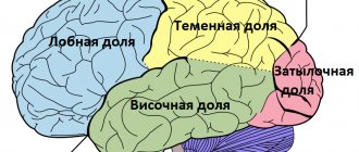
Among all these folds are certain very deep grooves that divide each hemisphere of the cortex into four areas called lobes. Each lobe performs one or more specific functions. The temporal lobes are associated with hearing and smell, the parietal lobes with touch and taste, the occipital lobes with vision, and the frontal lobes with human movement, speech, and complex thinking.
Within lobes there are special segments that receive sensitive impulses from any one part of the body. For example, the sense of touch in the parietal lobe is represented by a tiny zone that receives only sensations from the knee, and a large zone for the thumb. This is why areas of the body like the thumb are much more sensitive than areas like the knee. The same principle applies to other sensory as well as motor parts of the body. It is in the cerebral cortex that information received from the five senses—visual, auditory, tactile, gustatory, and olfactory—is analyzed and processed so that other parts of the nervous system can use it when needed. In addition, the premotor and motor cortex interact with other areas of the central nervous system and peripheral nervous system to provide the coordination of movements vital to all conscious activities of the human body.
Eyes
When people want to explain how they see, the eye is usually compared to a beautifully designed camera, but to fully understand how the outside world is reflected in the tiny camera of the eye, we need to go back to the basic principles of this process.
To understand the nature of light, it is best to consider it as a transmitting medium. Coming from any source, light reflects off objects in all directions, taking with it the ability for objects to be visible.
Another important factor regarding the characteristics of light is the ability of normally straight rays of light to be refracted when passing through a specific medium, such as a specially shaped glass lens in a camera or a tissue lens in the human eye.
Moreover, the degree of refraction can be adjusted using the shape of the lens. Beams of light can be concentrated to produce tiny but precise images of large objects.
Cornea
When a ray of light hits the eye, it first encounters this round, transparent window called the cornea: The cornea is the first of the two lenses of the eye. This is a strong fixed focus lens. The optical power of the cornea accounts for up to two-thirds of the total optical power of the eye. The cornea is only half a millimeter thick in the center and one millimeter thick where it meets the white of the eye, called the sclera.
The cornea consists of five layers. On the outside is a layer five cells thick called the epithelium, which corresponds to the skin of the body. Beneath this is an elastic, fiber-like layer known as Bowman's layer. Then comes the main layer (stroma), consisting of collagen. This is the densest part of the cornea. The stroma helps protect the cornea from infection due to the various anti-infective antigens it contains: the stroma is believed to control possible inflammation in the cornea.
Behind the stroma layer is another layer, one cell thick, called the endothelium. This thin layer ensures the transparency of the cornea and maintains the balance of water exchange between the eye and the cornea. Once formed, the cells in this layer cannot renew themselves, and therefore injury or disease to the endothelium can cause permanent visual impairment. The last layer, called Descemet's membrane, is elastic.
The tear film covers the epithelium . Without tears, the cornea would have no protection against bacterial microorganisms, pollution and dust . The tear film also creates an optical layer—without tears, the epithelium would lose its transparency and become cloudy.
After passing through the cornea, the light beam enters the first of two chambers inside the eye, the anterior chamber. It is filled with watery intraocular fluid, which is constantly exchanged.
Choroid of the eyeball
The choroid of the eyeball is an area that consists of three clearly distinguishable structures located in the center of the eyeball: the choroid itself, the ciliary body and the iris. These structures together are sometimes called the uveal tract .
The choroid itself is a thin covering of membranes between the outer protective sclera and the retina. This membrane is rich in blood vessels that nourish the retina and create a complex lattice structure throughout the eye. This lattice contains supporting tissue containing varying amounts of pigment, which prevents light from darting across the back of the eye, creating confused images.
The ciliary body consists of pointed sections of the uveal tract at the very front of the eye. Its role is to change the shape of the lens by moving the ciliary muscle, allowing a person to focus on nearby objects, and also to produce intraocular fluid that circulates in the chamber between the lens and the inner surface of the cornea.
The third specialized zone approaches the ciliary body - the iris, which forms the posterior part in the anterior chamber. This is the part of the eye whose pigment gives the eye its color. It acts like the aperture hole in a camera; its muscle fibers dilate or constrict the pupil, controlling the intensity of light entering the retina.
If strong light enters the pupil, the pupil becomes smaller without the person's conscious effort. In twilight light the pupil enlarges. Excitement, fear, and the use of certain medications also cause the pupil of the eye to dilate or constrict.
Just behind the iris is a soft, elastic, clear lens. It is relatively small, since the cornea does most of the work for it.
Vitreous body and retina
Behind the lens is the main - internal - chamber of the eye. It is filled with a substance called vitreous humor, which has a jelly-like structure; this substance makes the eye hard and elastic. A vitreous canal passes through the center of the chamber - the remains of the canal that carried the artery during the period of intrauterine development. The curved interior of the eyeball is lined throughout the inner chamber with a light-sensitive layer called the retina. It consists of two different types of light-sensitive cells, called rods and cones after their shape.
Rods are sensitive to low-intensity light and do not distinguish colors, which cones do for them. Cones are also responsible for transparency; they are especially numerous at the back of the eye, in an area known as the fovea, or macula. This is where the lens focuses the clearest image, and it is there that a person sees best.
| Supporting ligaments |
Focusing
The retina surrounding the fossa, or spot, produces clear images, but closer to its edges peripheral vision appears, when a person sees “halfway.”
Together, central vision and peripheral vision create a holistic picture of the world around us.
Optic nerve
Each light-sensitive cell in the retina is connected by a nerve to the brain, where all information about images, color and shape is collected and processed. All of these nerve fibers come together at the back of the eye and form one main “cable” known as the optic nerve. It exits the eyeball through a bony tunnel in the skull and re-emerges just below the brain in the area of the pituitary gland to join the second optic nerve.
Right: The right and left eyes have slightly different fields of vision. Each field of view is divided into right and left sides. When light rays reach the retinas, they change places and turn. These rays travel along the optic nerves to the optic chiasm, where decussation occurs. All information from the left side of each eye travels along the optic nerve through the lateral geniculate body and the area of optic radiation to the right visual cortex and vice versa. The images are then combined and interpreted by the brain.
The nerves on both sides then cross so that some of the information from the left eye goes to the right side of the brain and vice versa. The nerves of the temporal side of each retina do not cross and remain on the same half of the brain, while the fibers from the part of the eye that does the main work of vision go to different sides of the brain.
The optic nerve is nothing more than a bundle of nerve fibers carrying tiny electrical impulses along tiny cables, each of which is isolated from the next by a layer of myelin. In the center of the main cable there is a large artery running along its entire length. It is called the central retinal artery. This artery arises at the back of the eye, and its capillaries cover the entire surface of the retina. There is a corresponding vein that runs in the opposite direction along the optic nerve next to the central retinal artery and carries blood away from the retina.
The nerves coming from the retina are sensory nerves; Unlike motor nerves, which have only one connection on their route to the brain, optic nerves connect several times. The first encounter occurs just behind the point where sensory information from the different eyes switches places. This point is called the optic chiasm and is located close to the pituitary gland. Directly beyond this junction is the first junction, called the lateral geniculate body. Here the information from the left eye and the right eye is swapped once again. The function of this connection is related to the reflexes of the pupils.
From the lateral geniculate body, the nerves fan out on both sides around the temporal part of the brain, forming the optic radiation. They then turn slightly and come together to pass through the main "switchboard" - the internal capsule where all the motor and sensory information supplying the body is concentrated. From here, the nerves travel to the back of the brain to the visual cortex.
Ears
The ear not only provides us with a sense of hearing, but also a sense of balance. The ear is a complex organ; it is divided into three parts: the outer ear, which picks up sound like radar; the middle ear, in which a set of bones, like a mechanism, amplifies the resulting sound; and the inner ear, which converts sound vibrations into electrical impulses and determines the position of the head.
Impulses are transmitted to the brain by a pair of nerves that are located next to each other: the vestibular nerve for balance and the cochlear nerve for sound. The outer and middle ears are primarily responsible for hearing; The structures of the inner ear that interpret head position and sounds are different structures, although they are located in the same organ.
Hearing
What a person hears are sound waves produced by the vibration of air molecules. The length and strength of these waves determines the loudness of the sound; Loudness is measured in decibels (dB). The number of vibrations, or cycles, per second makes up the frequency: the more vibrations, the higher the sound. The frequency of sound is expressed in the number of cycles per second, or in hertz (Hz).
In young people, the amplitude of the audible frequency is from 20 to 20,000 Nz per second, although the ear is most sensitive to sounds in the average frequency from 500 to 4000 Ng. With age or prolonged exposure to noisy environments, a person's hearing becomes less sensitive to high frequencies. To measure the degree of hearing loss, normal hearing levels are defined by an international standard. A person's hearing level is the difference in decibels between the faintest pure sound heard by a person and a standard note produced by a special machine called an audiometer.
The ear acts as a receiver (outer ear), amplifier (middle ear) and transmitter (inner ear). The receiver is the "fleshy" part of the ear called the pinna (outer ear). In the center of the shell there is a bony canal leading to the eardrum. The walls of the canal secrete a waxy substance that protects the skin from drying out and peeling.
The amplifier is a system consisting of three bones called auditory bones. The first of these is the malleus, attached to the eardrum; the second is the stirrup, a really stirrup-like bone attached to the inner ear; and the anvil - a small bone connecting the first two. This transmitting device amplifies the movement of the eardrum by 20 times.
From the middle ear, a narrow tube called the Eustachian tube leads to the tonsils, which helps balance the air pressure on both sides of the eardrum. The clicking sound in the ears when a person descends quickly in an elevator is caused by small movements of the eardrum due to changes in air pressure in the middle ear.
The transmitting part of the ear is very complex. The mechanisms of hearing and balance form a common chamber filled with a fluid called endolymph, pressure waves are transmitted through this fluid from the middle ear to the stapes.
The hearing mechanism is located at one end of this chamber and is shaped like a curl, similar to a snail shell. It is called a snail; along its entire length there is a thin basilar membrane, from which thousands of nerve fibers extend to the cochlear nerve. Changes in the pitch or volume of sounds are picked up by tiny hairs on the basilar membrane as pressure waves, which are carried up and down the length of the cochlea by the endolymph. The cochlear (cochlear) nerve connects to a specialized part of the brain called the auditory center.
The way waves are converted into electrical impulses and how they are interpreted in the brain is not yet fully understood. Modern science believes that cochlear cells measure pressure waves in the endolymph and convert them into electrical impulses. It is also not clear how the ear distinguishes between the volume and pitch of sounds.
Equilibrium
As an organ of balance, the ear is responsible for constantly regulating the position and movements of the head. And if the exact position of the head is adjusted correctly, the body adapts to it, maintaining balance.
The delicate and highly sensitive organs of balance are located in the very depths of the ear, in that part called the inner ear and well protected by the bones of the skull. There is a labyrinth of tubes filled with liquid to different heights and at different angles. Of all these tubes, those directly involved in the control of balance are called the elliptical sac (uterus), spherical sac, and bony semi-laced canals. The elliptical saccule and the spherical saccule are involved in the process of determining the position of the head. Each of these two cavities contains a soft lining of cells, covered with a jelly-like substance interspersed with chalk granules.
When the body is upright, gravity causes these granules to press against the sensitive hairs in the jelly. The hairs send signals to the brain that say “vertical.”
When the head tilts forward, backward and sideways, the chalk granules push the hairs, bending them differently. This initiates new impulses to the brain, which, if necessary, can send commands to the muscles to adjust the body position.
The elliptical sac is also activated when the body begins to move forward or backward. If, for example, a child starts to run, the chalk granules are deflected back by hairs, as if the child were falling backwards. Once the brain receives this information, it sends signals to the muscles, which cause the body to lean forward, restoring balance. All these reactions occur in the other direction if the child leans back while sitting in a chair.
Start of movement and its end
Just above the elliptical sac in the ear are three fluid-filled semicircular canals. At the base of each canal there is an oval mass of gelatinous substance. This mass contains the endings of sensitive hairs, which bend due to the movement of fluid in the channels when the head moves.
The semicircular canals pick up information about when the head starts and stops moving, which is especially important during fast, complex movements.
When the head begins to move in any direction, the liquid in the channels, maintaining a state of rest by inertia, vibrates the sensitive hairs. The hairs send an impulse to the brain, which can respond with action. But when the head stops moving, especially when it stops rotating back and forth, the fluid continues to move inside the semicircular canals for a minute or more, causing the person to feel dizzy.
Control center
The part of the brain most responsible for directing muscle movement to maintain body balance is called the cerebellum. The eyes also play a significant role in maintaining balance, as they provide important information about the body's position in relation to the world around us. The eyes also have an important connection with the semicircular canals. When a person begins to move, for example, to the left, the movement of fluid in the semicircular canals causes the eyes to move to the right. But then the balance mechanism forces them to move to the left so that their position coincides with the position of the head.
This eye movement partly explains why people feel nauseous if they try to read in a moving vehicle, such as a car or bus. Reading turns out to be the opposite of natural eye movement, which provokes attacks of nausea and vomiting—signs of seasickness.
How to learn to maintain balance
This is a long process, which takes almost the first two years of a child's life, and another year is spent learning to stand on one leg. Before absolute balance can be achieved, both the brain and muscles must become sufficiently developed to provide the necessary strength and coordination of movements.
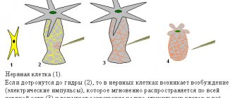
How does a motor neuron work?
In order for a bioelectric impulse to occur, a potential difference is required on the membrane of the nerve cell. This occurs as a result of changes in the concentration of potassium and sodium ions from the outer and inner surfaces of the membrane.
Subsequently, the impulse travels to the end of the long process, the axon, and reaches the junction with another cell. The place of such contact is called a synapse.
On the other side of the synapse, a short branching process, a dendrite, is adjacent to the point of contact. Signal transmission across a synapse is driven by active chemicals called transmitters.
Having arisen on the dendrite, the signal spreads along its membrane and passes further to the axon. To contract a skeletal muscle, the signal originates in the motor neuron of the cortex, passes along the pyramidal tract, passes to the interneuron and then to the region of the anterior horns of the spinal cord. This chain ends in muscle tissue.
The result of excitation of the motor center of the cortex will be a contraction of a group of muscle fibers.
Passage of information
Neurons communicate with each other using “nerve messages.” These “messages” are similar to electric current running through wires. Sometimes, when transmitted from one neuron to another, these impulses turn into chemical messages.
Nerve impulses
Information is transferred between neurons like electric current in wires. These messages are encoded: they are a sequence of completely identical impulses. The code itself lies in their frequency, that is, in the number of pulses per second. Impulses are transmitted from cell to cell, from the dendrite in which they originate to the axon through which they pass. But there is also a difference from electrical networks - impulses are transmitted not using electrons*, but using more complex particles - ions.
Medicines that affect the speed of impulses
There are many chemicals that can change the transmission characteristics of nerve impulses. As a rule, they act at the synaptic level. Anesthetics and tranquilizers slow down and sometimes even suppress the transmission of impulses. And antidepressants and stimulants, such as caffeine, on the contrary, contribute to their better transmission.
With great speed
Nerve impulses must travel quickly throughout the body. The surrounding glial cells help neurons speed up their passage. They form the sheath of the nerve fiber called myelin. As a result, the impulses travel at a mind-blowing speed - more than 400 km/h.
Chemical bonds
Messages transmitted from neuron to neuron must be converted from electrical to chemical form. This is due to the fact that, despite their large number, neurons never touch each other. But electrical impulses cannot be transmitted unless there is physical contact. Therefore, neurons use a special system called synapses to communicate with each other. In these places, neurons are separated by a narrow space called the synaptic cleft. When an electrical impulse reaches the first neuron, it releases chemical molecules called neurotransmitters from the synapse. These substances produced by neurons move across the synaptic cleft and land on receptors specially designed for them on another neuron. As a result, another electrical impulse occurs.
An impulse travels between neurons in less than a thousandth of a second.
Neurotransmitter differences
The brain produces about fifty neurotransmitters, which can be divided into two groups. The first consists of those that initiate the occurrence of a nerve impulse - they are called excitatory. Others, on the contrary, slow down its occurrence - these are inhibitory neurotransmitters. It is worth noting that in most cases, a neuron releases only one type of neurotransmitter. And depending on whether it is excitatory or inhibitory, the neuron affects neighboring nerve cells differently.
Artificial stimulation
An individual neuron or a group of neurons can be stimulated artificially using electrodes inserted into them, directing electrical impulses to precisely designated areas of the brain. This method is sometimes used in medicine, in particular for the treatment of patients suffering from Parkinson's disease. This disease, which manifests itself in old age, is accompanied by trembling of the limbs. This shaking can be stopped by continuously stimulating a specific area of the brain.
Neuron - microcomputer
Each neuron is capable of receiving hundreds of messages per second. And in order not to be overloaded with information, he must be able to judge the degree of its significance and make a preliminary analysis of it. This computational activity occurs inside the cell. There, excitatory impulses are added and inhibitory impulses are subtracted. And, in order for a neuron to generate its own impulse, it is necessary that the sum of the previous ones be greater than a certain value. If the addition of excitatory and inhibitory impulses does not exceed this limit, the neuron will be “silent”.
Information roads
In all this intricacy of neurons, there are beautifully defined pathways. Similar ideas, similar memories pass through, always firing the same neurons and synapses. It is still unknown how these circuit-like electronic communication circuits arise and are maintained, but it is clear that they exist and that the stronger they are, the more efficient they are. Frequently used synapses work faster. This explains why we remember faster things that we have seen or repeated several times. However, these connections do not last forever. Some of them may disappear if they are not used enough, and new ones will appear in their place. If necessary, neurons are always capable of creating new connections.
The small green dots in the photo are hormones inside the blood vessels.
Chemical doping
When an athlete is said to have used hormonal doping, it means that he took hormones either in pill form or by injecting them directly into the blood. Hormones can be natural or artificial. The most common are growth hormones and steroids, which make muscles bigger and stronger, as well as erythropoietin, a hormone that accelerates the delivery of nutrients to the muscles.
The brain is capable of performing millions of operations in a fraction of a second.
Hormones work in the brain
The brain also uses another tool to exchange information – hormones . These chemicals are produced in part by the brain itself in a group of neurons located in the hypothalamus. These hormones control the production of others produced in other parts of the body in the endocrine glands. They act differently from neurotransmitters, which bind directly to neurons and are carried in the blood to body organs distant from the brain, such as the breasts, ovaries, testes, and kidneys. By attaching to their receptors, hormones cause various physiological reactions. They, for example, promote the growth of bones and muscles, control feelings of hunger and thirst and, of course, affect sexual activity.
Symptoms of central motor neuron damage
Damage to central motor nerve cells most often occurs during stroke. When ischemia or hemorrhage occurs in the substance of the cerebral hemispheres, a section of tissue dies. Such lesions are almost always unilateral.
As a result, when central motor neurons are damaged, muscle dysfunction on one side is observed. The most noticeable symptom is unilateral paralysis, leading to the inability to actively move the arm and leg.
On the same side, muscle tone in the torso and facial muscles decreases. Damage to the central motor areas is accompanied by a number of changes in reflex activity.
Clinically, this is expressed in the appearance of various pathological reflexes. Their combination, decreased muscle tone and sensory disturbances allow the doctor to make a diagnosis.