The brain, along with the spinal cord, belongs to the central part of the human nervous system; it has a complex structure with many segments and zones responsible for certain functions. The main task of the nervous system is to form an appropriate adaptive response to external influences.
Content
- 1 Brain mass
- 2 Brain structure
- 3 Meninges of the brain
- 4 Structural parts of the brain 4.1 Medulla oblongata
- 4.2 Bridge
- 4.3 Cerebellum
- 4.4 Midbrain
- 4.5 Greater hemispheres
- 6.1 Prenatal development
Medulla
The medulla oblongata is responsible for coordination, motor activity and the body’s performance of certain reflex movements. Scientists have still not finally decided which part of the nervous system the medulla oblongata belongs to. In shape and location it is similar to the dorsal one, and in function - to the head one. But, since only what is inside the spinal column is considered spinal, the medulla oblongata is most often referred to as the brain.
Author of the article: Neurologist Konstantin Olegovich Makheev.
We recommend:
- Glioblastoma of the brain
- Violation of the emotional-volitional sphere in children
Brain mass
The mass of the human brain ranges from 1000 to more than 2000 grams, which on average represents approximately 2% of body weight. The brain of men weighs on average 100-150 grams more than the brain of women[2]. It is widely believed that a person’s mental abilities depend on the mass of the brain: the larger the brain mass, the more gifted the person. However, it is obvious that this is not always the case[3]. For example, the brain of I. S. Turgenev weighed 2012 g[4][5], and the brain of Anatole France - 1017 g[6]. The heaviest brain—2850 g—was found in an individual who suffered from epilepsy and idiocy[7][8]. His brain was functionally defective. Therefore, there is no direct relationship between brain mass and the mental abilities of an individual.
However, in large samples, numerous studies have found a positive correlation between brain mass and mental ability, as well as between the mass of certain brain regions and various indicators of cognitive ability [9][10]. A number of scientists [ who?
], however, cautions against using these studies to support inferences about low intelligence in some ethnic groups (such as Aboriginal Australians) who have smaller average brain sizes[11]. A number of studies indicate that brain size, which is almost entirely dependent on genetic factors, cannot explain most of the differences in IQ [12][13][14]. As an argument, researchers from the University of Amsterdam point to a significant difference in cultural level between the civilization of Mesopotamia and Ancient Egypt and their today's descendants in Iran and modern Egypt[15].
The degree of brain development can be assessed, in particular, by the ratio of the mass of the spinal cord to the brain. So, in cats it is 1:1, in dogs - 1:3, in lower monkeys - 1:16, in humans - 1:50. In Upper Paleolithic people, the brain was noticeably (10-12%) larger than the brain of modern humans [16] - 1:55-1:56.
Functions
Several departments and a complex structure control human life. We will look at the structure of the brain and the functions of individual parts below, but for now we will list the main tasks that the organ solves.
- Movement . The posterior regions of the brain are responsible for coordination. Human motor activity, the coordinated work of muscles, ligaments, bones and joints is the result of the interaction of nerve cells. The brain is responsible for maintaining balance and responding to external stimuli.
- Sense organs . Vision, hearing, smell and touch are made possible by the brain. Primary information in the form of impulses enters the departments of the organ, where it is subsequently transformed into a form familiar to us.
- Speech . Conversational skills and understanding of languages are the merit of the brain.
- Mental capacity . The ability to think logically, analyze information, and perform arithmetic operations distinguishes humans from animals. In mammals, the brain is small in relation to the body, so these functions are not available to them. The left side of the brain is responsible for mental activity.
- The work of internal organs . These processes in human life are innate, and he is no different from animals. Controlling the functioning of internal organs also includes a large set of reflexes: coughing, vomiting, sneezing, swallowing, etc.
Brain structure
Structure of the human brain
The volume of the brain of most people is in the range of 1250-1600 cubic centimeters and accounts for 91-95% of the capacity of the skull. The brain is divided into five sections: the medulla oblongata, the hindbrain, which includes the pons and cerebellum, the epophysis, the midbrain, the diencephalon and the forebrain, which is represented by the cerebral hemispheres. Along with the above division into sections, the entire brain is divided into three large parts:
- cerebral hemispheres;
- cerebellum;
- brain stem.
The cerebral cortex covers two hemispheres of the brain: right and left.
Midbrain
The range of functions of this department is small, but, nevertheless, they are very important. The structure of the human spinal cord is such that it is directly connected to the brain through the middle cord. It carries impulses that are caused by auditory and visual stimuli. In addition, it is responsible for the so-called hidden vision and turning the body towards noise.
Meninges of the brain
The brain, like the spinal cord, is covered with three membranes: soft, arachnoid and hard.
The soft, or vascular, membrane of the brain (lat. pia mater encephali) is directly adjacent to the substance of the brain, extends into all grooves, and covers all convolutions. It consists of loose connective tissue, in which numerous blood vessels branch that feed the brain. Thin processes of connective tissue extend from the choroid and penetrate into the mass of the brain.
The arachnoid membrane of the brain (lat. arachnoidea encephali) is thin, translucent, and has no blood vessels. It fits tightly to the convolutions of the brain, but does not extend into the grooves, as a result of which subarachnoid cisterns are formed between the choroid and arachnoid membranes, filled with cerebrospinal fluid, due to which the arachnoid membrane is nourished. The largest, cerebelloblongata cistern, is located behind the fourth ventricle, into which the median foramen of the fourth ventricle opens; the cistern of the lateral fossa lies in the lateral sulcus of the cerebrum; interpeduncular - between the cerebral peduncles; cistern crossroads - at the site of the visual chiasm (crossroads).
The dura mater of the brain (lat. dura mater encephali) is the periosteum for the inner cerebral surface of the skull bones. This membrane contains the highest concentration of pain receptors in the human body, while the brain itself has no pain receptors (see Headache
).
The dura mater is built of dense connective tissue, lined from the inside with flat, moist cells, and tightly fuses with the bones of the skull in the area of its internal base. Between the hard and arachnoid membranes there is a subdural space filled with serous fluid.
hindbrain
In the diagram of the structure of the cranium, the hindbrain is located in the back of the head. Compared to the large hemispheres, it is located below them in a separate recess. Consists of two parts. The pons is a small, dense bundle of nerve cells whose functions are limited to transmitting information between the main part of the brain and the spinal cord.
The cerebellum is a “miniature brain.” It consists of two hemispheres, although its mass makes up 10% of the brain’s weight. But the organ works much more than 10%. The cerebellum influences motor activity, coordination and balance. Deviations in work can be identified using tests carried out by a neurologist.
Structural parts of the brain
Computer tomography of the brain
Medulla
The medulla oblongata (lat. medulla oblongata) develops from the fifth cerebral vesicle (additional). The medulla oblongata is a continuation of the spinal cord with impaired segmentation. The gray matter of the medulla oblongata consists of individual nuclei of the cranial nerves. White matter is the pathways of the spinal cord and brain that extend up into the brain stem and from there into the spinal cord.
The anterior median fissure is located on the anterior surface of the medulla oblongata, flanked by thickened white fibers called pyramids. The pyramids taper downward due to the fact that part of their fibers move to the opposite side, forming an intersection of pyramids that form a lateral pyramidal path. The part of the white fibers that do not intersect form a straight pyramidal path.
Bridge
The pons (lat. pons) lies above the medulla oblongata. This is a thickened roller with transverse fibers. The main groove runs through its center, in which the main artery of the brain lies. On both sides of the furrow there are noticeable elevations formed by pyramidal tracts. The bridge consists of a large number of transverse fibers that form its white matter - nerve fibers. Between the fibers there are many accumulations of gray matter, which forms the nuclei of the bridge. Continuing to the cerebellum, the nerve fibers form its middle peduncles.
Cerebellum
The cerebellum (lat. cerebellum) lies on the posterior surface of the pons and medulla oblongata in the posterior cranial fossa. It consists of two hemispheres and a worm that connects the hemispheres to each other. The mass of the cerebellum is 120-150 g.
The cerebellum is separated from the cerebrum by a horizontal fissure, in which the dura mater forms the cerebellar tent, stretched over the posterior fossa of the skull. Each cerebellar hemisphere consists of gray and white matter.
The gray matter of the cerebellum is contained on top of the white matter in the form of the cortex. The nerve nuclei lie inside the cerebellar hemispheres, the mass of which is mainly represented by white matter. The cerebral cortex forms parallel grooves, between which there are convolutions of the same shape. The grooves divide each cerebellar hemisphere into several parts. One of the particles, a fragment adjacent to the middle cerebellar peduncles, stands out more than the others. It is phylogenetically the oldest. The flap and nodule of the worm appear already in lower vertebrates and are associated with the functioning of the vestibular apparatus.
The cerebellar cortex consists of two layers of nerve cells: outer molecular and granular. The thickness of the bark is 1-2.5 mm.
The gray matter of the cerebellum branches into the white matter (on the median section of the cerebellum you can see a branch of an evergreen thuja), which is why it is called the tree of life of the cerebellum.
The cerebellum is connected to the brain stem by three pairs of peduncles. The legs are represented by bundles of fibers. The lower (caudal) peduncles of the cerebellum go to the medulla oblongata and are also called the rope bodies. They include the posterior spinal-cerebellar tract.
The middle (pontine) cerebellar peduncles connect to the pons, through which transverse fibers pass to the neurons of the cerebral cortex. The corticopontine tract passes through the middle peduncle, through which the cerebral cortex acts on the cerebellum.
The upper cerebellar peduncles in the form of white fibers go in the direction of the midbrain, where they are located along the peduncles of the midbrain and are closely adjacent to them. The superior (cranial) peduncles of the cerebellum consist mainly of fibers of its nuclei and serve as the main pathways conducting impulses to the visual thalamus, subtuberculous area and red nuclei.
The legs are located in the front and the tire is located in the back. Between the tire and the legs lies the aqueduct of the midbrain (Aqueduct of Sylvius). It connects the fourth ventricle to the third.
The main function of the cerebellum is reflex coordination of movements and distribution of muscle tone.
Midbrain
The cover of the midbrain (lat. mesencephalon) lies above its lid and covers the aqueduct of the midbrain on top. The lid contains the tire plate (quadrigeminal). The two upper colliculi are associated with the function of the visual analyzer; they act as centers of orienting reflexes to visual stimuli, and therefore are called visual. The two lower tubercles are auditory, associated with orienting reflexes to sound stimuli. The superior colliculi are connected to the lateral geniculate bodies of the diencephalon using superior handles, and the inferior colliculi are connected to the medial geniculate bodies by inferior handles.
The spinal tract begins from the tegmental plate, which connects the brain with the spinal cord. Efferent impulses pass through it in response to visual and auditory stimuli.
Large hemispheres
The medial surface of the human cerebral cortex.
The brain has different hemispheres. The cerebral hemispheres include the lobes of the hemispheres, the cerebral cortex (cloak), the basal ganglia, the olfactory brain and the lateral ventricles. The hemispheres of the brain are separated by a longitudinal fissure, the recess of which contains the corpus callosum, which connects them. The following surfaces are distinguished on each hemisphere:
- superolateral, convex, facing the inner surface of the cranial vault;
- the lower surface, located on the inner surface of the base of the skull;
- the medial surface through which the hemispheres are connected to each other.
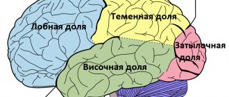
In each hemisphere there are parts that protrude most: in front - the frontal pole, behind - the occipital pole, on the side - the temporal pole. In addition, each cerebral hemisphere is divided into four large lobes: frontal, parietal, occipital and temporal. In the recess of the lateral fossa of the brain lies a small lobe - the insula. The hemisphere is divided into lobes by grooves. The deepest of them is the lateral, or lateral, also called the Sylvian fissure. The lateral sulcus separates the temporal lobe from the frontal and parietal lobes. From the upper edge of the hemispheres, the central sulcus, or Roland's sulcus, descends downwards. It separates the frontal lobe of the brain from the parietal lobe. The occipital lobe is separated from the parietal lobe only on the side of the medial surface of the hemispheres - the parieto-occipital sulcus.
The cerebral hemispheres are externally covered with gray matter, which forms the cerebral cortex, or cloak. There are 15 billion cells in the cortex, and if we consider that each of them has from 7 to 10 thousand connections with neighboring cells, then we can conclude that the functions of the cortex are flexible, stable and reliable. The surface of the cortex increases significantly due to grooves and convolutions. The phylogenetic cortex is the largest structure of the brain, its area is approximately 220 thousand mm2.
Functions of departments
The main function assigned to the brain structures is to ensure interaction with the environment. Here, teams are formed that are responsible for processes in the body such as thermoregulation, metabolism, selective development of structural formations (systems) that ensure the body’s adaptive abilities, and neurohumoral regulation.
The functions of the brain are determined depending on the location of its parts. Moreover, individual sections of the brain matter simultaneously perform several tasks, in parallel closely interacting with all other sections. If any part of the brain is damaged, its compensatory functions can be performed by another brain structure.
Oblong
The human brain is designed in such a way that it includes separate areas characterized by functional differences, for example, such as the oblongata, which continues the dorsal. The control center for cardiac activity is located in the medulla oblongata. Within the heart center, nuclei are distinguished from which the vagus nerves depart. The functions of the vagus nerve include autonomic regulation of the heart and the condition of blood vessels.
In the vasomotor center, in the structure of the nuclei of the vagus nerve endings, pressor and depressor zones are distinguished, which interact with each other. The pressor zone is responsible for the narrowing of blood vessels, the depressor zone is responsible for the expansion of the vascular lumen. The pressor zone reacts to the chemical composition of the blood. Information is transmitted through chemoreceptors.
The depressor zone responds to information coming from baroreceptors (they perceive the pressure that is exerted on the vascular walls). The hypothalamus is a section of the diencephalon that is responsible for neuroendocrine function and determines the effects of the cardiovascular system, taking into account the external environment and the current needs of the body.
The structure of the hindbrain provides for the presence of the pons, where part of the respiratory center responsible for respiratory function is located. The area responsible for inhalation is located in the pons, the area regulating the process of exhalation is in the medulla oblongata. Both areas interact with each other. During the process of inhalation, the external intercostal muscles are involved, which, by contracting, contribute to the expansion of the lungs and the entry of air into them.
Exhalation occurs due to contraction of the internal intercostal muscles. Commands that cause muscle contraction come from the corresponding zones of inhalation and exhalation through the structures of the spinal cord. The vagus nerve, hypothalamus and cortical structures can influence the frequency of respiratory movements. The medulla oblongata contains the nuclei of 4 pairs of cranial nerves (vagus, hypoglossal, accessory, glossopharyngeal).
The functions of the glossopharyngeal nerve include the motor activity of the pharyngeal muscles, the implementation of reflex reactions (sneezing, swallowing, coughing, gagging, the formation of speech sounds). It is believed that in humans, the medulla oblongata located in the brain contains centers such as cough-sneezing and vomiting.
Rear
As can be seen in the figure, the brain is structured in such a way that a person has several important brain regions, including the posterior one, consisting of the cerebellum and the pons. The fourth ventricle in the ventricular system expands like the spinal canal, forming a cavity in which the posterior section is located. The pons Varoliev is formed by a dense plexus of conducting pathways.
The cerebellum is the brain's motor center and contains pathways that connect it to many other areas of the brain and body systems. Bundles consisting of nerve fibers form the cerebellar peduncles - 3 pairs. The lower legs connect the cerebellum with the medulla oblongata segment, the middle ones provide interaction with the pons and then with the cortex, the upper ones with areas of the midbrain.
The mass of the cerebellum is equal to 10% of the weight of the entire brain matter. However, more than 50% of all neurons of the nervous system are localized in this area. The functions of the cerebellum include regulating the tone of skeletal muscles, maintaining body posture, maintaining balance, performing purposeful, voluntary movements, and coordinating motor activity. The cerebellum is involved in the planning and execution of movements:
- Ballistic (performed under the influence of gravity).
- Sports (ball throw, javelin).
- Musical (playing instruments).
General neuronal systems formed by the cerebellum and cortical regions are involved in thinking processes aimed at organizing complex movements. In the area of the bottom of the 4th ventricle there are nuclei of nerve branches - facial, vestibulocochlear, abducens, segmental trigeminal.
Average
The structure of the brain involves the separation of the middle section, which, in short, reflects the stability and immutability of the human nervous system. This department has undergone virtually no evolutionary changes. A horizontal section of the brain reveals the structures that make up the middle section, which is responsible for alternating sleep-wake cycles, concentration, reflex reactions (defensive, associated with orientation in space), and human functions such as hearing and vision.
The middle section contains structures that receive information from the organs of vision, regulate pain sensitivity and body temperature. The red nucleus of the department controls postural movements (formation and maintenance of posture), together with the substantia nigra it participates in the regulation of the extrapyramidal system (muscle tone, posture). The structures of the middle section form the body's reactions to external stimuli - visual, auditory.
The visual center is located in the area of the superior colliculus, and the auditory center is located in the area of the inferior colliculus. The aqueduct of Sylvius passes through the structures of the middle section, connecting the 3rd and 4th ventricles. In the middle section there are nuclei of the cranial nerves - trochlear, oculomotor, as well as one trigeminal nucleus. The trochlear and oculomotor nerve branches regulate the movements made by the eyes.
Intermediate
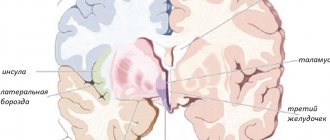
The human brain is designed in such a way that it contains an intermediate section that is responsible for functions such as sensory perception. The main part of the human diencephalon is the thalamus - the cerebral visual, sensory tubercle. The thalamus receives information from all receptors concentrated in the sensory organs.
Information flows to the thalamus from the organs of vision and hearing, from receptors - taste, olfactory, tactile, proprioceptors (receptors located in muscles, skin, ligaments and joint capsules), interoreceptors (receptors located in internal organs, tissues, elements of the vascular system) , vestibuloreceptors (receptors located in the labyrinth of the inner ear perceive changes in speed and direction when the body moves in space).
Appendages (superior, inferior, posterior) and the optic chiasm extend from the thalamus. The superior epididymis contains the pineal gland, which produces melatonin. The pineal gland also secretes histamine, norepinephrine, and serotonin, which are involved in controlling circadian rhythms. Regulatory activities are focused on the level of illumination.
In the posterior appendage, secondary centers are concentrated - auditory and visual, represented by the medial (located closer to the median plane) and lateral (lying away from the median plane) geniculate bodies. The functions of the lower appendage, the hypothalamus, are associated with its location in the limbic system, a brief description of which requires mention of its important role in controlling the work of internal organs, the formation of emotions and memory.
The hypothalamus is the highest regulatory brain center of the autonomic (autonomous) part of the nervous system. In this part of the human brain, the chemical composition of blood and cerebrospinal fluid (cerebrospinal fluid) is analyzed. The pituitary gland, which is part of the hypothalamus, is a gland that secretes hormones that regulate the activity of other glands of the endocrine system. Chemoreceptors (receptors that perceive chemical stimuli) transmit information about the concentration of substances contained in body fluids to the hypothalamus.
Based on the information obtained, the hypothalamus weakens or stimulates metabolic processes through a direct influence on the autonomic centers using nerve impulses or through the production of biological peptide substances - statins (inhibit the secretory activity of the anterior pituitary gland) and liberins (stimulate the secretory activity of the anterior pituitary gland). The functions of the hypothalamus include the control of processes based on biological motivation:
- Eating behavior (taste preferences, diet, diet features).
- Sexual behavior (behavioral reactions aimed at realizing sexual needs).
- Aggressive-defensive reactions.
The location of the hypothalamus within the limbic system of the brain suggests its participation in the interaction between somatic and autonomic functions. We are talking about motor reactions based on information received from the senses and the control of somatic functions taking into account the current needs of the body.
For example, if defensive behavior is activated due to biological needs, effective functioning of the skeletal muscles and sensory organs is necessary. A person needs to hear and see clearly and move dynamically. To increase the efficiency of muscle work, important parameters are the speed of transmission of nerve impulses, the provision of nerve structures and muscle tissue with nutrients and oxygen.
In conclusion, it can be argued that the hypothalamus provides internal resources for the implementation of external behavior. The nuclei of the thalamus are divided into groups - switching, integrative, modulating. The first group is an intermediate link in the afferent pathways through which information flows from organs to nerve centers. Afferent pathways extend from the head, limbs, and parts of the body.
The signals enter the analyzing zones of the cortical layer, where the received data is processed. Integrative nuclei provide communication between all structural divisions of the thalamus among themselves and with the associative zones of the cortical layer. An example is combining visual stimuli and verbal information into a single image. The functions of the thalamus support gnostic (cognitive) activity.
Modulating nuclei are the most ancient structure of the thalamus. This is the basis of the reticular formation, which stretches along the brain stem, consists of reticular nuclei and a branched neuronal network with numerous axons and dendrites. The reticular formation activates the cortical areas of the brain, which leads to concentration and timely processing of information coming through ascending pathways. The reticular formation controls the reflex functions of the spinal cord.
Sex differences
The adult male brain is on average 11–12% heavier and 10% larger in volume than a female brain[17][18]. No statistical difference was found between the ratio of body and brain size in men and women[19][20]. Tomographic scanning methods have made it possible to experimentally document differences in the structure of the brains of women and men[21][22]. It has been established that men's brains have more connections between zones within the hemispheres, and women's brains have more connections between the hemispheres. These differences in brain structure were most pronounced when comparing groups aged 13.4 to 17 years. However, with age in the brain of women, the number of connections between zones within the hemispheres increased, which minimizes the previously distinct structural differences between the sexes[22].
At the same time, despite the existence of differences in the anatomical and morphological structure of the brain of women and men, there are no decisive features or their combinations that allow us to speak of a specifically “male” or specifically “female” brain [23]. There are brain features that are more common among women, and others that are more often observed in men, however, both of them can also appear in the opposite sex, and there are practically no stable ensembles of such features observed.
Brain Development
Prenatal development
Development that occurs before birth, intrauterine development of the fetus. During the prenatal period, intensive physiological development of the brain, its sensory and effector systems occurs.
Natal state
The differentiation of cerebral cortex systems occurs gradually, which leads to uneven maturation of individual brain structures.
At birth, the child’s subcortical formations are practically formed and the projection areas of the brain are close to the final stage of maturation, in which the nerve connections coming from the receptors of various sense organs (analyzer systems) end and motor pathways begin [24].
These areas act as a conglomerate of all three brain blocks. But among them, the structures of the block regulating brain activity (the first block of the brain) reach the highest level of maturation. In the second (block of receiving, processing and storing information) and third (block of programming, regulation and control of activity) blocks, the most mature are only those areas of the cortex that belong to the primary lobes that receive incoming information (second block) and form outgoing motor impulses (3rd block)[25].
Other areas of the cerebral cortex do not reach a sufficient level of maturity by the time the child is born. This is evidenced by the small size of the cells included in them, the small width of their upper layers that perform an associative function, the relatively small size of the area they occupy and the insufficient myelination of their elements.
Period from 2 to 5 years
From the age of two
up to
five
years of age, the maturation of secondary, associative fields of the brain occurs, part of which (secondary gnostic zones of the analytical systems) is located in the second and third blocks (premotor area). These structures support the processes of perception and the execution of a sequence of actions[24].
Period from 5 to 7 years
The tertiary (associative) fields of the brain mature next. First, the posterior associative field develops - the parieto-temporo-occipital region, then the anterior associative field - the prefrontal region.
Tertiary fields occupy the highest position in the hierarchy of interaction between various brain zones, and here the most complex forms of information processing are carried out. The posterior associative area ensures the synthesis of all incoming multimodal information into a supramodal holistic reflection of the reality surrounding the subject in the entirety of its connections and relationships. The anterior associative area is responsible for the voluntary regulation of complex forms of mental activity, including the selection of necessary, essential information for this activity, the formation of activity programs on its basis and control over their correct course.
Thus, each of the three functional blocks of the brain reaches full maturity at different times, and maturation proceeds in sequence from the first to the third block. This is the path from bottom to top - from underlying formations to overlying ones, from subcortical structures to primary fields, from primary fields to associative fields. Damage during the formation of any of these levels can lead to deviations in the maturation of the next one due to the lack of stimulating effects from the underlying damaged level [24].
Diencephalon
The thalamus is directly responsible for communication with the outside world; it reacts to stimuli and transmits information about them to the hemispheres. The hypothalamus regulates autonomic functions and interacts with the nervous system. Below it is the pituitary gland, whose functions include regulation of sleep and wakefulness, metabolism, and control of body temperature.
Notes
- Evgenia Samokhina
“Burner” of energy // Science and life. - 2020. - No. 4. - P. 22-25. — URL: https://www.nkj.ru/archive/articles/31009/ - Whose brain weighs more? // samoeinteresnoe.com
- Sagan, 2005.
- Paul Browardel.
Procès-verbal de l'autopsie de Mr. Yvan Tourgueneff. — Paris, 1883. - W. Ceelen, D. Creytens, L. Michel (2015). "The Cancer Diagnosis, Surgery and Cause of Death of Ivan Turgenev (1818-1883)". Acta chirurgica Belgica 115
(3):241–246. DOI:10.1080/00015458.2015.11681106. - Guillaume-Louis, Dubreuil-Chambardel (1927). "Le cerveau d'Anatole France." Bulletin de l'Académie nationale de médecine 98
: 328-336. - Elliott GFS
Prehistoric Man and His Story. - 1915. - P. 72. - Kuzina S., Savelyev S.
Weight in society depends on brain weight.
Science: Secrets of the Brain
. Komsomolskaya Pravda (July 22, 2010). Retrieved October 11, 2014. - Neuroanatomical Correlates of Intelligence
- Intelligence and brain size in 100 postmortem brains: sex, lateralization and age factors. Witelson SF, Beresh H, Kigar DL Brain. 2006 Feb;129(Pt 2):386-98.
- Brain size and human intelligence (from R. Lynn’s book “Races. Peoples. Intelligence”)
- (2007) “Considerations relating to the study of group differences in intelligence.” Perspectives on Psychological Science 2
(2): 194–213. DOI:10.1111/j.1745-6916.2007.00037.x. - Brody, Nathan.
Jensen's Genetic Interpretation of Racial Differences in Intelligence: Critical Evaluation // The Scientific Study of General Intelligence: Tribute to Arthur Jensen. - Elsevier Science, 2003. - P. 397–410. - (January 2010) “Why national IQs do not support evolutionary theories of intelligence.” Personality and Individual Differences 48
(2): 91–96. DOI:10.1016/j.paid.2009.05.028. - (January 2010) “Evolution, brain size, and the national IQ of peoples around 3000 years BC.” Personality and Individual Differences 48
(2): 104–106. DOI:10.1016/j.paid.2009.08.020. - Drobyshevsky S.V.
Are we getting stupid? About the reasons for brain shrinkage. Archived from the original on September 5, 2012. - O'Brien, Jodi.
Encyclopedia of Gender and Society. - Los Angeles: SAGE, 2009. - P. 343. - ISBN 1-4129-0916-3. - Zaidi, Zeenat F (2010). "Gender Differences in the Human Brain: A Review." The Open Anatomy Journal 2
: 37–55. DOI:10.2174/1877609401002010037. - Kimura, Doreen (1999). Sex and Cognition
. Cambridge, MA: MIT Press. ISBN 978-0-262-11236-9 - (1980) “Analysis of brain weight. I. Adult brain weight in relation to sex, race, and age.” Archives of pathology & laboratory medicine 104
(12): 635–9. PMID 6893659. - “Male and female brains wired differently, scans reveal”, The Guardian, 2 December 2013
- ↑ 12
“How Men's Brains Are Wired Differently Than Women's” LiveScience, 02 December 2013 - Daphna Joel, Zohar Berman, Ido Tavor, Nadav Wexler, Olga Gaber.
Sex beyond the genitalia: The human brain mosaic (English) // Proceedings of the National Academy of Sciences. — 2020. — 30 November. - P. 201509654. - ISSN 0027-8424. — DOI:10.1073/pnas.1509654112. - ↑ 1 2 3 Mikadze Yu.V.
Neurophysiology of childhood. — Peter, 2008. - Luria A. R., 1973