| Brain stem | |
| Part | brain |
| Components | medulla oblongata, pons, midbrain |
| Catalogs | |
| |
| Media files on Wikimedia Commons | |
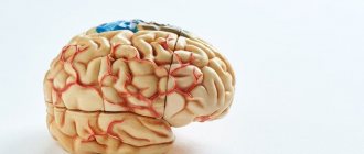
Brain stem
, or brain stem, is a traditionally distinguished section of the third brain, which is an extended formation that continues the spinal cord.
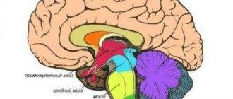
The trunk always includes the medulla oblongata, the pons, and the midbrain. Often it includes the cerebellum, sometimes the diencephalon.
The concept of the brain stem, which includes parts of the brain that are not of common origin, remains relevant due to its anatomical and morphological commonality.
Gross Anatomy of the Brainstem
1. Taenia choroidea (and lateral: Lamina affixa, Stria terminalis) 2. Thalamus, Pulvinar thalami - thalamus (visual thalamus), cushion of the thalamus 3. Ventriculus tertius - third ventricle 4. Stalk of Glandula pinealis - stem (?) pineal gland of the brain ( pineal body) 5. Habenula - leash 7. Colliculus superior - upper colliculus 8. Brachium colliculi superioris - handle of the upper colliculus (?) 9. Colliculus inferior - lower colliculus 10. Brachium colliculi inferioris - handle of the lower colliculus (?) 11. Corpus geniculatum mediale - medial geniculate body 12. Sulcus medianus - median sulcus 13. Pedunculus cerebellaris superior - superior cerebellar peduncle 14. Pedunculus cerebellaris inferior - inferior cerebellar peduncle 15. Pedunculus cerebellaris medius - middle cerebellar peduncle 16. Tuberculum anterius thalami - anterior thalami nuclei mustache (?) 17. Obex, Area postrema - valve, the most posterior field A: Thalamus (Thalamus), B: Mesencephalon (Midbrain), C: Pons (Bridge), D: Medulla oblongata (Medulla oblongata)]]
Ten pairs of cranial nerves exit from the ventral (facial) side, another pair (IV nerve) exits from the dorsal side, but immediately bends and exits to the ventral side.
Anatomical features
The brain is a complex organ that acts as the center of the human nervous system. Scientists estimate that it may contain more than 20 billion different neurons that transmit signals to other parts of the body. The brain stem includes several sections, each of which is responsible for specific functions. There are 5 of them in total:
- Oblong;
- Intermediate;
- Rear;
- Average;
- Finite.
Anatomy also involves the identification of several equally important parts: the cerebral cortex, the cerebellar cortex, the vermis with nuclei, the pons, the thalamus, the hypothalamus, the pituitary gland, the basal ganglia.
The structure itself presents the following picture:
- The medulla oblongata acts as a continuation of the spinal cord, emerging from the vertebral region. It contains two types of substance: white and gray. The function of the first is to conduct information between body systems. The second is the nerve nuclei, which mature by age 7.
- Valoriev Bridge. It is the next section emerging from the medulla oblongata, located in the middle part of the trunk, formed by the base, quadrigeminal, components of the cranial ventricles and tegmentum. Consists of longitudinal and transverse fibers. The former are built from neural clusters, presented in the form of nuclei, from which the latter pass. The latter include the upper and lower layers, through which pyramidal paths are laid.
- Cerebellum. It consists of small hemispheres that are covered with white and gray matter. Reaches its maximum size by the age of 15.
- Midbrain. Attached to the cerebellum by two peculiar legs, it includes 2 visual and 2 auditory sections in the form of separate tubercles through which nerve fibers pass.
- Hemisphere cortex. Between the hemispheres is the corpus callosum, which provides communication between all parts. All thought processes take place in the cortex.
The structure of the brain stem includes another significant section. It is called the reticular formation, which includes dendrites and axons that form the reticulum, which is a special network. The main function of this area is to manage information transmitted from the brain to other parts of the body. There are 2 types of information conduction: afferent, which directs data to the formation, and efferent, which performs the opposite effect.
Diamond fossa
The structure belonging to the trunk is a diamond-shaped fossa (fossa rhomboidea). Its lower part belongs to the medulla oblongata, the upper part to the pons. The rhomboid fossa is the bottom of the fourth cerebral ventricle (ventriculus quartus) and contains on its surface such components as medullary (brain) stripes (striae medullares), triangles of the hypoglossal and vagus nerves, and the vestibulo-cochlear area (area acustica). The diamond-shaped fossa is divided longitudinally by the median groove (sulcus medianus posterior).
Cranial nerve nuclei
One of the most important components of the brain stem is the cranial nerve nuclei, which extend from its base. They are located between the rear and oblong parts, with a small number of them located on the bridge. The nuclei consist of nerve endings that have a direct effect on the trunk. They are presented in the form of branches that penetrate through its most important parts.
Each core has its own purpose. The following nerves emerge from this zone:
- Olfactory;
- Visual;
- Oculomotor;
- Facial;
- vestibulocochlear;
- Block;
- Discharge;
- Trigeminal;
- Glossopharyngeal;
- Sublingual;
- Additional;
- Wandering.
Their full functioning is very important for the human body. Dysfunction of any nerve can have serious consequences that impair quality of life and even lead to death.
Functional components of the barrel
In the brain stem, the same principle of localization of afferents and efferents is preserved as in the spinal cord. The role of sensory and motor roots is taken over by the cranial nerves. In addition to the original four components, the so-called also appear in the trunk. “special” afferents and efferents serving derivatives of the branchial arches. Special afferents (SSA) are represented by the VIII pair - n. vestibulocochlearis, serving specific receptors of the inner ear. Special visceral afferents (SVA) in the brainstem are represented by fibers from the taste receptors (VII, IX and X nerves, and the nucleus of the solitary tract common to them).
Special visceral efferents (SVE) are represented by nerves innervating muscles that are phylogenetically derived from the muscles of the branchial arches of the protozoan. For humans, these are: chewing muscles (innervation by the V nerve), facial muscles (VII), muscles of the larynx and pharynx (IX, X), as well as the sternocleidomastoid and trapezius muscles of the neck (XI nerve).
In the lower part of the trunk (medulla oblongata), the dorsoventral orientation of the components remains, as in the spinal cord (GSA, GVA, GVE, GSE), higher up the trunk the orientation changes to lateral-medial and ceases to be linear. GSA is displaced more ventrally by SSA, and the SVE component is also displaced more ventrally.
Functions
All parts of the brain stem are equally necessary. They provide people with the opportunity to smell, hear sound, understand speech, think about any serious things. If not for them, humanity could have remained in the Stone Age forever.
The functions of the brain stem are reduced to the distribution of information between the brain and the central nervous system. They are provided by nuclei and nerve endings. In this case, the trunk is a physiological connecting step between the spinal cord and the brain. If it is damaged, then signals from the brain will not be able to reach the end point, which will completely eliminate the normal functioning of the human body.
There are several groups of functions that are characteristic of the brain stem. Among them:
- Motor. This includes all actions associated with the muscles of the eyes and eyelids. The function is also responsible for the reflexes of the eyeballs and controls the chewing muscles.
- Sensitive. Ensures the functioning of taste buds, as well as all reflexes that affect the digestive system. Helps transmit signals for swallowing and many other actions, including even vomiting. Additionally responsible for sneezing.
- Parasympathetic. Affects movement and dilation of the pupils, controls the ciliary muscles. Managed by cores, ensuring the execution of a block function.
- Upper salivary. It affects the salivary glands, ensuring timely and necessary formation of saliva.
- Vestibular. Responsible for the functioning of the vestibular apparatus, which helps control body balance and stay on your feet.
- Swallowing. Ensures the swallowing reflex works. Complements the work of the sensitive function.
- Auditory. Transmits information to the cerebellum, is responsible for hearing, as well as recognizing heard sounds.
- Sensory. Gives sensitivity to the skin on the face, analyzes taste and sound, and recognizes vestibular stimuli.
The brain stem has the most important functions. It gives every person the opportunity to hear, feel, see, move, think. All of them are necessary for a full life.
| Brainstem region | Functions |
| Midbrain | · Functioning of visual and auditory organs; · Management of relevant bodies; · Orientation in space. |
| Medulla | · Reflexes associated with coughing, vomiting, sneezing; · Breath control; · Cardiovascular system management; · Functioning of the digestive tract. |
| Pons | · Providing blood supply to the brain; · Fast transmission of signals between the brain and the central nervous system. |
| Cerebellum | · Coordination of movements, balance; · Muscle tissue tone. |
| Diencephalon | · Function of the thyroid gland; · Control of the adrenal glands. |
The importance of such functions makes us take the condition of the brain stem more seriously. He is no exception and may be susceptible to various life-threatening diseases.
The brainstem is a concentrated bundle of conducting tissues, gray and white matter, which form various nuclei. Each of which has its own function and allows you to control various actions of parts of the human body.
The motor nuclei help the eyes and eyelids work properly, ensuring they display reflexes in a timely manner. This part of the trunk also helps the chewing muscles work properly. This core is located in the bridge. The trochlear nerve, called the parasympathetic nucleus by experts, helps the motor function by influencing the functioning of the pupil and ciliary muscles.
Also, together with the motor function, the salivary system works, helping in the consumption of food and the secretion of saliva. It is poorly controlled by human willpower, but actively functions in any state of the body. Along with it, the sensitive core simultaneously performs its work - it guarantees the functionality of the taste buds that are located on the surface of the tongue, and also ensures the correct functioning of digestive reflexes.
Auditory receptors are controlled by the cochlear nucleus, which, together with the vestibular nucleus, helps keep the body in balance and not fall from the effects of earth gravity. This part of the brain is an amazing organ that allows a person to be “alive”: touch, hear and understand sounds, see, move and most importantly think.
Descending Paths
Thanks to the research of such scientists as R. Magnus and I.F. Klein, it was established that there is a complex system of reflex centers in the medulla oblongata that ensure a certain position in the body due to static and static-kinetic reflexes. These reflexes, in fact, are mechanisms for the redistribution of muscle tone in such a way that a position that is comfortable for the animal is maintained (posture-tonic reflexes) or a return to this position from an uncomfortable one (righting reflexes), and also ensures the preservation of balance during acceleration (stato-kinetic reflexes) . The implementation of these reflexes occurs with the participation of such formations of the trunk as the reticular formation, the red nucleus and the vestibular nuclei.
The reticular formation is a formation that runs from the spinal cord to the thalamus in a rostral (towards the cortex) direction. In addition to participating in the processing of sensory information, the reticular formation has an activating effect on the cerebral cortex, thus controlling the activity of the spinal cord. With the help of this mechanism, the tone of skeletal muscles, sexual and autonomic functions of a person is controlled. For the first time, the mechanism of the effect of the reticular formation on muscle tone was established by R. Granit: he showed that the reticular formation is capable of changing the activity of γ-motoneurons, as a result of which their axons (γ-efferents) cause contraction of muscle spindles, and, as a result, consequence, increased afferent impulses from muscle receptors. These impulses, entering the spinal cord, cause excitation of α-motoneurons, which is the cause of muscle tone.
It has been established that two clusters of neurons take part in performing this function of the reticular formation: neurons of the reticular formation of the pons and neurons of the reticular formation of the medulla oblongata. The behavior of neurons in the reticular formation of the medulla oblongata is similar to the behavior of neurons in the reticular formation of the pons: they cause activation of α-motoneurons of the flexor muscles and, therefore, inhibit the activity of α-motoneurons of the extensor muscles. Neurons of the reticular formation of the pons act exactly the opposite, excite α-motoneurons of the extensor muscles and inhibit the activity of α-motoneurons of the flexor muscles. The reticular formation has a connection with the cerebellum (some of the information from it goes to the neurons of the medulla oblongata (from the nuclei of the cortical and spherical cerebellum), and from the tent - to the neurons of the pons) and with the cerebral cortex, from which it receives information. This suggests that the reticular formation is a collector of nonspecific sensory flow, possibly involved in the regulation of muscle activity. Although the need for a reticular formation that duplicates the functions of neurons in the vestibular nuclei and red nucleus remains unclear.
The vestibular nuclei (from the Latin vestibulum - vestibule) are an organ that records changes in the position of the body in space and is located in the inner ear. Excitation of the vestibular nuclei occurs under the influence of an adequate stimulus acting on the vestibular apparatus. Starting from the Deiters nucleus - one of the main nuclei - as well as from the superior and medial nuclei, the vestibulospinal tract influences the alpha motor neurons of the spinal cord: the neurons of the vestibular nucleus excite the extensor α motor neurons, and mainly on the axial muscles (muscles of the spinal column) and at the same time inhibit α-motoneurons of the flexors through the mechanism of reciprocal innervation. During experimental transection of the vestibulospinal tract, a predominance of tone in the flexor muscles is observed.
Also, from the vestibular nuclei of the medulla oblongata there is a path to the so-called medial fasciculus, directed towards the spinal cord. This bundle performs an important function: it connects together all the nuclei of the nerves involved in regulating the activity of the muscles of the eyeball. Signals coming from the vestibular nuclei enter the longitudinal medial fasciculus, due to which the phenomenon of nystagmus occurs when the vestibular apparatus is activated.
Thus, when the vestibular apparatus is irritated, a redistribution of muscle tone occurs and a change in the activity of the muscles of the eyeball occurs, as a result of which the animal is able to maintain balance and direct its gaze in the desired direction.
The red nucleus is located in the midbrain region. The neurons of this nucleus receive information from the cerebral cortex and cerebellum, that is, all information about the position of the body in space, the state of the muscular system, and skin. The influence on alpha motor neurons of the spinal cord is carried out using the rubrospinal tract. The rubrospinal tract begins from the cells of the red nucleus located in the cortex of the cerebral peduncles. Activation of red nucleus neurons produces excitatory postsynaptic potentials in flexor motor neurons and inhibitory postsynaptic potentials in extensor motor neurons. In this respect, the rubrospinal tract is similar to the corticospinal tract.
Stem section
The first living beings to appear on Earth had only a medulla oblongata.
It was he who provided them with all the necessary instincts that helped them survive. But this is not enough, because... they needed to constantly develop their reflexes and thinking. After some time, new organisms began to be born with larger brains. The brain stem, which appeared during evolution, was responsible for ensuring respiratory function and blood supply to all necessary parts of the body. As it developed, it began to consist of a huge number of different centers, which began to form a very complex system. Now this section is a necessary part of the brain, without which life is impossible.
It is located between the large opening of the head in the occipital region and the slope of the inner part of the skull. The trunk extends the spinal cord, connecting it with the main one located inside the head. Its length is about 7 cm, and it includes several separate parts that are very important for the body.
Diseases
Like any other organ, the brain can fail. The same applies to its trunk. Most problems become the consequences of injuries or other diseases, and sometimes simply manifestations of age. There are several diseases:
- Stroke;
- Tumor;
- Cysts;
- Chordomas;
- Ischemia;
- Malformation;
- Aneurysms;
- Epidermoids;
- Meningiomas.
Most of them appear extremely rarely. The bulk of reported cases of brainstem lesions are stroke and various tumors. They are the most dangerous and require the highest quality and fastest treatment. But why do they arise?
Causes
A particular disease can develop for many reasons. Those most at risk are those who already have serious brain diseases, lead an unhealthy lifestyle, or suffer from regular stress. But even healthy people can get problems with the brain stem. Violations occur for the following reasons:
- Diseases associated with blood vessels, as well as their damage;
- Traumatic brain injuries;
- Poor circulation;
- Nervous breakdowns, severe stressful situations;
- Extreme sports, as well as extreme sports in everyday life;
- Eating junk food or untreated water;
- Alcohol abuse, smoking;
- Congenital diseases associated with the brain stem.
When any diseases appear, they must be treated immediately. Lack of necessary medical intervention can lead to severe irreversible consequences or death.
Literature
- Great Medical Encyclopedia / Ch. ed. B.V. Petrovsky. — 3rd edition. - Soviet Encyclopedia, 1982. - T. 13. - P. 351.
- Agadzhanyan N. A., Tel L. Z., Tsirkin V. I., Chesnokova S. A. Human physiology. - St. Petersburg: Sotis, 1998. - pp. 54-57.
- Antonen E. G. Motor pathways // Spinal cord (anatomical, physiological and neurological aspects).
| Nervous system | |
| Normal human anatomy | |
| Central | Brain | Brain structures | Spinal cord |
| Peripheral | Somatic Nervous System Autonomic Nervous System: Sympathetic Nervous System | Parasympathetic Nervous System | Enteric nervous system |
Stroke
The most common disease affecting the brain stem is stroke. It is always associated with disturbances in the functioning of blood vessels. As the body ages or certain diseases occur, their walls become thinner and more inelastic, and may become covered with plaques or become completely clogged.
There are two types of stroke: ischemic and hemorrhagic. The first is an infarction of the brain stem and is considered extremely dangerous due to blockage of blood vessels and subsequent oxygen starvation of nerve cells. The second manifests itself in the form of hemorrhage in the brain tissue. There is a risk of death in both cases.
Mechanism of action
In most cases, a hemorrhagic stroke occurs like this: first, a blockage of a vessel occurs, and then, under increased pressure, it bursts. When thinning, the vessels can immediately burst or become damaged without the formation of blood clots or any plaques. Immediately after the rupture, severe bleeding occurs in the brain, after which a hematoma appears, limiting the access of oxygen to the neurons. This becomes a failure, the consequence of which is a disruption of the functioning of all body systems.
With an ischemic stroke, severe damage to brain tissue also occurs, which greatly complicates the survival of the patient. After damage, the tissue gradually begins to die. Therefore, it is important to provide medical assistance to the victim as quickly as possible.
Causes
You can prevent a stroke if you try to exclude from your life all the moments that lead to this dangerous phenomenon. Doctors have been able to identify several main factors that increase the risk of cerebral infarction. Among them:
- Diabetes;
- Rheumatism;
- Hypertension;
- Atherosclerosis.
Everyone who is affected by at least one point needs to be as attentive to their health as possible and consult a doctor at the first alarming sensation.
Symptoms
A stroke is always sudden. A person can feel great throughout the day, and at one point hemorrhage occurs. Occasionally, shortly before a stroke, discomfort or pain may appear in the head. Symptoms indicating a cerebral hemorrhage are as follows:
- Dizziness;
- Increased sweating;
- Pale skin color;
- High body temperature;
- Pressure interruptions;
- Cardiopalmus;
- Breathing problems;
- Muscle paralysis.
The brain stem may be significantly damaged, making full recovery impossible. In this case, the development of severe complications associated with other diseases or characteristics of the body cannot be ruled out.
Treatment
Providing prompt assistance is the most important condition for preserving the life of the patient. But even she does not give any guarantees. Approximately 60% of victims die in the first days after a major stroke. In some cases, a person may die within two weeks. Only 20% of stroke survivors survive. If help is provided in the first hour after an attack, there is a chance of successful therapy. However, all consequences can be treated with great difficulty.
Transfer to a hospital is a prerequisite. It will not be possible to treat the victim at home; refusal to hospitalize will lead to death. Treatment involves constant monitoring by doctors and taking medications aimed at:
- Elimination of the formation of blood clots in blood vessels;
- Thinning of blood and existing blood clots;
- Reduced pressure;
- Normalization of cholesterol levels.
Additionally, physiotherapy is prescribed. In severe cases, emergency surgery may be performed. It is required to stop hemorrhage when conventional means do not have the desired effect.
Tumor
The second most common tumor is the brain stem tumor. Some of them can be very dangerous, but most do not require any medical intervention. There are several types of tumors:
- Primary. Appear when brain tissue is damaged.
- Secondary. They are a consequence of other diseases.
- Deforming. They negatively affect the shape of the brain stem, deforming it. They may be located on the stem or some other parts.
- Diffuse. They merge with the brain matter, which creates serious difficulties in treatment. Cases of successful therapy are very rare.
- Parastem. They grow to the trunk, causing deformation.
- Diamond-shaped. Appear in the occipital part of the skull.
- Cerebellar. The cerebellum is affected along with the trunk.
- Exophytic. They form on the cerebellum, then reach the trunk.
Neoplasms develop gradually, increasing in size. Sometimes their growth may slow down or stop completely, eliminating the need for treatment. The reasons for their appearance are various injuries and complications after serious illnesses.
Symptoms
Identifying neoplasms that affect the brain stem is not so easy. If they are small in size, they may not cause any symptoms at all, which creates certain difficulties in diagnosis. By the time a tumor is discovered, as a rule, it has already grown to a large size.
Symptoms that may indicate the growth of a tumor are as follows:
- Headache;
- Dizziness;
- Coordination problems;
- Problems with vision or hearing;
- Disorientation in space;
- Tremor of the hands or head;
- Unstable mood.
If such symptoms occur, you should consult a doctor. The patient will be scheduled for an MRI, which will determine the presence of a tumor.
Treatment
The prognosis always depends on what kind of tumor the patient has. Its growth rate, size and exact location are of great importance. Benign neoplasms are easily removed surgically, for which an incision is made through which the tumor itself is excised. Malignant tumors cannot be removed using this method, so you will have to give preference to radiation therapy or other methods.
Methods for treating tumors:
- Surgical removal. Excision of the tumor using physical pressure with a knife; it requires making an incision. Suitable only for benign tumors.
- Radiation therapy. X-ray exposure to the tumor through all other structures of the head. Effectively slows down the growth of tumors.
- Stereotactic. A combination of several types of exposure is used, including radiation. It is characterized by the absence of any painful sensations for the patient.
If necessary, doctors can combine several treatment methods at once. This will increase the chances of successful tumor removal.