03/20/2020 506 Anatomy
Author:ProSpinu
How does the human spinal cord work, where is it located and how does it function? In short, this is the main organ of the central nervous system. With its help, signals from the periphery enter the central part and vice versa. Its anatomy is quite complex; it has many nerve endings, substances and membranes. To better study the features and role played by this body, we suggest staying with us and reading the article.
Anatomical features
A rather thick tourniquet, white in color, located in the spinal canal - this is the human spinal cord. Its diameter is about 1-1.5 cm, and its length almost reaches half a meter (up to 45 cm). This organ weighs about 38 g.
The narrow spinal canal is not only the location of an important organ, but also its protection. The core of the organ consists of a gray substance. It is covered by a white substance, which is also covered with protective and nourishing shells for the core. This is the general plan of the structure of the spinal cord.
Topography
The structure and functions of the spinal cord are quite complex. Neurosurgeon students study it in detail. Experts very carefully consider the development of the spinal cord. Ordinary people are interested in the question of what its topography is and familiarity with the leading role of this organ.
So, it is quite simple to describe the essence and goals that this body serves. The cervical spinal cord at the level of the back of the head in the area of the foramen passes into the cerebellum. The spinal cord ends at the level of the first 2 lumbar vertebrae. The conus spinal cord is located where a pair of vertebrae are located in the lumbar region. Next is the well-known “terminal thread”.
But this fragment is considered atrophied. It is called the “end” region. Nerve endings called “roots” are distributed along the entire circumference of the thread. The filum terminale is equipped with a substance containing a small proportion of nervous system tissue. But the outer part is not even equipped with a similar fabric.
The topography of the organ includes a pair of thickenings where the innervating processes emerge (cervical thickening of the spinal cord and lumbar). The outer and rear surfaces of the bundle are separated by slits called “middle”. The one in front is deeper, the back one is smoothed.
External structure
The general structure of the spinal cord suggests its division into a number of surfaces: posterior, anterior and two lateral. The spinal cord has faint grooves on the lateral surface. They are located longitudinally, and nerves extend from the grooves. They are also called “roots”. In the lumbar area, together with the terminal filament, they form a tail, which is commonly called a horse's tail. The grooves divide half of this cord into the following structures:
- front;
- lateral;
- posterior (cords).
The grooves of the spinal cord extend along the canal. The roots are divided into anterior ones - they are formed by efferent neurons, and posterior ones, created by afferent neurons. Their bodies converge into a knot. The roots unite and form a nerve. So, on all sides of the tourniquet there are over 30 nerve endings, forming exactly the same number of pairs. This is the external structure of the spinal cord.
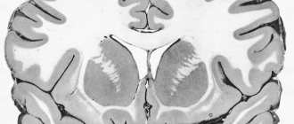
Anatomically, it consists of 2 types of substances: white and gray. The first is the processes of the neural type, and the gray is their bodies.
White matter
All cords are made entirely of white matter of the spinal cord. They consist of longitudinal nerve fibers. These threads converge, forming peculiar conductors. Based on their functional purpose, fibers are divided into 3 types:
- motor;
- associative;
- sensitive.
The first are represented by short bundles and combine all parts into a single system. The second ones are called ascending. They give signals to the centers. Still others are descending. They provide signals from central structures to areas of the horns.
Gray matter
It structurally resembles grouped longitudinal plates consisting of homogeneous neurons. It contains not only neuronal bodies, but also neuropil, glial cells and capillaries. Along the entire spine it forms 2 columnar types, left and right. They are connected by gray adhesions.
The anterior horns contain the largest neurons. They form the motor nuclei of the spinal cord and inhibitory neurons. The structure of the gray matter of the background horns is not the same. It contains a huge number of intercalary type neurons.
The lateral horns of the spinal cord fill the centers of the ANS, the dilation of the pupil, the bases of innervation of the digestive system and other important organs of the human body. In the nucleus of the gray matter of the spinal cord there is a canal that neurosurgeons call “central.” It is filled with liquor. In adults, in some places it is filled with cerebrospinal fluid, and in others it is overgrown.
Shells
Anatomy of the spinal cord describes the membranes of the spinal cord:
- vascular soft;
- hard;
- avascular or arachnoid.
The characteristics of shell 1 are as follows: soft, penetrated by vessels and nerves. It is enveloped by the avascular part. There is some space here called “subarachnoid”. The cerebrospinal fluid generated in one of the systems flows into this niche. The last shell is made up of connective tissue; it is strong and flexible. The membranes of the spinal cord and brain are identical and form a single structure.
Spinal cord reflexes
Knowledge of the functions of this important element of the nervous system is impossible without understanding the simple reflex arc. At the single segment level, it has a fairly short path:
People have spinal cord reflexes from birth and they can be used to determine the functional viability of a particular part of this organ.
The reflex arc can be represented as follows:
- This path begins from a special nerve link called a receptor. This structure receives impulses from the external environment.
- Further, the path of the nerve impulse lies along centripetal sensory fibers, which are the axons of peripheral neurons. They carry information to the central nervous system.
- The nerve impulse must enter the nerve cord, this occurs through the dorsal roots to the nuclei of the dorsal horns.
- The next element is not always present. It is the central link that transmits impulse from the rear to the front horns.
- The most important link in the reflex arc is the effector link. Located in the anterior horns. From here the impulse goes to the periphery.
- Along the anterior horns, irritation from neurons is transmitted to the effector - the organ that carries out direct activity. Most often it is skeletal muscle.
The impulse from neurons travels through this complex path, for example, when tapping the tendons of the knee with a hammer.
Segmental structure
A segment of the spinal cord is a piece of a tourniquet along with associated nerves. There is no morphological separation of one segment of the spinal cord from another. It is extremely functional. Each of the segments innervates a certain region. The designation of spinal cord segments is represented by alphanumeric indices, oriented to a part of the spinal cord and containing segment numbers.
The spinal cord consists of about 33 segments. The spinal cord segments have 4 roots, a pair of anterior and posterior. The spinal column is significantly longer than the cord, so it should be remembered that the segments are not numbered in the same way as the vertebrae. Any nerve consists of motor-sensitive roots. They come out in bunches from this bundle to the openings between the vertebrae.
The nerve ending located behind forms a ganglion and merges with the nerve ending in front. In this case, a mixed nerve is formed, which is divided into branches:
- The meningeal branch innervates in accordance with the nature of the spinal cord membrane and the canal wall.
- Dorsal - the skin in the corresponding areas, as well as deep muscle tissue.
- The connective tissue branch is the connecting link between the tourniquet and the ganglia.
- The abdominal branch is responsible for innervation of the limbs, lateral surfaces of the body and tissue of the abdominal part of the body.
What is the composition of gray matter?
Gray matter is formed from nerve cells, of which there are more than thirteen million (including glia and processes). All neurons grouped into nuclei or plexuses that make up the gray matter of the SC are divided into three main groups:
Internal cells with numerous processes that do not extend beyond the boundaries of the substance itself and provide interaction (forming synapses) with other neurons of the SC.
Root cells (the largest motto- and multi-neurons of the ANS), the neurons of which protrude beyond the boundaries of the gray matter and innervate skeletal muscles.
Tufted neurons forming many pathways leading to the brain. These cells got their name due to their ability to form bundles, which are a kind of switches connecting SC segments.
So, the spinal cord includes neuroglia, ganglion nerve cells, pulpal and non-pulpate nerve fibers. These cells are grouped into nuclei and are located in segments along the entire length of the spinal cord and its primary three-membered reflex arc.
Each segment of gray matter is separated by barriers in the form of Rexed plates that perform specific functions:
- Reception of impulses from the dorsal roots and their transmission to the spinothalamic tract.
- Accompaniment of impulses (afferents) of temperature and pain sensitivity.
- Processing information from the muscular system.
- Receiving and transmitting brain impulses.
- Innervation of the muscles of the limbs, body and internal organs.
Disturbances in the functioning of any of the links in this system lead to serious consequences, including paralysis, partial or complete.
Blood supply
The tourniquet is supplied with blood through the adjacent arteries. Through the fusion of the branches of the vertebral arteries, the anterior artery is formed. It is designed to be located along the front slit of the tourniquet. The blood supply to the spinal cord is also provided by the arteries located there. They are located behind the tourniquet.
They connect to the neck and arteries, which are called the “posterior intercostal, lumbar and lateral sacral arteries.” Between them there is a network of anastomoses, due to which the tourniquet is literally entangled in the branches of the arteries. To supply blood to the spinal cord, in addition to arteries, veins are needed, which also provide blood outflow.
Functions and role in the body
The human spinal cord has two main functions: one normalizes the brain-body connection. It is reflexive, it puts everything into action not without the participation of the will. The second conducts impulses to the main brain in an ascending manner and transmits them back from it. The descending or efferent pathways of the spinal cord are responsible for this activity.
The ascending tracts of the spinal cord are represented by the following tracts:
- spinothalamic;
- spinocerebellar;
- wedge-shaped and thin beams.
The pyramidal tracts, vestibulospinal, tectospinal and red nuclear spinal tracts are classified as special efferent pathways.
The reflex function is aimed at maintaining posture (position reflexes) and the ability to consistently alternate actions (motor programs), for example, walking. This function also provides a reflex defense mechanism (quickly moving the limbs away from hot objects).
Autonomic reflexes of the spinal cord are control signals that ensure the smooth functioning of internal organs. Myomatic reflexes are designed to provide contractile activity of muscles in response to their burning.
Anatomy and physiology of the spinal cord is a whole field of knowledge that describes its structure and features of functioning. It helps to understand how important the organ is and how the spinal cord and brain are connected. Thanks to this description, people receive the necessary ideas about an important organ.
Functions
The activity of the spinal cord is continuous. It provides motor activity of the body. There are two main functions of the human spinal cord - reflex and conduction.
Each department ensures the functioning of a completely specific area of the body. Segments (cervical, thoracic, for example) provide the functions of the organs of the sternum and arms. The lumbar segment is responsible for the proper functioning of the muscles and digestive system. The sacral segment is responsible for the functions of the pelvic organs and legs.