A person, as an organism, is constantly in interaction with the environment. All living organic systems exist according to a fundamental phenomenon - the principle of irritability and reactivity, which is ensured by the nervous system of the brain. This means that information, interacting with the body, causes the latter to react and respond to it - this is the basis of life and existence.
To survive, our biological species must perceive information about external reality and analyze the information received: color, temperature of the material, movements and actions of other objects, and the formation of our own behavioral patterns. All these processes are provided by the nervous system of the brain. The exchange of information between the internal state of the body and the environment occurs due to the structures of the nervous system: receptors, pathways and higher analyzers of sensory information.
- Structure
- Functions
- Recovery
Structure
Initially, the body had enough elementary information and simple reactions - unconditioned reflexes - unconscious nervous responses that did not require comprehension. These physiological mechanisms are provided by the spinal cord.
Over the course of evolution, the structure and mechanics of the structures became more complex: the medulla oblongata was formed above the spinal cord, the hindbrain and midbrain were formed above it, then the intermediate brain and the cerebral cortex - the highest complexity known biological structure on the planet. This complication of the nervous system allowed a person to perceive and process information of a more complex order: subtle tactile feelings, overtones of sound and shades of colors.
The highest department - the cortex - allowed a person to form speech and gave him the ability to interact complexly between people. Thanks to the cortex, people have developed a social structure, morality, ethics, knowledge, the ability to receive and transmit experience and, what distinguishes humans from other animals, self-awareness.
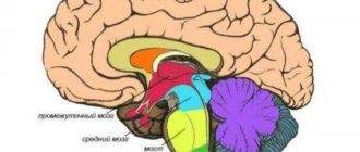
The central nervous system is divided into the spinal cord and the brain. These structures were formed in an evolutionary way from lower simple to higher and complex structures.
The nervous system of the brain consists of neurons, processes and glia. The spinal cord and brain are in constant continuous communication with each other through pathways - a set of specific structures that transmit information from one section to another. Paths can be thought of as wires that transfer energy from power plants to homes.
The brain consists of the following sections (from lower to higher structures):
- The medulla oblongata is a continuation of the spinal cord.
- Hindbrain: cerebellum and pons.
- Midbrain: subcortical centers of hearing, vision, transitory pathways between the spinal cord and cortex.
- Diencephalon: thalamus, pituitary gland, hypothalamus.
- The telencephalon is the cerebral cortex. The following zones are distinguished: frontal, parietal, occipital and temporal.
The medulla oblongata is the transition of the spinal cord to the brain. The olive nuclei, reticular formation, and cranial nerve nuclei are located here. Nerves come from here in 4 branches. The centers of respiration and circulation are also located here.
The nerves of the brain emerge from the structures of the cerebellum and the pons: the trigeminal, abducens and facial nerves. Their fibers are directed to the facial muscles, mouth, tongue and inner ear.
The basis of the midbrain is the quadrigemale, on which the centers of vision and hearing lie. These are mixed structures: they receive information and send impulses back, that is, they consist of sensitive and motor centers. Conventionally, the midbrain is divided into three tiers: the roof, the tire and the legs. Inside it runs the cerebral aqueduct - a channel connecting the ventricles of the brain.
The diencephalon, or diencephalon, consists mainly of regulators that maintain the constancy of the internal environment: the thalamus, hypothalamus and lower part of the pituitary gland. The thalamus is in contact with the quadrigeminal region and consists of anterior, lateral and posterior nuclei. The epithalamus or pineal body “hangs” above them. Below the thalamus is the hypothalamus. Its main structures are the optic chiasm (where the optic nerves come from, transmitting excitation to the occipital cortex), gray tuberosity and lamina terminalis. Adjacent to the diencephalon is the third ventricle, which appears as a vertical slit and bathes neighboring structures with blood.
The cortex occupies approximately 45% of the entire brain. Externally, it looks like convolutions and grooves, each of which is responsible for a separate function. The nerve fibers of the cortex are conventionally divided into three balls:
- Associative - responsible for connecting areas of the cortex within the boundaries of one hemisphere.
- Commissural - connects the cortical layer of the hemispheres.
- Projection is a “regulator” of information: it is responsible for connecting different parts of the nervous system with each other.
The structural and functional unit of the cortex is the module. This is a vertical column consisting of a layer of associative and commissural fibers.
There is a topographic map of the hemispheres compiled by the German explorer Brodmann. In his work, the scientist identified 52 zones, which are called Brodmann’s cytoarchitectonic fields. The map shows all the cortical zones, indicated by a number. Each zone is responsible for a specific function. For example, field 24 is an error detector located in the anterior cingulate cortex of the brain.
The peripheral part is the cranial fibers. 12 - this is how many pairs of cranial nerves extend from the brain stem.
The point of contact between two neurons is called SYNAPSE.
It is formed either by an axon branch and the neuron body, or by an axon and a dendrite. Synapse structure (Fig. 27):
A. Presynaptic membrane (membrane covering the axon terminal at the point of contact). Presynaptic endings form plaques containing vesicles containing the transmitter. With the help of a mediator, excitation is transmitted through the synaptic cleft to the postsynaptic membrane.
B. Synaptic cleft (distance between pre- and postsynaptic membranes 20 - 40 n/m.). During the period of excitation, adhesive proteins appear in the synaptic cleft, which fix the position of the pre- and postsynaptic membranes and contribute to the accurate transmission of the transmitter.
B. Postsynaptic membrane (part of the neuron membrane containing receptors for the transmitter). Mediators can be different and excitatory and inhibitory. The postsynaptic membrane is part of the postsynaptic density, which includes 1000-1500 proteins (reinforcement proteins, cytoskeletal proteins, actomyosin).
Functions
Tasks of the medulla oblongata:
- elementary defensive reactions: blinking, coughing, sneezing, vomiting and tearing;
- reflexes of the alimentary tract: swallowing, sucking, secretion of gastric juice;
- cardiac reflexes that regulate heart function and vascular tone;
- respiratory center, regulating the uninterrupted sequence of inhalation and exhalation. Physiologically, this is an unconscious reflex, but breathing is the only subcortical function that can be controlled by consciousness, that is, a person is able to independently regulate breathing movements.
The medulla oblongata includes the vestibular tract, which is involved in the reflex formation of posture. Here a redistribution of muscle tone occurs.
Hindbrain. The main function of the pons is to ensure the transit of information from the spinal part of the nervous system to the brain. Descending and ascending pathways are laid through the bridge, connecting parts of the nervous system. Here begins the reticular formation, which is responsible for the activation of the cortical layer. It is this formation that is responsible for morning awakening and evening falling asleep - it regulates the processes of excitation and inhibition of consciousness.
The cerebellum is the center that regulates the coordination of movements. Control of motor reactions occurs reflexively, without the participation of consciousness. Functions of the cerebellum:
- body balance in space;
- stability of muscle tone;
- muscle memory and coordination.
The midbrain is responsible for sensory information at an unconscious level. Functions:
- orientation of hearing and vision;
- reaction to a sharp sound or flash (a person sharply turns his head towards the source of irritation, this reflex is called “What is it?”);
- coordination of the eye muscles and eyelids (the eyelids reflexively close when a potential threat to the eyes approaches);
- regulation of static posture;
The structures of the midbrain are part of the antinociceptive system complex - a set of structures that reduce pain in response to a strong stimulus. For example, the antinociceptive system is activated in women giving birth, partially relieving pain.
Diencephalon. The nuclei of the hypothalamus are responsible for:
- internal thermoregulation;
- feeling of hunger;
- rage and fear;
- sexual attraction.
The connection between emotions and the hypothalamus is explained by the nerve messages of the latter with the limbic system (a set of structures responsible for the emotional sphere of a person). In addition, the hypothalamus is responsible for metabolism, lactation and the physiological mechanisms of pregnancy.
The thalamus is responsible for:
- all types of visual sensitivity;
- analysis of tactile sensations;
- processing of audio information;
- maintaining balance.
The telencephalon is represented by the cerebral cortex. It is responsible for the higher mental functions of a person, for his socialization and self-awareness. Functions of the frontal lobe:
- voluntary regulation of behavior, integration of surrounding reality into actual experience;
- abstract and concrete thinking;
- motivation, formation of higher needs (self-realization, creativity);
- control over one's own behavior;
- development of an action program and behavior strategy;
- voluntary attention;
- human socialization, acquisition and use of experience, concepts of morality and spirituality;
- awareness and voluntary formation of speech.
When the functions of the frontal cortex are damaged, the patient experiences difficulties in making decisions, lack of motivation, apathy and abulia (pathological lack of will), and antisocial behavior.
The parietal cortex is responsible for general sensitivity: temperature, pain, tactile sensations, a sense of body position in space, a sense of body weight and stereognosis (the ability to recognize an object by touch). This zone takes on the function of analyzing and understanding the received sensory information. In combination with the frontal and occipital cortex, a healthy person is able to regulate volitional acts: he understands and sees what he is doing. The parietal region also processes taste and smell. In some insects and sharks, the crown senses electrical and magnetic signals, which is inaccessible to humans.
The occipital region is a zone of the visual cortex. This place is responsible for the perception and processing of visual information.
The temporal cortex is responsible for the perception of sound of the highest complexity. If the subcortical centers process simple sounds, then thanks to the temporal region a person isolates a set of sounds and converts them into speech or music. Also, the temporal zone partially “helps” the visual cortex in processing visual information - it recognizes faces. Wernicke's center is located on the posterior temporal gyrus, a center responsible for understanding human speech.
What state should the nerve centers be in? Physiological processes in the nervous system operate on the foundation of excitation and inhibition processes. It is their relationship that determines the rate of physiological and biochemical events. In addition, the nerve centers must be in anatomical integrity and in relationship with other parts of the NS.
Mechanism of excitation transmission in synapses
(using the example of the mediator acetylcholine). An excitation impulse that approaches the presynaptic membrane increases its permeability to calcium ions, which enter the synaptic plaque, bind to the protein, and a release of the transmitter from the plaque occurs. The transmitter quantum passes through the synaptic cleft, contacts the receptor of the postsynaptic membrane, partially increases its permeability to sodium ions and causes partial depolarization by 3-5 mV. (excitatory postsynaptic potential or EPSP). To obtain excitation in the nerve center, it is necessary to sum up 3-5 such EPSPs to achieve a critical level of depolarization (Fig. 28). To do this, it is necessary to deliver at least three excitation impulses to the presynaptic membrane and knock out 3 quanta of the transmitter.
An action potential arises in the axon hillock of a neuron.
The generation of an action potential in a neuron is stopped by retrograde signaling (Fig. 29). During neuron excitation, metabotropic receptors are additionally activated, which increase the permeability of the postsynaptic membrane for calcium ions. Calcium, once in the cytoplasm of the neuron, activates phospholipase, which releases arachidonic acid from the membrane. The mediator 2-AG is formed from it
(2-arachidonoylglycerol), which is transported across the synaptic cleft back to the presynaptic membrane, finds a CB-1 (cannabinoid) receptor. Excitation of these receptors blocks the flow of calcium in the presynaptic membrane and the transmitter is not released from the vesicles. The generation of action potential in the neuron stops. This mechanism regulates the frequency of nerve impulses in a neuron.
PROPERTIES OF THE NERVE CENTER
- One-way transmission of excitation in the synapse (from pre to postsynaptic membrane). Transmission in the opposite direction is impossible, because the transmitter is contained only in presynaptic plaques, and the receptor for it is only on the postsynaptic membrane.
- Synaptic delay. At the synapse, the transmission of excitation is delayed by 0.6 - 0.8 sigma (1 sigma = 0.001 sec.). Time is spent on the release of the transmitter, its transport through the synaptic cleft, contact with the receptor and summation of EPSPs.
- Summation of EPSPs.
A . Sequential , when the successor in time is summed up by EPSPs.
B. Spatial (simultaneous). Several axons can be in contact on one neuron. From each, a quantum of ACh will be released simultaneously, which will immediately cause the membrane potential in the neuron to drop to a critical level (Fig. 30).
- High fatigue of the nerve center.
With prolonged exposure to the stimulus, ACh is consumed in the presynaptic region and the postsynaptic membrane is not excited. The functionality of a tired nerve center is restored through rest. It can be active or passive . Passive rest. When doing nothing, it is expected that a sufficient amount of ACh will be synthesized in the presynaptic plaque . Leisure . To do this, you need to do another type of work and connect a parallel reflex arc to the excitation. From it, collateral excitation will approach the tired nerve center and release the missing amount of ACh into the synapse. The EPSP will be summed simultaneously on a neuron from several synapses. This type of rest is more productive, restoration of working capacity occurs faster than with passive rest (Fig. 31).
- Rhythm TRANSFORMATION . The nerve center can change the number of nerve impulses that approach it in the area of presynaptic plaques, increasing or decreasing the frequency (transforming). Therefore, the transformation can be either upward or downward. DECREASING – occurs due to the summation of EPSPs
- CONSEQUENCE – the ability of the nerve center to generate excitation after the action of a stimulus. This may be due to the action of humoral stimuli that can cause depolarization or to the presence of recurrent collaterals. Through them, impulses return to the neuron and excite it (Fig. 34).
- HIGH SENSITIVITY of the nerve center to biologically active substances (BAS). There are compounds in the blood that can increase the permeability of the neuron membrane to sodium ions, causing depolarization. At a small concentration, partial depolarization occurs, which determines the tone of the neuron and its readiness to respond. This is important for maintaining homeostasis in the body. At a high concentration of biologically active substances in the blood, self-excitation of neurons may occur without the action of stimuli.
The nerve center is highly sensitive to lack of oxygen. Neurons of the cerebral cortex are able to exist without oxygen for no more than 3-5 minutes, and this determines the duration of clinical death. Neurons of the underlying parts of the central nervous system can exist without oxygen a little longer.
- LOW LABILITY of the nerve center. Lability is functional activity. Up to 500 impulses approach the nerve center, but it can transmit 100-120 impulses. This is due to the sequential summation of EPSPs when the firing rate is lost. Low lability protects the working body from overload.
Disruption of GABA receptors after drinking alcohol (intoxication, withdrawal syndrome, hangover)
- Intoxication (stage 1). If a person starts drinking alcohol, the alcohol binds to the same receptors that GABA acts on. It acts like a master key. As a result, the scales tip sharply to the right. The person begins to slow down, fall into a state of anesthesia, and anesthetized sleep sets in. In some people, the body begins to recalibrate the GABA receptors to return to balance. The body either reduces sensitivity or reduces the number of GABA receptors. Thus, a person smoothly moves into the second stage of alcohol dependence.
- Alcohol withdrawal syndrome (stage 2-3). During alcohol withdrawal, the scales sharply tilt to the left, towards activation by glutamate, because the number of GABA receptors in alcoholism decreases compensatoryly. This leads to hyperactivation of the entire body, the pulse quickens, blood pressure increases, sweat breaks out, sleep is disturbed, fear and anxiety appear, it is impossible to sleep and eat.
- Relief of withdrawal syndrome (stage 2-3). A person notices that if he gets a hangover in this state, his condition will improve for a while. This happens because even with a small number of GABA receptors, we can compensate for this with more alcohol. This can really help for a while, but after a few hours alcohol withdrawal will set in again and the binge cycle will repeat.
To contents
Brain and thinking
The higher functions of the cerebral hemispheres are associated with the associative cortex. Associativity here means that it unites many information flows. And on the lateral surface of the hemispheres we see primarily the associative parietal cortex and the associative frontal cortex. The first occupies mainly the posterior part of the parietal lobe; it is located between the two main sensory centers. As a result, visual, auditory, tactile, taste information and other information flows are collected here. A holistic sensory picture of the external world is formed.
Here a holistic perception of a certain object arises. If it is an orange, then we understand that it is a fruit called "orange", that it is round in shape, has an orange color, smells a certain way, and so on. In this zone, images of this kind arise, and with them a person associates the verbal names of one or another environmental object. It is the associative parietal cortex that acts as the location of our speech centers. When there are many of these centers, they gather into a single system, which can be called an “information model of the external world.” With its help we think, create, dream... This is a very important part of our mental activity.
The frontal cortex is the main behavioral control center. Here decisions are made to launch certain programs. And the first thing she does is assess the severity of various needs. This part of the brain selects a dominant need, and then it must launch a program that would allow this need to be satisfied. In this case, the frontal cortex takes into account signals from the associative parietal cortex, as well as from memory centers: from the hippocampus, from those neural networks that were modified during long-term learning. She runs the program and monitors its implementation. Such monitoring is especially important if the program is long, if you need to look at each stage to see whether it was possible or not to achieve a certain current goal.
Damage to this area leads to the fact that such functions of the human brain as will and initiative are greatly affected. In addition, the properties of the associative frontal cortex determine such features of our temperament as impulsiveness and persistence.
Difference between intrinsic neurotransmitters and drugs (alcohol, etc.)
Now let’s talk about the difference between internal neurotransmitters and external “neurotransmitters” that can deceive our body and pretend to be internal substances. All of these external substances are drugs and alcohol is one of them. In the figure you can see on the left internal, endogenous neurotransmitters: endorphin, dopamine, GABA, etc. On the right are external, exogenous “neurohormones”, they are also drugs: alcohol, heroin and others.
They replace their own neurotransmitters. This is how alcohol begins to replace its own endorphins and GABA. Opiates (heroin, morphine, opium) replace internal endorphin. Cocaine and amphetamine replace their own dopamine, directly affecting dopamine neurons. Alcohol affects dopamine indirectly through GABA neurons. Marijuana, hashish, and spice replace their own neurotransmitter, anandamide.
Fundamental differences:
- The duration of action of neurotransmitters is short, seconds or minutes. The duration of action of drugs can be several hours. Determined by the rate at which alcohol is eliminated from the body.
- The brain very clearly independently regulates concentration and those areas of the brain where the neurotransmitter should act. The concentration of the drug is determined by the consumer himself, how much alcohol the person drinks. Alcohol does not target drug delivery to specific areas of the brain. Alcohol spreads evenly throughout the brain and affects all receptors at once. Because of this, the cerebellum suffers, the person staggers, the respiratory center suffers, the person can go into anesthesia and die from respiratory arrest.
- Its own neurotransmitters support normal brain physiology. Drugs, including alcohol, destroy the brain and all internal organs.
With you was Dr. Zvyagin Alexander Viktorovich, psychotherapist, narcologist, medical psychologist, specialist in the treatment of alcohol addiction. I wish everyone good health and speedy independence.
To contents
Disruption of opiate receptors after drinking alcohol
Alcohol is a very simple chemical molecule, but the breakdown products of alcohol act not only on GABA receptors, but also on opiate receptors too. All opiates (heroin, morphine, opium) act on these same receptors. Normally, the neuron releases its own endorphins into the bloodstream, for example, after orgasm, and finds opiate receptors in the human body. Immediately after endorphin connects with opiate receptors, the cell begins to work differently. We experience subjective sensations in the body: warmth, spreading of heaviness, softness of muscles, decreased pain, sleep. Alcohol can cause a very similar sensation.
And just like with heroin addiction, if you inject yourself with alcohol, then over time the number of opiate receptors on the nerve cells of the body will decrease. It is for this reason that at the time of alcohol and especially heroin withdrawal a person experiences pain.
To contents
Brain and needs
The brain's key task is to guide behavior, which in most cases is aimed at satisfying a specific need. There are a number of basic needs that are built into the brain from birth and are the basis of our behavior.
The list of needs primarily includes vital programs responsible for human survival: nutrition, security, homeostasis, and so on. The role of social programs responsible for life within the community is great. And there are special programs that make you strive for freedom, novelty, and imitation.
The center of each biological need can be found in one or another area of the brain and analyzed what factors this center reacts to. As a rule, firstly, external signals are significant, say, some painful stimuli. Secondly, internal signals, for example, the chemical composition of the blood. Hormonal levels are of great importance for certain types of behavior.
The innately given activity of the center of a particular need largely determines our temperament and the individual structure of our personality. The genetic basis and individual experience are also very significant, especially those acquired at the beginning of life, in the early period of ontogenesis. In animal psychology, events of this kind are referred to as “imprinting.”
Each specific behavioral act can lead either to the satisfaction of a need or to the fact that it is not satisfied. If a need can be satisfied, positive feelings are generated in the brain. They force the brain to remember successful behavior algorithms. With frustration, negative feelings arise. On their basis, forgetting occurs and the ratings of those programs that ended in failure are reduced.