, arachnoidea mater cranialis (encephali)
. A thin, vascular membrane that is held relative to the hard shell only by the force of surface tension, and is attached to the soft shell using connective tissue strands. Rice. G .
Subarachnoid space
, spatium subarachnoideum
. Located between the arachnoid and soft membranes. It is penetrated by connective tissue trabeculae and filled with cerebrospinal fluid. Rice. G
Cerebrospinal fluid
, liquor cerebrospinalis
. It is characterized by a low amount of protein and contains from 2 to 6 cells per 1 mm. It is secreted by the choroid plexuses and enters the subarachnoid space through holes in the wall of the fourth ventricle.
Subarachnoid cisterns
, cisternae subarachnoideae
. Local expansions of the subarachnoid space containing cerebrospinal fluid.
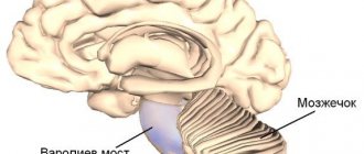
Cerebellomedullary (large) cistern
, cisterna cerebellomedullaris (magna)
. Located between the cerebellum and medulla oblongata. It communicates with the fourth ventricle through the median aperture and continues into the subarachnoid space of the spinal cord. Rice. B.
Cistern of the lateral fossa cerebri
, cisterna fossae lateralis cerebri
. Determined in the lateral sulcus between the insula, parietal, frontal and temporal lobes. Contains branches of the middle cerebral and insular arteries. Rice. IN .
Interpeduncular cistern
, cisterna interpeduncularis
. Located behind the chiasm cistern on the lateral side of the temporal lobe and cerebral peduncles. It contains the oculomotor nerve, basilar, superior cerebellar and posterior cerebral arteries. Rice. B.
Covering tank
, cisterna ambiens
. Located on the lateral side of the cerebral peduncle. Contains the posterior cerebral, superior cerebellar arteries, basal (Rosenthal) vein and trochlear nerve. Rice. E. eleven.
Cerebellopontine cistern
, cisterna pontocerebellaris
. It is located in the region of the cerebellopontine angle and communicates with the fourth ventricle through the lateral aperture. Rice. D.
12.
Arachnoid granulations
, granulationes arachnoidalis
. Avascular, villous-shaped outgrowths of the arachnoid membrane, penetrating the sagittal sinus or diploic veins and filtering cerebrospinal fluid from the subarachnoid space in the blood. Intensive formation of these structures begins after 10 years.
The cisterns of the brain are areas, the space located between the structures of the brain. In general, the human brain is an organ of the central nervous system, consisting of an incredibly large number of neurons that are interconnected.
Brain structure
The cranial cavity, which is the “storage” of the brain matter, also protects the bones from mechanical influences coming from the outside. It must be said that the brain is covered with several membranes:
- Cobweb;
- Soft;
- Solid.
They are all responsible for certain processes. And their consideration should be given special attention.
Meninges of the brain and their characteristics
So, the dura mater is a dense cranial periosteum, which has a particularly close connection with it. On its inner surface there are several processes that penetrate into the crevices of the brain in order to delimit the departments. One of the largest of these processes is located in the middle of the two hemispheres. It forms a kind of sickle. Its posterior section is connected to part of the cerebellum, thus limiting it from the occipital lobes.
On the upper part of the shell there is another small process - it is located near the sella turcica, thereby forming the diaphragm. This provides the pituitary gland with a high level of protection against too high pressure from the brain mass. In certain areas there are special sinuses - they are called sinuses. Venous blood flows through them.
Arachnoid and soft shells
The arachnoid membrane is located inside the dura mater. It is quite transparent and thin, however, despite this, it is highly durable. The arachnoid membrane completely covers the medulla, flowing from one part to the second. It is separated from the vascular space by a special subarachnoid space. It is not empty - it contains cerebrospinal fluid.
In those places where the shell is located above deep grooves, the so-called subarachnoid space is much wider. As a result, brain cisterns are formed. And that is why in these places the space forms a capillary gap, as it narrows. And since we are talking about this, we should note something about the arachnoid membrane.
The cisterns that form in it have the following names: cerebellocerebral and cistern of the intersection. The first is characterized by the fact that it is located between the cerebellum and the place where the medulla oblongata is located, and the second is responsible for functioning directly at the base of the brain. By the way, the cerebellomedullary is also called the large cistern of the brain.
And the meninges are the connective tissue structures that cover the spinal cord. And the most important thing that should be mentioned is that without tanks neither the brain nor the nervous system will work. The cerebellum will not receive all the necessary substances, and this is very important, since they feed the brain.
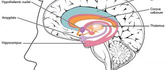
The human brain acts as a coordinating organ, which also ensures the regulation of all functions and systems of the body. Leading experts from various countries have been studying the anatomy of this main functioning organ for many years.
The brain consists of 85 billion nerve cells that form gray matter. The weight of the brain depends on gender and some characteristics of the human body. For example, in men, its average weight is 1350 g, and in women – 1245 g.
The weight of the brain is 2% of the total mass of the forehead.
It is worth noting that the brain mass can be more than 500 g larger than the average, but this does not in any way affect intellectual abilities. It has been found that people with a more developed brain structure, as well as a higher number of connections produced by this organ, have some intellectual advantage.
The main components of the brain are nerve and glial cells. The former form and then organize the transmission of impulses, while the latter perform executive functions. Inside the brain there are cavities (ventricles).
The brain is covered by 3 main membranes:
- Solid
- Soft
- Arachnoid
Between these membranes there is a free space that is filled with cerebrospinal fluid. The study of the anatomy of each shell made it possible to distinguish individual structural features and the number of vessels. Also, these shells additionally protect against the consequences of traumatic brain injury.
Dura mater
The dura mater (DRM) covers the cranial cavity from the inside and also acts as the internal periosteum. In the area of the large foramen and the back of the head, the dura mater is directed to the spinal cord. In the area of the cranial base, the shell adheres tightly to the bone tissue. A particularly strong connection can be seen in the area where the elements perform the connecting function and the release of nerves from the cranial cavity.
The entire internal area of the dura mater is covered with endothelium, due to which the shell takes on a smooth surface and a nacreous hue.
In some areas, division of the shell is noted, after which its processes begin to form in this place. In the areas where the processes extend, channels are formed, which are also covered with endothelium.
These tubules are the sinuses of the dura mater.
Sinuses of the brain: anatomy
The formation of dura mater sinuses occurs due to their separation into two plates, which are represented by channels. These channels distribute venous blood from the brain, which is then sent to the jugular veins.
The dura mater leaves that form the sinus appear as tight, stretched cords that do not subsequently collapse. allows blood to circulate freely from the brain, regardless of the state of a person’s intracranial pressure.
The following types of dura mater sinuses are distinguished:
- Superior and inferior sagittal. The first runs along the upper edge of the falx and ends in the area of the occipital protrusion, and the second along the lower edge of the falx and passes into the straight sinus
- Straight. Passes along the area in which the process of the falx communicates with the cerebellar tentorium
- Transverse (paired). Located in the transverse groove of the skull, located along the posterior edge of the tentorium of the cerebellum
- Occipital. Located in the thickness of the cerebellar falx, and then moves to the foramen magnum
- Sigmoid. Located in a groove in the ventral part of the skull
- Cavernous (paired). Located on the sides of the formation in the body of the sphenoid bone (sella turcica)
- Sphenoparietal sinus (paired). Subject to the lesser edge of the sphenoid bone and eventually breaks off into the cavernous sinus
- Rocky (paired). Located near the superior and inferior edges of the pyramidal temporal bone
The sinuses of the meninges begin to generate anastomoses with the external venous vessels of the brain using emissary veins. The sinuses also begin to communicate with the diploic branches, which, in turn, are located in the cranial vault and are further directed to the vessels of the brain. Next, the blood begins to flow through the choroid plexuses and then flows into the sinuses of the dura mater.
Venous sinuses
The following venous sinuses of the brain are distinguished:
- Upper. It passes along the falciform process and ends at the level of the occipital protuberance, where it passes into the right sinus.
- Lower. If the previous structure ran along the upper edge of the falciform process, then this one ran along the lower edge. It opens into the straight sinus.
- Straight. Located between the cerebellum and the falx process.
- Transverse sinus of the brain. This cavity is a pair, and was located in the cranial groove of the same name.
- Occipital. Distributed around the foramen magnum. Later it becomes sigmoid.
- Cavernous. Also paired. It is located and surrounds the sella turcica - the place in which the pituitary gland lies. This sinus differs from others in that the internal carotid artery, abducens, oculomotor, ophthalmic and trochlear nerves pass through it.
- There are also intercavernous, wedge-shaped, superior petrosal and inferior petrosal sinuses.
Vascular MO
The main number of pigment cells is observed at the base of the brain. This shell also includes:
- Lymphoid and mast cells
- Fibroblasts
- Neuron fibers and their receptors
Each part of the membrane is accompanied by arterial vessels, which further reach the arterioles. Between the walls and shells there are Virchow-Robin spaces, which are filled with cerebrospinal fluid. Ropes pass through them - fibrils, on which vessels are suspended, creating conditions for their displacement during pulsation, without affecting the medulla.
Spider MO
This type of meninges is separated by the subarachnoid space from the subdural, and appears as a tight rope between the gyri, but is not connected directly to the sulci themselves. The composition of the arachnoid MO includes various types of sections that belong to channels and cells.
The areas above the channels are distinguished by high permeability, through which various types of substances contained in the cerebrospinal fluid pass with a current.
In the areas where the shell is located, the subarachnoid space forms cisterns of various sizes (subarachnoid). Above the convex areas of the brain and on the surface of the convolutions, the arachnoid and vascular MOs are tightly connected to each other. It is in these areas that the subarachnoid space narrows significantly and ultimately turns into a capillary gap.
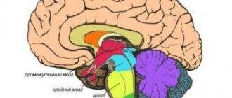
The largest cisterns in size are the brain cisterns, the anatomy of which varies quite a lot. The following types are distinguished:
- Cerebellocerebral, which is located between the medulla oblongata and the cerebellum. In the rear part, this tank is limited by the arachnoid membrane. Is the largest tank
- The lateral fossa cistern is located in the cranial fossa
- Cistern chiasm, located at the base of the cerebrum, in front of the optic chiasm
- Interpeduncular, formed in the fossa of the skull between the cerebral peduncles, in front of the posterior perforated substance
The subarachnoid space in the area of the foramen magnum is connected with the subarachnoid space of the spinal cord. The cerebrospinal fluid that fills the subarachnoid space is produced by the vascular plexuses of the cerebral ventricles.
From the lateral ventricles, the cerebrospinal fluid is directed to the 3rd ventricle, where the vascular plexus is also located. From the 3rd ventricle, through the plumbing system of the brain, the cerebrospinal fluid is directed to the 4th ventricle, and then joins the cerebellocerebral cistern of the subarachnoid space.
What should I do if I have a similar but different question?
If you did not find the information you need among the answers to this question, or your problem is slightly different from the one presented, try asking an additional question to the doctor on the same page, if it is on the topic of the main question. You can also ask a new question, and after a while our doctors will answer it.
We answer 96.81% of questions.
Hydrocephalus is a pathological process characterized by an increased content of cerebrospinal fluid in the cerebrospinal fluid spaces.
Which leads to their subsequent expansion and the development of atrophic changes in the brain tissue from compression.
CSF spaces include:
- 1) Cistern (especially the large cistern of the brain should be highlighted)
- 2) Ventricles (this is why during prenatal ultrasound examination the width of the bodies and horns of the lateral ventricles of the brain is measured)
- 3) Subarachnoid fissures located under the arachnoid membrane.
The main mechanisms that can lead to the development of hydrocephalus are the following, and they can be isolated or combined with each other:
- increased formation of cerebrospinal fluid
- decreased absorption
- dysregulation of cerebrospinal fluid balance.
Hydrocephalus primarily develops in childhood, including in newborns. The prevalence of this pathology is 0.01-1% among all children born under one month. Therefore, prenatal diagnosis occupies a special place, which is very relevant for severe degrees of hydrocephalus with almost complete atrophy of the brain substance.
Vessels and nerves of solid MO
The dura mater covering the anterior fossa of the skull is supplied with blood from this artery. In the posterior cranial fossa, the posterior meningeal artery branches, which goes from the carotid artery to the pharyngeal branch and then penetrates into the cavity of the cranium.
Also included in this area are the meningeal branches from the vertebral artery and the mastoid branch from the occipital artery. The veins of the choroid are connected to the adjacent sinuses of the solid myocardium, including the pterygoid venous plexus. In the area of the anterior cranial fossa, branches from the optic nerve (tentorial) enter it.
This branch, in turn, supplies the cerebellum and the medullary falx with the necessary substances. The middle meningeal branch, as well as a branch from the mandibular nerve, is directed to the area of the middle cerebral fossa.
Age-related features of the membranes of the brain and spinal cord
The anatomy of the hard mass in a newborn appears to be thin, tightly fused with the bone structure of the skull. The processes of this shell are poorly developed. The sinuses of the dura mater appear as thin walls, with a relative width. Also, the sinuses of a newborn’s brain are marked by greater asymmetry than in adults. However, after 10 years of development, the topography and structure of the sinuses are identical to adults.
The arachnoid and choroid of the brain in newborns are thin and delicate. The subarachnoid space is distinguished by its relatively large size, the capacity of which reaches about 20 cm 3 and subsequently increases rapidly. By the end of 1 year of life up to 20 cm 3, by 5 years up to 50 cm 3, by 9 years up to 100-150 cm 3.
The cerebellocerebral, interpeduncular and other cisterns at the base of the brain in a newborn are quite large. Thus, the height of the cerebellocerebral cistern is approximately 2 cm, and its width (at the upper border) is from 0.8 to 1.8 cm.
In order to function normally and maintain the vital functions of the body, the brain must be protected from external negative factors that can damage it. The role of protection is played not only by the bones of the skull, but also by the membranes of the brain, which represent a so-called protective case with numerous layers and structure. The layers of the meninges form, which contribute to the normal activity of the vascular plexuses, as well as the circulation of cerebrospinal fluid. We will look at what tanks are and what role they play below.
Meninges of the brain
The membranes have several layers: hard, which is located near the bones of the skull, arachnoid or arachnoid, and also a choroid, called a soft leaf, which covers the brain tissue and fuses with it. Let's take a closer look at each of them:
- The hard shell has a close connection with the bones of the skull. On its inner surface there are processes that enter the brain fissures to separate the sections. The largest process is located between the two hemispheres and forms a falx, the posterior part of which connects to the cerebellum, limiting it from the occipital parts. At the top of the dura there is another process that forms the diaphragm. All this helps to provide good protection against the pressure of brain mass on the pituitary gland. In some areas of the brain there are so-called sinuses through which venous blood drains.
- Inside the hard shell is the arachnoid membrane, which is quite thin, transparent, but strong and durable. It tears the brain matter. Under this membrane there is a subarachnoid space, which separates it from the soft sheet. It contains cerebrospinal fluid. Above the deep grooves, the subarachnoid space is quite wide, resulting in the formation of.
The meninges are structures of connective tissue that cover the spinal cord. Without the tanks, the brain and nervous system will not function.
Types of tanks and their location
The main volume of cerebrospinal fluid (CSF) is located in the cisterns, which are located in the area of the brainstem. Below the cerebellum in the posterior cranial fossa is called the greater occipital or cerebellocerebral fossa. Next comes the prepontine or pontine cistern. It is located in front of the bridge, bordering the interpeduncular cistern; behind it borders the cerebellocerebral cistern and the subarachnoid space of the spinal cord. Next are located. They are pentagonal in shape and contain cisterns such as the interpeduncular and intersection. The first is located between the cerebral peduncles, and the second is between the frontal lobes and the optic chiasm. The bypass or bypass cistern has the appearance of a distorted canal, which is located on both sides of the cerebral peduncles, bordered in front by such cisterns as the interpeduncular and pavement, and behind by the quadrigeminal. Next, let's look at where the quadrigeminal or retrocerebellar cistern of the brain is located. It is located between the cerebellum and the corpus callosum. In its area, the presence of arachnoid (retrocerebellar) cysts is often noted. If the cyst increases in size, then a person may experience increased pressure inside the skull, impaired hearing and vision, balance and orientation in space. The cistern of the lateral fossa is located in the cerebrum, in its lateral sulcus.
The cerebral cisterns are located primarily in the anterior part of the brain. They communicate through the foramina of Luschka and Magendie and are filled with cerebrospinal fluid (CSF).
Types of neoplasms
The classification is based on the localization and pathogenesis of formation.
- A retrocerebellar cyst is a fluid-filled cavity formation located behind the cerebellum.
- Arachnoid - the walls of the formation with liquid are the arachnoid membranes and parts of the brain tissue.
- A choroid plexus cyst is a cavity formation with fluid located among the plexuses of blood vessels.
- Pineal - a cystic formation with fluid in the area of the epiphysis.
Tumors, which are usually benign, are classified based on location:
- Liquor retrocerebellar cyst - located deep in the brain, containing liquid exudate. It often appears as a result of damage to the skull and intracranial formations, operations, and cerebral hemorrhage.
- Arachnoid retrocerebellar cerebrospinal fluid cyst - forms more often, in contrast to cerebrospinal fluid cyst. It is formed on the surface of the hemispheres of the main organ of the central nervous system and is filled with cerebrospinal fluid. Most often diagnosed in childhood due to intracranial pressure, inflammation, brain and cranial injuries.
What do arachnoid and retrocerebellar cysts look like?
Treatment options depend on the location of the retrocerebellar cysts. Moreover, the following subtypes of cystic formations are distinguished:
- Congenital (primary) - appears in the fetus during embryogenesis.
- Acquired (secondary) - occurs as a result of damage to brain structures, traumatic brain injury.
Movement of cerebrospinal fluid
The circulation of cerebrospinal fluid occurs continuously. It should be. It fills not only the subarachidal space, but also the central cerebral cavities, which are located deep in the tissue and are called cerebral ventricles (there are four in total). In this case, the fourth ventricle is connected to the spinal fluid canal. The liquor itself plays several roles:
Surrounds the outer layer of the cortex;
Moves in the ventricles;
Penetrates into brain tissue along blood vessels;
Thus, they represent part of the line of circulation of cerebrospinal fluid, are its external storage, and the ventricles are its internal reservoir.
Diagnosis and treatment of arachnoid cyst
Cysts that occur without any manifestations can only be detected by chance. In the case of neurological manifestations, the doctor first analyzes the patient’s complaints. However, the manifestations can only indicate that there are some malfunctions in the functioning of the brain, but do not allow us to classify the problem.
Hematomas, brain tumors, cysts located inside the brain have the same symptoms. For a more accurate diagnosis, the doctor may prescribe electroencephalography, echoencephalography or rheoencephalography. The disadvantage of these methods is that they do not provide information about the exact location of the formation or its nature.
Today, the most accurate diagnostic method, which allows a high degree of accuracy to distinguish an arachnoid cyst from a tumor or hematoma, is computed tomography (CT) and magnetic resonance imaging (MRI).
This can be achieved by various methods including:
- Shunting. With this method, the surgeon installs a tube (shunt) into the cyst through which fluid is drained to other parts of the body (for example, the abdominal cavity), where it is absorbed by other tissues.
- Fenestration. In this case, holes are created in the patient's skull and the walls of the cyst to drain and ensure normal flow of cerebrospinal fluid.
- Needle aspiration and connection using holes of the inner part of the cyst with the subarachnoid space to drain fluid into it.
Peculiarities
The circulation of cerebrospinal fluid has different directions of movement, it occurs slowly, depends on the pulsation of the brain, breathing rate, and the development of the spine as a whole. The main part of the cerebrospinal fluid is absorbed by the venous system, the rest by the lymphatic system. Liquor is closely connected with the meninges and tissue, ensuring the normalization of metabolic processes between them. Liquor provides an additional outer layer, which protects the brain from injuries and disorders, and also compensates for the distortion of its size, moving, depending on the dynamics, maintains the energy of neurons and the balance of osmosis in tissues. Through the cerebrospinal fluid, waste and toxins are released into the venous system, which appear in the cerebral tissue during metabolism. Liquor serves as a barrier at the border with the bloodstream; it retains some substances that come from the blood and allows others to pass through. In a healthy person, this barrier helps prevent various toxins from entering the brain tissue from the blood.
Second opinion for arachnoid cyst
Despite the fact that MRI diagnostics using a contrast agent provides the doctor with the necessary information, there is still a risk of error. It is associated primarily with the doctor’s lack of residual experience in interpreting MRI results and identifying cysts. Not a single patient is immune from such errors, and they happen both in large cities and in small towns.
The National Teleradiological Network (NTRS) offers you the opportunity to receive consultations from the country's leading specialists in the field of MRI diagnostics, who have extensive experience in analyzing tomographic images of various diseases. To get a consultation, you just need to upload the scan results to our server and within a day you will receive an alternative opinion to the opinion of your doctor.
Perhaps it will be the same as the first medical opinion, perhaps it will differ from it, but the second opinion will definitely allow you to reduce the risk of misdiagnosis and incorrect treatment to almost zero.
Deformation of tanks
Tanks play a special role in the movement of cerebrospinal fluid. The expansion of the cerebral cistern signals a disorder in the activity of the cerebrospinal fluid system. An increase in the size of the large tank, which is located in the small posterior cranial fossa, leads to deformation of the brain structure quite quickly. Usually people do not experience discomfort with mild enlargement of the cisterns. He may be bothered by minor headaches, mild nausea, and blurred vision. If the disease continues to progress, it can lead to serious health risks. Therefore, the synthesis and absorption of cerebrospinal fluid must remain in balance.
If a large amount of cerebrospinal fluid collects in it, they speak of a disease such as hydrocephalus. Let's consider this issue in more detail.
Complications of the disease
Hydrocephalus is a dangerous enough disease to ignore the symptoms of its manifestation. Ignoring treatment of pathology can cause disability or even a threat to life.
If treatment is not treated in a timely manner, a person’s ability to work and importance are lost. Impaired mental functioning, problems with movement and urination, blurred vision, epileptic seizures - all these are possible complications that result from delays in seeking medical help.
The greatest danger, perhaps, is the development of cerebral edema, characterized by a gradual loss of consciousness, which is accompanied by drowsiness, unilateral dilation of the pupil, fever and pyramidal insufficiency.
Chronic hydrocephalus in adults has a more favorable prognosis with appropriate and timely treatment.
Hydrocephalus
This disease occurs when the circulation of cerebrospinal fluid is disrupted. The reason for this may be increased synthesis of cerebrospinal fluid, difficulties in its movement between the ventricles and subarachnoid space, failure of the absorption of cerebrospinal fluid through the walls of the veins. Hydrocephalus can be internal (fluid forms in the ventricles) or external (fluid accumulates in the subarachnoid space). The disease occurs due to inflammation or metabolic disorders, congenital defects of the pathways that carry cerebrospinal fluid, as well as as a result of brain injuries. The presence of cysts also leads to the appearance of symptoms of pathology. A person complains of headaches in the morning, nausea, and vomiting. There may be congestion at the bottom of the eye or swelling of the optic nerve. In this case, a brain tomography is performed to make the correct diagnosis.
Pathologies and diseases
Venous discirculation is a pathology characterized by impaired outflow of venous blood from the sinuses. The causes of the disease are as follows:
- traumatic brain injuries;
- fractures of the skull bones;
- strokes;
- tumors;
The actions of all these factors come down to one phenomenon - external compression of the walls of the venous sacs. Sooner or later, the patient will begin to be bothered by the following symptoms:
- Constant headaches, especially in the morning.
- Migraine that appears after minor irritants - stress, fatigue, lack of sleep.
- When rising, a person feels darkening in the eyes and dizziness.
- Noise in ears.
- Constant fatigue, asthenia, muscle weakness.
- Insomnia is a sleep disorder.
- Memory deterioration, general inhibition of mental processes.
- Paresthesia on the arms and legs (crawling “goosebumps”, numbness).
Thrombosis of the cerebral sinuses is a serious disease that is manifested by the presence of blood clots (thrombi) in the sinuses. As a result, local blood flow deteriorates. This disease most often appears after:
- past infectious diseases: otitis media, sinusitis, tonsillitis;
- acute bacterial conditions: tuberculosis.
- fungal infections;
- excessive use of hormonal drugs;
- systemic autoimmune diseases: lupus erythematosus, sarcoidosis.
This disease usually develops acutely – within a few days. In a minority of patients, symptoms peak at 30 days. Signs of thrombosis are:
- Severe headache, nausea and vomiting, dizziness, double vision.
- Local seizures.
- Sensory and motor dysfunction. These people may experience sudden numbness or lack of strength in their arm.
In the case when the development of thrombotic disease develops rapidly, septic thrombosis is formed, accompanied by sudden changes in body temperature, extreme sweating and various disturbances of consciousness - from mild delirium to complete loss of consciousness - coma.
Fetal brain cistern
From the eighteenth to the twentieth week of a woman’s pregnancy, according to the results of an ultrasound, we can talk about the state of the fetal cerebrospinal fluid system. The data makes it possible to judge the presence or absence of brain pathology. The cistern magna is easily identified when using the axial scanning plane. It gradually increases in parallel with the growth of the fetus. So, at the beginning of the sixteenth week, the cistern is about 2.8 mm, and at the twenty-sixth week its size increases to 6.4 mm. If the tanks are larger, they speak of pathological processes.
Prevention of violations
Most brain development disorders occur during fetal development. You must adhere to the following recommendations:
- try to avoid infectious diseases, especially in the first trimester of pregnancy;
- Take medications with caution.
To prevent the development of hydrocephalus in children, it is necessary to avoid traumatic brain injuries and infectious diseases of the nervous system, since these factors are considered to provoke the development of hydrocephalus.
To maintain the viability of a patient with cistern deformities, doctors prescribe medications and regular examinations. If a deterioration of the condition is suspected, urgent surgical intervention is performed.
Diagnostics
In case of disturbances in the cerebrospinal fluid system, the following diagnostics are performed: MRI, CT, fundus examination, examination of brain cisterns using radionuclide cisternography, as well as neurosonography.
It is very important to know how the liquor system works, how its pathology arises and manifests itself. In order to undergo full treatment in case of detection of pathologies, it is necessary to consult a specialist in time. In addition, ultrasound results at different stages of pregnancy make it possible to study the development of the fetal brain in order to make a correct prognosis and plan treatment in the future.
Between the pia mater and the arachnoid there is a slit-like subarachnoid (subarachnoid) space of the brain, which directly passes into the same space of the spinal cord. The space between the membranes is filled with cerebrospinal fluid, which is similar in composition to blood plasma, is produced in the intracerebral cavities (ventricles of the brain) and circulates in the brain and spinal cord, supplying it with nutrients and other factors necessary for life.
Treatment of cerebral hydrocephalus in adults
If deformation processes are detected early, drug therapy is carried out. If the amount of accumulated fluid is very large, the patient may require urgent surgery. To do this, a small hole is made in the patient's skull into which a tube is placed. With its help, excess liquid is pumped out.
First of all, before determining therapeutic measures, it is necessary to identify the root cause of the development of hydrocephalus of the brain. In case of pathology of a tumor nature, first of all, the tumor formation itself is eliminated. When choosing a type of treatment, a specialist should be based on indications such as:
- stage of disease development;
- pathogenesis;
- age category of the patient;
- cerebrospinal fluid pressure level and many others.
If the disease is at the initial stage of formation, then the use of medications is possible. However, we must remember that medications may not eliminate the pathological process, but only slow down its development.
Surgery is considered the only effective method. A hastily performed operation and accompanying favorable factors can lead to a complete recovery and normal lifestyle.
Additional treatment methods include:
- physiotherapy;
- massage;
- microcurrent reflexology;
- treatment with medications.
The main objective of a neurosurgical operation is to create an artificial pathway that facilitates the removal of excess fluid to those areas where it will be freely absorbed.
For these purposes, the bypass method is used, which includes three components:
- venticular crater - it is installed in the area of the lateral ventricles of the brain;
- valve – regulates the outflow of fluid;
- peripheral catheter - is installed in the area whose task is to absorb outflow.
Be sure to read:
Schizophrenia: causes, symptoms, diagnosis and treatment
In modern medicine there is such a method as endoscopy. Its main goal is to create pathways for the outflow of cerebrospinal fluid from the ventricles into the cisterns of the brain, where fluid absorption will take place. This method has some advantages compared to bypass surgery, since it eliminates the presence of foreign bodies in the body, contributes to less trauma and reduces the risk of adverse consequences.