MRI is especially informative in detecting vascular pathologies, tumor processes and complications of traumatic brain injuries. Common indications for magnetic resonance imaging of the brain include chronic headaches, dizziness and syncope, numbness of the face, neck and upper extremities. During the procedure, the attending physician determines the condition of the bone tissues of the skull, soft tissues of the neck, spinal, cerebellar and cerebral arteries.
When studying the brain, anatomical and physicochemical disorders of its activity, structural changes in tissues, stenosis and occlusion of blood vessels, the quality and nature of neoplasms, and post-traumatic bone deformations are studied. In some cases, patients are prescribed MRI with a contrast agent, especially when it comes to differentiating tumors.
MRI of the brain - what is it?
MRI of the head is a non-invasive, painless procedure. Magnetic resonance imaging will show what changes have occurred in the brain, identify lesions, injuries, cysts, and determine the condition of blood vessels and the pituitary gland. The study is safe and can be used during breastfeeding and pregnancy, excluding the first trimester.
An image of the head is called a tomogram. After the examination, the patient is immediately given a series of images on film that show different sections of the brain. If necessary, the complete study is recorded on electronic media.
Pros and cons of MRI
MRI with contrast is considered the most effective method for diagnosing various pathologies. The human head is a vulnerable part of the body, so in most cases there is a high chance of getting head or brain injuries.
The advantages of MRI with contrast are that this procedure is painless, safe for health and is the most informative. This type of brain examination allows us to identify the scale, structure and parameters of the emerging tumor or metastases in the early stages of the disease.
The person undergoing this procedure will not feel any effects, and the exposure in this form is minimal and completely safe.
This type of research is not only the most effective, but also the safest for the human body.
But there is also a drawback to this procedure. It consists in the fact that there are some contraindications, that is, not every person is given permission to undergo the study. Such people are provided with an alternative research method, namely computed tomography.
What will a head MRI show?
A complete list of pathologies and conditions that a brain MRI can show:
- stroke;
- brain development disorders;
- tumor formations;
- foci of demyelination;
- consequences of TBI;
- vascular abnormalities;
- eye diseases;
- pathologists of the inner ear;
- traumatic injuries;
- pituitary diseases;
- signs of infectious diseases.
For better visualization, intravenous contrast is used. Preparations based on gadolinium are used, which, unlike iodine-containing contrast agents, is safer and causes fewer side effects.
Using contrast, abnormalities in the functioning of blood vessels are detected, as well as the activity/inactivity of lesions in multiple sclerosis.
How to prepare for the procedure
Nothing plays such a role in a brain MRI procedure as proper preparation. The reliability of the result will depend on it. It begins several days and sometimes weeks before diagnosis, and includes:
- Collecting the patient's medical history and complaints - the doctor must know what exactly is bothering his patient, in what situations discomfort occurs, and whether there is a possibility of hereditary diseases. The doctor also needs to find out whether the patient has previously had serious head injuries or head surgery.
- Laboratory diagnostics, the results of which can provide the doctor with additional information about the state of the patient’s body and the problems he has - chronic diseases and pathologies that are among the contraindications to this type of diagnosis.
- Consultation with specialized specialists who will add additional points to the patient’s history, which will help narrow the range of possible problems. Based on their findings, a radiology specialist will be able to select a highly specialized program before starting the procedure, as he will assume what an MRI of the brain can show.
During a consultation with a radiologist (doctors in this specialization usually examine the brain using MRI), the patient is explained in detail what he will encounter during the diagnosis. The doctor explains what an MRI of the brain is and how the process will go. Details such as the power of the installation in Tesla units are usually not covered, but the patient may wonder why an MRI should be done in his situation, and how the results will help in eliminating problems with his health.
During preparation, most of the examinees are interested in how long the brain examination procedure takes, how the results are interpreted, and what they need to be prepared for after the diagnosis. Answers to these questions are also obtained during a preliminary consultation with a radiologist. How long an MRI lasts depends on its diagnostic purposes and the power of the machine. The average duration of the procedure does not exceed half an hour.
The preparatory stage is completed before placing the patient on the magnetic resonance imaging table. The examinee should remove metal objects from the area being examined, including removable dentures, piercings, hearing aids, and accessories made of metal. It is advisable to remove keys, coins, phone and bank cards from your pockets (the latter fail when exposed to a magnetic field).
A special approach should be taken when examining children, mentally ill people and people suffering from claustrophobia, since they can only prepare for an MRI of the brain with the help of sedatives. In case of general long-lasting nervousness or a high risk of panic attacks, MRI diagnostics are performed under anesthesia, the use of which requires separate studies (ECG, a number of laboratory tests).
Important! If you plan to use a contrast solution during the procedure, an allergy test and a check of the functional state of the kidneys are needed on the eve of the procedure.
Technique
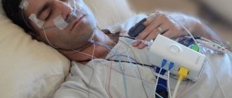
During magnetic resonance imaging, the patient will be asked to lie on a special mobile table. Later he will enter a tunnel, where the influence of the magnetic field will begin.
It is recommended that you lie still so that doctors get the clearest pictures possible. Diagnostics is accompanied by unpleasant clicking and noise, so it is recommended to wear headphones.
Using MRI of the brain, it is possible to detect an increase in the geometric dimensions of the ventricles of the brain, primary and secondary tumors, and thinning of tissue structures. The doctor is in the next room and monitors the progress of the examination and the patient’s condition.
Diagnostic time takes from 20 to 60 minutes. When using contrast, a regular scan is first performed, after which the examination is repeated, but with the administration of the drug.
The optimal power of a tomograph for brain research is considered to be 1.5 Tesla. Such magnets are located in closed devices.
How does the procedure work?
Before undergoing an MRI with drug administration, the patient will first have to undergo an MRI without drug administration. This is necessary in order to evaluate and compare the two results, after which it will be possible to draw accurate conclusions and prescribe treatment.
After the procedure, the person will have to temporarily leave a magnetic tunnel called a tomograph and will be injected with a substance called contrast. Contrast is injected into the vein only under the supervision of a resuscitator. This is an important step in the procedure. Therefore, despite the fact that such a substance is safe, it is important to listen carefully to your condition.
If dizziness, nausea or other signs of the body’s rejection of the drug appear, you should immediately inform a specialist. When no negative reactions are observed, the MRI procedure is performed again. During the examination, the person is periodically given a new dose of the drug without interrupting the procedure.
In order to conduct research, a person is placed in a tunnel specially equipped with all the necessary sensors. The patient puts on headphones, and a special bulb is placed in his hand, with the help of which he can give a signal about any deviations in his condition.
Since the procedure is absolutely painless, there is no need to worry, but the burden on the psyche at this moment is very large. The patient still remains partially outside, and there is enough air in the device. But if anxiety affects the patient too much, he may give a signal that he needs time to calm down a little. In some cases, he is given sedatives.
Contrast MRI of the brain is performed when it is necessary to conduct angiography of blood vessels in the brain. This method provides the maximum amount of information and is a reliable method for determining the presence of pathological conditions in a patient.
After conducting a brain examination with contrast, doctors will receive a three-dimensional picture of blood flow in the vessels, as well as the tissues that are nearby.
Then the doctor deciphers all the data in the images, and then the neurologist will be able to begin treatment, but this is only if no tumors were found in the patient. In the latter case, the treatment will be carried out by an oncologist.
If the images revealed pathologies related to blood vessels, the neurosurgeon will decide on additional examinations and treatment.
results
Usually, immediately after an MRI, the doctor sees changes that have occurred in the brain. The description is completed within an hour, but may take longer if there are many patients or a complex case is being studied.
Remember, the conclusion is not a final diagnosis. Only a specialized doctor can diagnose the disease and prescribe treatment, since many factors and the results of other examinations need to be taken into account.
Using tomography, specialists assess the extent of the lesion and give an objective assessment, which helps to accurately draw up a treatment plan or plan surgery.
Indications for the study
There must be a compelling reason to perform a brain MRI scan with contrast agent. Such an examination is prescribed in cases where accurate data is needed to confirm the diagnosis and determine the presence or absence of tumors in the brain.
Indications for MRI of the brain include injuries, vascular diseases, and epilepsy. It is carried out before surgery to assess the condition of the organ, as well as for persistent headaches, adenoma, if there is a suspicion of infectious diseases such as encephalitis, HIV, etc.
They may prescribe such a procedure to a patient if he has tumors, metastases, or suspected stroke. An MRI of the brain is usually prescribed after an initial examination by a doctor and tests.
There are quite a lot of symptoms that can lead to the appointment of an MRI of cerebral vessels. The main ones are:
- tinnitus;
- vegetative-vascular dystonia, previously established;
- suspicion of an aneurysm;
- frequent nosebleeds.
If a patient is suspected of having a concussion or is experiencing partial loss of consciousness, it is necessary to undergo the procedure. Also, reasons for undergoing an examination may be dizziness, sudden loss or impairment of hearing and vision, or previous injuries to the vertebrae and skull.
MRI is often indicated for those who have already had surgery, in order to assess the state of the brain after surgery, as well as determine the presence or absence of deterioration.
MRI in combination with a contrast agent can detect any disease in the brain even at the very initial stage of pathology development.
If there is at least one of the above reasons why you need to undergo an MRI, you need to consult a doctor and discuss with him the issue of referral for this procedure. An MRI scan done on time can not only simplify the task of treatment and determine the presence of pathology, but also save life or prolong it.
When a patient has doubts about how often an MRI of the brain can be done, the doctor will be able to reassure him, since this examination can be done as many times as necessary for the patient’s complete healing.
Price
Magnetic resonance imaging is an expensive diagnostic, so patients are prescribed procedures after the initial examination.
The price of the examination will depend on the type of equipment and the prestige of the clinic.
At rumrt.ru you can sign up for an MRI of the brain at a convenient diagnostic center.
Be sure to ask the doctor who gives the referral for recommendations on the type and power of the device. Since for some pathologies up to 1 Tesla may be enough, for others the minimum power should be 1.5 Tesla.
Doctors provide consultations in the following clinics:
"SM-Clinic" on the street. Novocheremushkinskaya (metro station “New Cheryomushki”)
- Bikineeva Dina Ilyinichna
"SM-Clinic" on Volgogradsky Prospekt (metro station "Textilshchiki")
- Atyasova Elena Viktorovna
- Babaeva Renata Magamedovna
- Bikineeva Dina Ilyinichna
- Blinova Elena Nikolaevna
- Kobriseva Yulia Alexandrovna
- Muratkhodzhaeva Malika Farkhadovna
- Mukhin Vyacheslav Yurievich
"SM-Clinic" on the street. Yaroslavskaya (metro station "VDNH")
- Bashirova Naima Magomedovna
- Muratkhodzhaeva Malika Farkhadovna
- Panfilov Oleg Nikolaevich
"SM-Clinic" on the street. Clara Zetkin (metro station "Voikovskaya")
- Azaryan Samvel Amirkhanovich
- Ivanova Irina Vladimirovna
- Ikonnikova Nina Yurievna
- Kiseleva Elizaveta Alexandrovna
- Muratkhodzhaeva Malika Farkhadovna
- Nikonorova Tatyana Alekseevna
- Panfilov Oleg Nikolaevich
"SM-Clinic" on Simferopol Boulevard (metro station "Sevastopolskaya")
- Atyasova Elena Viktorovna
- Brindar Nina Ganovna
- Gudimov Yakov Alexandrovich
- Makarov Artem Mikhailovich
Children's department on Volgogradsky Prospekt (Textilshchiki metro station)
- Atyasova Elena Viktorovna
- Babaeva Renata Magamedovna
- Bikineeva Dina Ilyinichna
- Blinova Elena Nikolaevna
- Kobriseva Yulia Alexandrovna
- Muratkhodzhaeva Malika Farkhadovna
- Mukhin Vyacheslav Yurievich
Children's department on the street. Novocheremushkinskaya (metro station “New Cheryomushki”)
- Bikineeva Dina Ilyinichna
Children's department on Simferopol Boulevard (metro station Sevastopolskaya)
- Atyasova Elena Viktorovna
- Brindar Nina Ganovna
- Gudimov Yakov Alexandrovich
- Makarov Artem Mikhailovich
Children's department on the street. Yaroslavskaya (metro station "VDNH")
- Bashirova Naima Magomedovna
Diagnostic results
Regardless of how long an MRI of the brain takes, the doctor interprets the results within 24 hours. In private clinics, this process takes less time, and the patient learns the diagnosis in 2-3 hours. In municipal clinics, you can find out the diagnosis in at least a day, and a quick result is possible only in emergency situations.
Closed and open MRI images of the brain look like monochrome black and white pictures, which clearly show the structures inside the skull. Pathology can be determined by characteristic patterns, painted in different shades of gray, white and black, and their localization. Thus, tumor processes look like bright white spots with an asymmetric edge, and multiple sclerosis is detected by MRI by the presence of small brightened areas in the white matter of the brain.
Important! Only a radiologist in tandem with a highly specialized specialist can interpret the result of a brain diagnosis, as shown by a separate image, and confirm or refute a preliminary diagnosis.
When MRI should not be used
The first thing doctors warn about before undergoing an MRI of the brain is that this procedure, like other methods of radiation diagnostics, has contraindications. Fortunately, only a small proportion of patients are not concerned about whether an MRI can be done in their situation. This method is relatively new, and everything new scares people and forces them to weigh the pros and cons.
The list of contraindications to MRI of the head is small and includes conditions and diseases that can be complicated when a person enters a strong magnetic field. These include:
- heart rhythm problems that have been resolved in the past by having a pacemaker installed;
- problems with cerebral vessels, which were eliminated with the help of hemostatic clips;
- Hearing problems that were solved in the past by placing an implant in the middle ear.
Patients with dental implants, crowns and braces made of ferromagnetic alloys, as well as after gunshot wounds, if there is a possibility of metal fragments being present in the body, are not allowed to undergo MRI diagnostics.
These contraindications are due to the fact that the magnetic field created during MRI of the brain can cause metal fragments to move. If implanted devices act as electronic stimulators (pacemakers) or repeaters (implants in the middle ear), then they may fail.
In some cases, an MRI procedure in patients with implanted ferromagnetic alloy devices can lead to sudden death, so it is important to inform the doctor about their presence.
There are also relative contraindications to diagnostics using magnetic fields:
- pregnancy up to 12 weeks;
- constant use of nervous system stimulants;
- heart failure in the stage of decompensation;
- insulin pumps implanted into the body of the subject;
- artificial heart valves.
When conducting examinations using contrast, special caution is exercised if the patient is diagnosed with renal failure. The problem is that contrast solutions are excreted through the urinary system, and the kidneys may suffer from the increased load on them.
Relative contraindications to MRI include tattoos made with inks containing metals. They do not pose a threat to life, but can affect the diagnostic results, distorting them.
Menstrual bleeding, IUD, breastfeeding and pregnancy over 12 weeks do not prevent head MRI.