Rathke's pouch (also called the pituitary tract) is an anatomical structure that appears during development in the embryo and is subsequently transformed into the anterior lobe of the pituitary gland. That is, normally, at the time of birth, Rathke’s pouch is absent, but in some people, some parts of it may remain.
If Rathke's pouch fragments are filled with colloid, a cyst is formed. This formation can have dimensions from several millimeters to several centimeters, oval or round in shape and is localized mainly between the anterior and posterior lobes of the pituitary gland.
Features of the pathology
The exact causes of Rathke's cyst have not yet been established, but some reasons are known that create an increased threat to its manifestation. This may include:
- genetic predisposition,
- acquired inflammations of various etiologies,
- intrauterine hypoxia,
- work in production facilities with increased doses of ultraviolet radiation, radiation,
- complicated childbirth.
Rathke's pouch cyst and pregnancy are considered by doctors to be quite close and related concepts, although they are not often encountered, only 1% of all cases of tumors. This formation can grow in the human body throughout his life completely asymptomatically. However, if favorable conditions arise for its growth, it will begin to actively develop.
Those who already drive in a high-risk group include:
- survivors of meningitis, encephalitis and stroke,
- cancer patients after chemotherapy,
- elderly.
Who is at high risk?
Rathke's pouch cysts rarely appear in humans. The congenital disorder occurs in 1% of the total number of benign neoplasms. Doctors were able to identify the category of the population susceptible to developing such a disease.
These include:
- Patients of mature age, in whom the tendency to form a cyst appears after 40 years.
- People treated for meningitis.
- Patients after strokes with cerebral hemorrhages.
- Those who have had encephalitis.
- Workers in hazardous conditions.
- People after chemotherapy or radiation.
- Patients whose relatives have such a disease or other brain tumors.
The congenital pathology described in the article is a rare occurrence. It accounts for only 1% of cases among other benign neoplasms. Nevertheless, experts were able to identify a group of people whose representatives are more susceptible to developing the disease. It includes:
- Women of mature age. A tendency towards an increase in the size of the cyst and the appearance of its symptoms is observed after 40 years.
- Patients after meningitis or encephalitis of any etiology.
- Patients who have had a stroke.
- Workers of hazardous chemical production.
- People whose relatives have been diagnosed with a brain tumor.
A match on one or more points from the list above means an increased risk of developing a neoplasm. Such people should pay special attention to their own health and be sure to undergo a body examination.
Rathke's pouch cyst: what is it?
It is localized in the head at the base of the skull in the area of the pituitary gland, which is why it is called a pituitary cyst.
It appears during intrauterine development of the fetus. This formation resolves in newborns. But in some children it remains and a cyst forms in its place. As long as it is not large, nothing bothers the patient. But as soon as its cavity is filled with colloidal fluid and reaches a significant size, it begins to put pressure on the pituitary gland, optic nerves and other organs, and then problems begin.
What complications can there be?
Rathke's pouch cyst does not pose a health threat. Complications can be caused by:
- Accelerated growth. Sometimes the patient becomes completely blind, mental abnormalities occur, and a clinical picture of destruction of the pituitary gland develops. Such situations are possible due to the development of a cyst.
- Sometimes a Rathke's pouch cyst develops into a tumor. If the neoplasm is benign, its development can be stopped with a therapeutic method.
- Formation of a malignant tumor, in which the disorder becomes unfavorable.
Transformations of such a cyst are rare; often the disease is at one stage of development.
Compressed blood vessels and brain tissue by a cyst can cause the following complications:
- Problems with intracranial pressure.
- Loss of vision.
- Hemorrhage.
- Pituitary adenoma.
- Formation of malignant formations.
To prevent the consequences of cyst formation from being complex, it is necessary to regulate hormonal levels and avoid head injuries. Go to the doctor at least once a year for an examination. Do not work in dangerous hazardous conditions, stop using tobacco and alcoholic beverages.
In most examples, pituitary developmental cysts do not reveal specific symptoms. Pathological transformations appear in a situation when the size of the formation becomes too large. High pressure is placed on the brain and tissues near the pituitary gland.
The work of the optic nerves and the dura mater of the brain, which helps regulate blood circulation in the head, becomes difficult. Patients develop the following pathological signs: disruption of the endocrine system, vision problems, migraines.
Several hormones important for the body appear in the anterior part of the pituitary gland. Such substances control metabolism and the functioning of various glands that affect the development of reproductive organs and other parts of the body.
Reasons for development at different ages
Based on many years of practical experience, doctors have come to the conclusion that the basis for the development of the disease at an early age are:
- infectious diseases suffered by the pregnant mother,
- heredity,
- oxygen starvation of the fetus,
- taking certain medications, poisoning with toxins,
- exposure to radiation and ultraviolet radiation (solarium, sun).
The formation of a Rathke cyst in the pituitary gland tissue occurs during the embryonic period of fetal development, when the body of the future person is formed. In the event that a malfunction occurs in the body of the mother and child, the cells from which the pituitary gland was built remain and continue their work according to a given program, creating a cavity inside the organ itself, which subsequently begins to fill the walls of the pocket with mucoid epithelium and, ultimately, an opuole is formed.
What factors provoke pathology?
The exact causes of the occurrence and development of the neoplasm have not been established. Doctors identify a number of factors, the presence of which increases the likelihood of a cyst. These include:
- hereditary predisposition;
- viral diseases with aggravated course;
- inflammatory processes;
- hypoxia;
- intoxication of the body of a pregnant woman;
- irradiation, radiation;
- difficult childbirth;
- excessive exposure to UV rays.
The neoplasm is formed in the embryonic period. Doctors suggest that from its remains another structure is born - the adenohypophysis. Rathke's pouch cyst and endocrine gland cyst have the same origin. Therefore, doctors tend to believe that a pathological neoplasm and a pituitary adenoma can develop in parallel.
Symptoms
One of the unpleasant properties of the disease is that it can be asymptomatic for a long time. Rathke's cyst is often discovered accidentally during an MRI. Obvious symptoms appear with its rapid growth.
Manifestations:
- insomnia,
- headaches,
- decreased vision,
- noise in the ear,
- loss of consciousness,
- nausea,
- mental disorders, memory loss,
- loss of coordination of movements,
- disruptions of the endocrine system.
The symptoms become more pronounced the larger the size of Rathke's tumor.
Dysfunction of the pituitary gland is also a rather unpleasant symptom, as it leads to an increased level of the hormone prolactin in the body, which, in turn, leads to various disorders in both men and women.
In the latter, first of all, menstrual cycles are disrupted, infertility develops, masculinization occurs, expressed in male-type hair growth, and a decrease in the timbre of the voice.
In men, hyperprolactinemia leads to feminization: female-type fat deposition and breast growth begin, and gradual impotence occurs. In both sexes, bone density decreases with the subsequent development of osteoporosis, diabetes and obesity develop.
Symptoms of Rathke's opuoli also include:
- decrease in saar in the blood,
- loss of appetite and weight loss,
- chronic fatigue syndrome,
- hypotension,
- leaching of calcium and potassium from bones.
All these symptoms are associated with a decrease in the level of the hormone cortisol in the body.
Cyst during pregnancy
During pregnancy, a woman must undergo a series of examinations. Sometimes diagnostics reveal disorders that were not previously thought of. If a Rathke's pouch cyst is detected in pregnant women, you will have to come to the doctor regularly for examination. The problem may be that all cysts tend to increase in size during pregnancy.
If the tumor is too large, the blood supply to certain parts of the brain is disrupted, the work of the pituitary gland is hampered, and hormonal disruptions occur. The functioning of the endocrine system is important for pregnant women, since the fetus will develop normally if the mother’s hormones are in order.
Diagnostic methods
Considering the insidiousness of the disease, if the above symptoms appear, you should undergo diagnostics to identify a Rathke cyst. There are three main diagnostic methods:
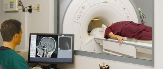
- Magnetic resonance imaging – MRI. The method is effective and accurate.
- CT – computed tomography. Using this method, a specialist classifies education and receives information about the process of its degeneration. When examining the brain, computed tomography is extremely necessary.
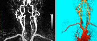
- Angiography study of cerebral vessels and conditions.
To accurately establish the final diagnosis of Rathke's cyst, a combination of all the data obtained is used: radiographic, clinical and bioimical. Sometimes a repeat MRI is required to clarify the diagnosis. It is especially important to obtain accurate diagnostic data when surgical intervention is required.
Treatment
The vast majority of doctors are inclined only to a surgical solution to the problem, believing that conservative treatment or drainage of a Rathke cyst does not give the desired result. Moreover, attempts at aspiration, that is, sucking out the contents of the cyst, can lead to its degeneration into a malignant tumor.
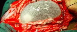
The removal operation is performed at the neurosurgery center, where today robots and high-precision instruments are used to carry it out. It is performed through the nasal opening, where a fiber optic endoscope with a microscopic video camera is inserted, so the surgeon sees the entire operation on the monitor screen.
The entire capsule with its contents is removed and sent for histological examination. After such operations, there is no open wound left, infection is excluded, and after 2–3 days the patient can be discharged home, and a week later full recovery begins.
If the symptoms of the disease are minor, treatment with hormonal drugs is prescribed, selected strictly individually for each patient. And the purpose is to stop the growth process of the tumor. If hormones cannot stop its growth, then surgical removal is prescribed.
There are two types of operations:
- The classic type, when removal is made through an incision in the area of the nasal concha through the sphenoid sinus. A binocular microscope is used.
- Laparoscopy is the most popular method. The operation is performed using an endoscope through the nasal cavity. In this case, the formation is completely removed. This type of surgical intervention is advantageous because it eliminates damage to the blood vessels of the brain and the pituitary gland itself.
Symptoms
A Rathke's pouch cyst can develop without symptoms over a long period of time. Often the disorder is detected during the diagnosis of other diseases. Clinical signs are often detected when the tumor expands and mechanical pressure is applied to nerve tissue and the pituitary gland.
In this case, problems arise with the blood supply to the brain, poor functioning of brain structures, and the outflow of cerebrospinal fluid becomes difficult. In addition, when cystic formations form, the following symptoms appear: dizziness, sleep problems, the appearance of hallucinations, migraines, vision problems, epileptic seizures, nausea, vomiting, noise in the ears, problems with coordination, loss of consciousness, mental disorders, problems with memory, endocrine system disorders, fine motor skills are difficult.
In many ways, the symptoms and level of their manifestation are determined by the volume of cystic formations and the pressure that is exerted on nerve cells, arteries, and veins.


