Diffuse axonal damage is observed in almost half of patients with severe traumatic brain injury, in which sudden acceleration or deceleration of the head, for example, at the time of an accident, leads to tension and rupture of axons.
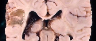
Pathomorphologically, diffuse axonal damage to the brain is characterized by widespread primary and secondary axonal ruptures (with retraction balls, accumulations of microglia, a pronounced astroglial reaction) in the centrum semiovale, subcortical formations, corpus callosum, brain stem, as well as pinpoint and small focal hemorrhages in the same structures.
Diffuse axonal damage to the brain is characterized by a long-term, multi-day coma from the moment of injury. Against this background, brainstem symptoms are expressed (different positions of the eyes along the vertical axis, severe paresis of the reflex upward gaze, suppression or loss of pupillary response to light on both sides, etc.).
Gross changes in the frequency and rhythm of breathing are often observed.
postural reactions are typical: coma is accompanied by symmetrical or asymmetrical decerebration or decortication, both spontaneous and easily provoked by painful and other stimuli. At the same time, muscle tone is extremely variable, which mainly manifests itself in the form of hormetonia or diffuse hypotension. Paresis of the limbs of a pyramidal-extrapyramidal nature, including motor tetrasyndromes, is often detected. Autonomic disorders are clearly expressed: arterial hypertension, hyperthermia, hyperhidrosis, hypersalivation, etc.
A characteristic feature of the clinical course of diffuse axonal brain damage is the transition from a long coma to a persistent or transient vegetative state, the onset of which is indicated by the opening of the eyes spontaneously or in response to various irritations, but there are no signs of tracking, fixation of gaze and execution of basic instructions. Vegetative states last from several days to several months with the development of a new class of neurological signs - symptoms of functional (or anatomical) separation of the cerebral hemispheres and subcortical-stem structures of the brain. Disinhibition of subcortical, oral-stem and spinal mechanisms is observed. The chaotic and mosaic autonomization of their activity causes the appearance of unusual, diverse oculomotor, pupillary, oral, bulbar pyramidal and extrapyramidal phenomena. The vivid reaction of the pupils to light is restored; although anisocoria may persist, constriction of the pupils on both sides predominates, often with variable spontaneous or (as a reaction to light stimulation) paradoxical dilation. Painful and especially postural irritations sometimes lead to tonic contraction of the eyes and the appearance of large converging nystagmus.
Facial synkinesis is often pronounced - chewing, sucking, smacking, grinding teeth, squinting, blinking. Swallowing and yawning automatisms are observed.
In the clinical picture of persistent vegetative states due to diffuse axonal damage to the brain, along with the activation of spinal automatisms, signs of polyneuropathy of spinal and radicular origin also appear (fibrillation of the muscles of the limbs and trunk, wasting of the muscles of the hand, widespread neurotrophic disorders).
As the vegetative state emerges, the neurological symptoms of dissociation are replaced by a predominance of symptoms of loss. Among them, extrapyramidal syndrome dominates with severe stiffness, incoordination, bradykinesia, oligophasia, hypomimia, minor hyperkinesis, and ataxic gait. At the same time, mental disorders clearly manifest themselves. Among them, the most characteristic are spontaneity (with indifference to the environment, untidiness in bed, lack of any active activity), amnestic confusion, dementia; at the same time, gross affective states are observed in the form of anger, aggressiveness, irritability.
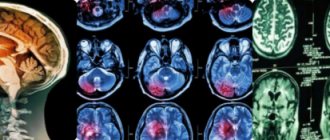
CT scan for diffuse axonal damage to the brain shows a general increase in brain volume (due to its edema and swelling), narrowing or complete compression of the lateral and third ventricles, subarachnoid convex spaces and cisterns of the base of the brain. Against this background, small focal hemorrhages can be detected in the white matter of the cerebral hemispheres, the corpus callosum, as well as in the subcortical and brainstem structures.
With the development of a vegetative state, characteristic dynamics of computed tomographic data are often noted: after 2-3 weeks. after injury, the phenomena of edema and swelling of the brain regress, small foci of increased density (hemorrhages) are either not visualized or their density becomes reduced, the cisterns of the base of the brain and convexital subarachnoid fissures begin to clearly appear, and there is a tendency to expand the (previously narrowed) ventricular system. Typically, this coincides in time with the transition of patients from a coma to a vegetative state.
Source: StudFiles.net
Course of the pathological process
Along with diffuse axonal damage, a prolonged coma occurs, which is accompanied by acute changes in muscle tone (from a sharp decrease to a spontaneous, paroxysmal increase in the limbs).
When the brain stem is damaged, the following symptoms of diffuse axonal brain damage are noted:
For injuries resulting from acceleration and deceleration forces, the impact of the skull on external structures is not necessary. Since the brain and cranial cavity have different densities, when exposed to the same inertial forces, they react unevenly. This movement discrepancy may contribute to rupture of the cerebral veins leading to the dural sinuses, as well as removal and rupture of parenchyma against rigid structures of the skull. In addition to this mechanism, because the central region of the brain is relatively fixed due to the presence of the brainstem, the peripheral regions of the brain and cerebellum tend to have a greater range of motion.
- fixed upward gaze;
- weakening or absence of reflex in response to irritation of the ocular membrane;
- inhibition of the oculocephalic reflex, etc.
The condition is almost always accompanied by meningeal syndrome (inelasticity of the neck muscles, intolerance to irritants, paresis).
Vegetative disorders are clearly expressed:
This difference in range of motion between the central and peripheral regions of the brain causes stretching of the axons and blood vessels of the brain, which may be a consequence of temporary dysfunction of the rupture of these structures 9, 10. Secondary lesions occur due to aggressions that begin after the moment of the accident, resulting from the interaction intra- and extra-frontal factors that add up to prevent the survival of encephalic cells freed from the initial injury. At the accident scene, the main factors of secondary injury are clinical relationships such as hypotension, hypoglycemia, hypercarbia, respiratory hypoxia, anemic hypoxia and hydroelectrolytic disturbances.
- overheating caused by the accumulation of excess heat in the body;
- increased sweating;
- increased salivation;
- impairment of respiratory function requiring connection to a ventilator.
The clinical course of DAP is characterized by frequent transitions from a comatose state to a stable or temporary vegetative state. This is evidenced by spontaneous or forced opening of the eyes (in response to a stimulus). There are no signs of tracking or gaze fixation.
Finally, there are mechanisms of cell, neuronal, endothelial and glial death due to ionic and biochemical disturbances that are associated with both primary and secondary lesions. Understanding these mechanisms has important implications for exploring the clinical and pharmacological approaches that have been used in recent years.
There are currently two known mechanisms of cell death, apoptosis and necrosis. Methods for identifying these structures in histological sections have been widely used in recent years. Apoptosis is a morphological manifestation of programmed death used in physiological situations, such as during the maturation of the nervous system, in which neurons that do not develop appropriate synapses are discarded. 16 Cell death by apoptosis has the advantage of causing little inflammatory response. which minimizes tissue damage; on the other hand, since it is a physiological mechanism, it expends energy.
Autonomic disorders with DAP can last for several days or months, in some cases even for years. They are gradually being joined by new classes of neurological signs - symptoms of functional or anatomical isolation of the cerebral hemispheres and subcortical-stem structures. This causes the occurrence of chaotic, uncontrolled oculomotor, glossopharyngeal and other phenomena.
On the other hand, all these factors can also lead to neuronal necrosis. Unlike apoptosis, in which the cell actively controls the process of its destruction, in necrosis an energetic breakdown occurs and death occurs due to the cell's inability to maintain its homeostasis. Thus, it is a less organized process and involves a higher intensity inflammatory response around it. The most well-known stimuli leading to necrosis are excitotoxicity and oxidative stress.
Excitotoxicity is the mechanism by which glutamate and other excitatory neurotransmitters cause cellular damage. 20 Glutamate levels increase during injury through multiple pathways. Second, other factors such as hypoxia, oxidative stress, and excitotoxicity-induced depolarization itself can disrupt membrane permeability control and further increase the presence of extracellular glutamate. Ionotropic receptors are the most studied in the mechanisms of excitotoxicity. Cell damage during excitotoxicity occurs in two stages.
Against the background of hyperreflexia (increased reflexes), postural-tonic reactions (“position reflexes”) and uncoordinated reflexes are observed spontaneously or in response to stimuli:
- tonic;
- turning the head, bending;
- paroxysmal tension of the abdominal muscles;
- flexion of the leg at the hip and knee joint in response to forced flexion of the toes (“triple shortening”);
- high-amplitude movements;
- broken hand poses;
- aimless repetition of movements;
- hand trembling.
Over a short period of time, the form and combination of such reactions in the patient change many times.
At the second moment, the opening of long-lasting Ca channels leads to excessive accumulation of intracellular calcium. The accumulation of calcium, in turn, can stimulate the activation of proteases, phospholipases and nitric oxide, which trigger a series of events that lead to cell death 22.
Activation of phospholipase A increases membrane permeability, generating the formation of arachidonic acid and free radicals. Oxidative stress results from the action of free radicals on tissue. Free radicals cause cell damage through various mechanisms. These actions lead to increased membrane permeability, which promotes the accumulation of intracellular calcium and glutamate release.
With DAP, facial synkinesis (involuntary muscle contractions accompanying other movements) may be observed:
- chewing;
- yawn;
- swallowing;
- smacking, etc.
Upon exiting the vegetative state, neurological reactions replace the symptoms of prolapse:
- extrapyramidal syndrome (excessive or insufficient motor activity, in this case there is severe stiffness);
- impaired coordination of movements;
- bradykinesia (slowing of friendly movements);
- impoverishment of speech;
- sudden uncontrolled movements of muscle groups;
- gait characteristic of ataxia.
At all stages of the course of DAP, mental disorders are present. The main ones are confusion, clouding of reason, amnesia. Physical and mental exhaustion are clearly expressed, there is no motivation for motor, speech and other forms of activity, (neuropsychic weakness) is observed.
Nitric oxide is produced by increased intracellular calcium concentrations associated with cellular aggression mechanisms present in injury. Diffuse lesions are those that affect the brain as a whole and usually result from kinetic forces that cause the encephalon to rotate within the cranial cavity. Dysfunctions can be detected by stretching or tearing both axons and vascular structures in specific areas of the brain.
Mild concussion covers cases with completely reversible neurological impairment, without loss of consciousness, and can be divided into three subgroups. The first subgroup consists of patients who experience confusion and mental disorientation within seconds, without amnesia.
Course of the disease
Simultaneously with diffuse axonal damage, long-term comatose states are detected, accompanied by changes in the functioning of muscle tissue. The tone quickly decreases or increases sharply. When damage to the brain stem occurs, the following signs of diffuse axonal damage to the head are revealed:
- Fixed focus of vision at the top point.
- The reflex responding to the absence of irritation of the eye shell is absent.
- Decreased oculocephalic response.
This condition always occurs in conjunction with meningeal syndrome. Disorders of the autonomic system are well distinguished:
- Overheating can occur due to a high concentration of heat.
- Intense sweating.
- Salivation.
- Problems with respiratory function. Patients are often placed on mechanical ventilation devices.
With this disease, the patient often falls into a coma and comes out of this state. Stable or periodic vegetative is determined. This is indicated by the voluntary opening of the eyes. Doctors do not observe signs of gaze fixation in such situations; the patient does not follow moving objects.
The duration of autonomic disorders can last several months or more than a year. Associated symptoms occur. The cerebral hemispheres are gradually isolated in terms of functionality or anatomical features. This provokes the appearance of chaotic, uncontrolled processes in the area of the pharynx and tongue.
Description of the disease
In general, a traumatic brain injury is a serious injury to the brain that has dangerous (sometimes irreversible) consequences for the body.
There are three degrees of severity of the lesion:
The last type is presented in the form of large-scale axonal damage, accompanied by cerebral hemorrhage. The clinical picture is characterized by a lesion at the junction of the white and gray matter. Small hemorrhages are found in the corpus callosum, semioval section, and upper part of the trunk.
In the second case, patients develop retrograde amnesia within five to ten minutes, and in the third subgroup, patients develop retrograde and anterograde amnesia. However, trauma develops and begins only after a sequence of events that can last for hours. There is a blockage in axonal transport caused by damage to intracellular structures, leading to local swelling and finally axonal rupture 31.
In diffuse lesions, the brain swells over many years due to loss of control of vascular toners. It was thought that this would lead to an uncontrolled increase in cerebral blood flow, which could cause unilateral or bilateral hemispheric filling. Thus, there would be no true edema, since there would be no accumulation of fluid in the interstitial space 32.
Diffuse axonal brain damage is a severe, extremely dangerous condition that poses a threat to life. Against the background of injury, hematomas form and accumulations of cerebrospinal fluid (CSF) form in the brain due to impaired outflow. Swelling and depressed fractures are observed. The listed phenomena precede DAP as such.
A brain tumor can be hemispherical or bihemispheric, also called diffuse. Focal lesions consist of hematomas—internally—or extracerebral or localized ischemic areas that affect only one area of the brain. In purely focal lesions, the remainder of the encephalium is assumed to retain preserved tissue and vascular properties.
Among focal lesions, cranial fractures may be linear or associated with focal subsidence. They arise from low kinetic energy injuries where there is contact of the head with rigid external structures 35. Linear fractures usually result from injuries that distort the cranial storage, and failures reflect more punctual impact. Isolated linear fractures do not cause brain damage, but they may be associated with lesions of vascular structures that cause potentially fatal complications such as extradural hematomas.
The main clinical manifestation is a prolonged coma, which over time turns into a vegetative state. With the latter, there is no cortical activity. This may continue for more than one year.
It is almost impossible to restore the previous state of the brain; few people manage to return to normal life. If the patient has experienced trauma, he is indicated for lifelong observation by a specialist and careful monitoring of the damage.
In cases where the dura is opened, the patient is also at risk of cerebrospinal fluid leak, meningitis and cerebral abscess. Children younger than one year of age may have a special type of immersion fracture, a ping-pong fracture. In this type of immersion, the area of the skull cap receiving impacts becomes concave, without the formation of bony spicules.
Extradural hematoma, in most cases, is the result of a cranial impact with low kinetic energy. For bleeding to occur, the vascular structures located in the epidural space must be ruptured, which usually occurs along fracture lines.
Diagnostics
Diagnosis of diffuse axonal brain injury is based on the biomechanics of traumatic brain injury and a description of the clinical picture. Analysis of the biomechanics of TBI makes it possible to suggest various variants and degrees of severity of primary brain damage, as well as to predict the development of secondary damaging mechanisms.
However, acute subdural hematoma is associated with accelerating and decelerating mechanisms of high kinetic energy injuries and, like diffuse cerebral lesions, does not require contact of the skull with external structures that need to be generated 41. The presence of blood in the subdural space, in close contact with the cortex, can cause secondary lesions associated with excitotoxicity 42.
Brain contusion consists of hemorrhagic areas around small vessels and necrotic brain tissue. Bleeding usually begins on the surface of the spinal cord, where the greatest friction occurs between the brain and the rigid structures of the skull. The mechanisms of contusion formation may be due to direct aggression of the parenchyma, as in the case of fractures with cranial current, or movement of the encephalon within the cranial box, which can lead to fragmentation of the parenchyma to the base of the skull or other rigid structures. In a piamara rupture, the contusion is called a rupture.
Computed tomography data in the acute stage of DAP are characterized by a moderate or severe increase in brain volume, narrowing or compression of the lateral spaces and the base of the brain. Tissue density often remains normal, but it may decrease or increase.
Finely focal accumulations of blood are often found in the white matter, corpus callosum and brainstem regions. The dynamics of computed tomography in patients with DAP is characterized by the rapid development of symptoms of the degenerative process.
3-4 weeks after the injury, clots of cerebrospinal fluid are detected in the frontal regions and between the hemispheres, which may disappear as the psychoneurological state normalizes.
Electroencephalography in DAP is characterized by persistent or temporary changes in subcortical or brainstem structures. Electroencephalography often reveals signs of diencephalic syndrome.
Somatosensory evoked potentials are the response of nerve structures when stimulated by current. With DAP, they are correlated with the severity of the injury. Recovery of CVD can take from several months to a year (or more), indicating a regenerative process.
A characteristic feature of the neurotransmitter response during DAP is a prolonged increase in serotonin concentration with inhibition of dopamine release and activation of adrenaline. This may be the basis for the use of drugs aimed at inhibiting the reactions of the sympathetic-adrenal system in complex therapy.
The results of a computed tomography scan can roughly suggest the presence or absence of intracranial pressure. The likelihood of intracranial pressure is high if there is no visualization during the examination of the third ventricle and basal cistern of the brain, or if there are signs of compression. In this case, a sensor can be connected to detect and correct intracranial pressure.
If a CT scan shows intact cerebrospinal fluid outflow tracts (third ventricle and basal cisterns), intracranial pressure is likely to remain normal. The likelihood of benefit from measures aimed at reducing it is then low. The information value of the study may be reduced in the presence of complications.
Acute respiratory distress syndrome: symptoms, diagnosis, treatment
- Acute onset (within 1 week)
- Bilateral opacities on chest x-ray
- PaO₂/FiO₂ (arterial to inspired oxygen) ratio ≤300 on positive end-expiratory pressure (PEEP) or continuous positive airway pressure (CPAP) ≥5 cmH₂O.
If there are no risk factors for ARDS, then acute pulmonary edema due to heart failure should be excluded.
Etiology
Many different conditions can lead to ARDS, although sepsis is the most common cause, usually of pulmonary origin (eg, pneumonia). Other conditions associated with ARDS include aspiration, inhalation injury, acute pancreatitis, trauma, burns, pulmonary contusion, lung injury after blood transfusions, cardiopulmonary bypass, fat embolism, widespread intravascular coagulation, and evidence of drug overdose.
Pathophysiology
The pathophysiology of ARDS is complex and incompletely understood. At the beginning of the development of ARDS, the primary pathological link is diffuse alveolar damage, although this is not expressed equally in all patients. Diffuse alveolar damage leads to damage to the alveolar-capillary membrane, consisting of alveolar type I and II pneumocytes and capillary endothelial cells. The alveolar air spaces are then filled with a proteinaceous milieu, inflammatory cells (neutrophils and activated alveolar macrophages), and inflammatory mediators, including proinflammatory cytokines, lipid mediators, and oxidants. Epithelial injury can be severe, with necrosis and sloughing of type I cells exposing the basement membrane.
Fibrin deposition occurs along the exposed basement membrane, resulting in hyaline membranes that are characteristic of diffuse alveolar damage. Type II cell damage and alveolar flooding contribute to surfactant dysfunction. Further, high-pressure, high-volume mechanical ventilation can harm the lungs by promoting a cascade of proinflammatory cytokines. At an early stage, ARDS clinically manifests as hypoxemic acute respiratory failure with an increased alveolar-arterial oxygen gradient and poor pulmonary compliance.
Multiple organ failure may occur simultaneously, especially if sepsis is the underlying cause of ARDS. Right ventricular dysfunction is also common and associated with worse outcome. Following the acute onset of alveolar edema and inflammation, some patients experience rapid resolution and return to normal lung histology and function.
The fluid that constitutes pulmonary edema is cleared by active transport of sodium and chloride across the alveolar epithelium. In other patients, the early exudative inflammatory stage progresses into the fibroproliferative phase. At this later stage, organized fibrous tissue develops in the lung and collagen deposition occurs, leading to irreversible and sometimes catastrophic pulmonary fibrosis. This phase is characterized by persistent respiratory failure, high minute ventilation, and poor pulmonary compliance.
Diagnostics
Because the diagnosis of ARDS is based on clinical criteria rather than a pathological diagnosis, ARDS should be considered in all patients with severe illness. About 40% of patients who meet criteria for ARDS are never diagnosed.
If patients develop bilateral infiltrates on chest x-rays, they may have or develop ARDS. The importance of assessing patients for the development of ARDS is primarily due to the survival benefits obtained with low tidal volume ventilation and pressure-limited ventilation.
Anamnesis
The history should be aimed at determining the presence of an underlying condition associated with ARDS, such as sepsis, pneumonia, gastric aspiration, pancreatitis, blood transfusion, or major trauma. The root cause may be an important determinant of the outcome; patients with ARDS due to sepsis tend to have the highest mortality. Specific treatments directed at the underlying cause are warranted, with particular attention to identifying and treating the source in the context of sepsis.
Symptoms that indicate ARDS include acute onset of shortness of breath and hypoxia leading to acute respiratory failure, and cough with expectoration of frothy sputum (a sign of pulmonary edema). The history should also contain information that would help suggest an alternative diagnosis to ARDS, such as pulmonary edema secondary to heart failure, diffuse alveolar hemorrhage due to pulmonary vasculitis, collagen vascular disease, or acute eosinophilic pneumonia.
Study
On physical examination, patients with ARDS have acute hypoxic respiratory failure that requires high oxygen concentrations and/or PEEP to maintain oxygen saturation >90%. Peak inspiratory pressure and positive end-expiratory pressure are also increased. Auscultation of the lungs may reveal basal or diffuse crackles. Particular attention should be paid to testing to identify the source of infection if sepsis is suspected to be the underlying cause of ARDS.
Basic tests include arterial blood gas analysis to calculate the PaO₂/inspired oxygen ratio. When screening for ARDS, the ratio of oxygen saturation to fraction of inspired oxygen (SpO₂/FiO₂) can also be used as long as SpO₂ is less than 97% (below the plateau on the oxyhemoglobin dissociation curve). SpO₂/FiO₂ 315 has been shown to correlate with PaO₂/FiO₂ 300.
Using the SpO₂/FiO₂ ratio identifies patients with similar clinical outcomes to those using the PaO₂/FiO₂ ratio.
A chest x-ray should be performed to look for bilateral infiltrates that are consistent with pulmonary edema and not fully explained by atelectasis or pulmonary effusion. Brain natriuretic peptide (BNP) levels should be considered if heart failure is a potential cause of bilateral radiographic infiltrates. BNP levels less than 100 ng/L (100 picograms/mL) indicate a low likelihood of heart failure, while BNP levels >500 ng/L (>500 picograms/mL) indicate it is probable. An echocardiogram should be done if heart failure is still a possible diagnosis after the BNP level is known, especially if there are no risk factors for ARDS. If BNP and echocardiogram are inconclusive, pulmonary artery catheterization (to assess left ventricular end-diastolic pressure) may be useful in differentiating heart failure from ARDS. However, routine pulmonary artery catheterization is not indicated in all patients.
Cultures of blood, sputum and urine should be performed to investigate the presence of sepsis. Bronchoalveolar lavage (BAL) or endotracheal aspiration for Gram stains and cultures is also recommended in patients with ARDS if pneumonia is suspected and in those without a specified predisposing condition.
BAL may also be useful in identifying other causes of acute respiratory failure with bilateral pulmonary infiltrates that mimic ARDS, such as diffuse alveolar hemorrhage or acute eosinophilic pneumonia. The best diagnostic test is an open lung biopsy. It is not routinely performed in critically ill patients due to the high risk of morbidity and mortality, but may be useful in resolving prolonged diagnostic uncertainty.
Serum lipase and amylase tests should be performed in patients with suspected acute pancreatitis.
A computed tomography (CT) scan of the chest is not usually needed to diagnose or treat ARDS. CT scanning is more sensitive than plain X-rays and can be useful in some cases to diagnose pneumonia or lung disease. CT scanning has shown that ARDS affects the lung parenchyma heterogeneously, with the dependent parts of the lung being most affected. However, routine chest CT scanning in ARDS to assess the heterogeneity of infiltrates is not currently indicated.
Risk factors
- Sepsis Sepsis is the most common underlying cause of ARDS and is usually of pulmonary origin. The prevalence of ARDS in patients with sepsis is between 6% and 7%, but is significantly higher in patients with septic shock. It is believed that systemic activation of inflammation and coagulation leads to indirect damage to the alveolar-capillary membrane.
- Aspiration of gastric contents is a common cause of ARDS. Approximately one third of hospitalized patients with previous aspiration are prone to ARDS. Aspiration is thought to cause direct damage to the alveolar epithelium and alveolar-capillary membrane.
- Pneumonia from any source (bacterial, viral, fungal, parasitic) is a common cause of ARDS. Direct injury by the pathogen and the inflammatory response to the pathogen are believed to be the responsible mechanisms.
- Approximately 7-10% of patients with severe trauma develop ARDS. Potential mechanisms include indirect injury from early hemorrhagic shock or late onset of multiple organ failure. Pulmonary contusions increase the risk of ARDS, as do long bone fractures, aspiration, and multiple blood transfusions.
- Multiple transfusions of blood components are associated with ARDS.
- ARDS, also known as primary graft dysfunction, occurs in 10% to 25% of lung transplant patients. The mechanism is thought to be injury due to ischemic reperfusion.
- Although ARDS is not well studied, it probably occurs in 10% to 20% of patients with acute pancreatitis. In one study, treating patients with acute pancreatitis with octreotide reduced the incidence of ARDS.
- Alcohol abuse is associated with an increased incidence of ARDS in adults with septic shock.
- ARDS occurs after burns and smoke inhalation, with an incidence of 40% among mechanically ventilated burn patients in one study.
- ARDS occurs after significant episodes of drowning (grades 3–6). These patients usually recover much more quickly than with other causes of ARDS.
Differential diagnosis
| Disease | Differential signs/symptoms | Differential examinations |
|
|
|
|
|
|
|
|
|
|
|
|
|
|
|
Diagnostic criteria
Berlin modification of the American European Consensus Committee (AECC) definitions
In 2012, minor changes were made to the AECC definitions of ARDS (called the Berlin Definition). A diagnosis of ARDS can be made if the patient meets all of the following criteria:
- Acute onset (within 1 week of known clinical event)
- Bilateral opacities on chest x-ray (not explained by effusion, collapse, or nodules)
- Respiratory failure not fully explained by heart failure or fluid overload (objective evaluation such as an echocardiogram is recommended unless there are risk factors).
Severity of ARDS
- Mild: PaO₂/FiO₂ 200-300 with PEEP or CPAP ≥5 cmH₂O
- Medium: PaO₂/FiO₂ 100 to 200 with PEEP ≥5 cmH₂O
- Severe: PaO₂/FiO₂ ≤100 with PEEP ≥5 cmH₂O.
Treatment
The goals of treatment for patients with ARDS are supportive care and a protective ventilation strategy using low tidal volumes to limit terminal plateau pressures. If the suspected underlying cause of ARDS is infection, the source should be identified and controlled, and antibiotics should be started immediately. Otherwise, the immediate goals are support and prevention of complications.
Mortality from ARDS is not primarily due to respiratory failure. Most patients die from the underlying cause of ARDS, secondary infections, other organ failure, underlying comorbidities, or complications of prolonged hospitalization.
Oxygenation and ventilation
Oxygen saturation must be maintained between 88% and 95%, which usually requires mechanical ventilation with titration of the fraction of inspired oxygen (FiO₂). Occasionally, patients can be treated with noninvasive ventilation, but failure rates in such cases are high and most will require endotracheal intubation and mechanical ventilation. Early use of high-flow oxygen via nasal prongs in patients with acute hypoxemic respiratory failure may also improve clinical outcomes, but this has not been studied in the context of ARDS. Ventilator-associated lung injury may be limited by the use of tidal volumes and plateau pressure restrictions with a protective ventilation strategy. This therapy has shown a reduction in mortality. A tidal volume of 4 mL/kg to 8 mL/kg per predicted body weight should be used to maintain inspiratory pressure <30 cmH₂O.
The predicted body weight for men is calculated as 50 + 0.91 × (centimeters of height minus 152.4), and for women - 45.5 ± 0.91 × (centimeters of height minus 152.4). If plateau pressure is > 30 cmH₂O, tidal volume should be reduced to 5 ml/kg or 4 ml/kg if necessary.
Positive end expiratory pressure (PEEP) and FiO₂ should be titrated using established PEEP titration tables. Although the use of PEEP titration tables with higher PEEP levels does not improve survival, available evidence suggests that higher PEEP levels are safe and may improve oxygenation in some patients.
Individual PEEP titration (rather than using a PEEP titration chart) is not recommended. A large randomized trial of individualized PEEP titration and recruitment maneuver versus titration of PEEP and FiO₂ using established protocols showed an increase in mortality with an individualized approach to titration of PEEP/recruitment maneuver. Prone positioning can improve oxygenation in patients with ARDS and has been shown to reduce mortality in patients with severe ARDS (PaO₂/FiO₂ < 150).
Given the potential complications of prone positioning, including facial swelling, pressure ulcers, and migration of catheters and endotracheal tubes, prone positioning should only be considered in patients with severe ARDS (PaO₂/FiO₂ < 150).
Respiratory acidosis, which is a common complication of ventilation with insufficient tidal volume, is corrected by increasing the respiratory rate. Although it is unknown what level of respiratory acidosis is harmful in patients with ARDS, permissive hypercapnia is often caused by inadequate airway ventilation. However, severe hypercapnia is independently associated with higher ICU mortality.
Normocapnia often cannot be achieved (and should not be the goal). Clinical guidelines recommend maintaining arterial pH between 7.30 and 7.45, but studies show that patients who undergo permissive hypercapnia can tolerate blood pH as low as 7.15. Bicarbonate infusions may be given when the pH is below 7.15.
Refractory hypoxemia
In patients with refractory hypoxemia despite an FiO₂ of 1.0 and a high PEEP level, oxygenation techniques should be considered for treatment.
- 1. Neuromuscular paralysis
Neuromuscular palsy improves the patient's ventilator synchrony and often improves oxygenation. In a randomized trial in patients with severe ARDS (PaO₂/FiO₂ ratio < 150), 48 hours of neuromuscular palsy with cisatracurium improved oxygen supply and reduced mortality in an adjusted analysis without increasing hospitalization-related paresis.
Neuromuscular palsy should be initiated when adequate oxygenation (oxygen saturation greater than 88% to 95%) cannot be achieved despite low tidal volume ventilation and adequate sedation, especially if there is ventilator-patient asynchrony. Intermittent doses of paralytics can be used as effectively as continuous intravenous infusion. If the patient is on a continuous intravenous infusion of muscle relaxants, neuromuscular monitoring of muscle twitch response to the drug should be used.
- 2. Inhaled nitric oxide and inhaled prostacyclin
Inhaled nitric oxide can improve oxygenation in patients with ARDS, but does not increase mortality and is associated with acute renal failure, and should therefore only be used as salvage therapy for refractory hypoxia. Inhaled prostacyclin is easier to administer than inhaled nitric oxide and also has the potential to improve oxygenation in ARDS through better ventilation-perfusion matching. However, there are currently no published large randomized controlled trials of inhaled prostacyclin; thus, it should be used cautiously and only as salvage therapy.
- 3. Extracorporeal membrane oxygenation
In some centers, extracorporeal membrane oxygenation (ECMO) is used in patients with profound refractory hypoxia. In one randomized controlled trial, transferring patients with severe ARDS to a tertiary care center that could provide ECMO along with a standardized approach to clinical management of ARDS resulted in improved outcomes, although not all transferred patients received ECMO.
- 4. High frequency oscillatory ventilation
Although routine use of high-frequency oscillatory ventilation (HFOV) for moderate to severe ARDS has not been shown to be beneficial or possibly harmful, HFOV may still have a role as a life-saving therapy for patients with severe ARDS and refractory hypoxia, as the use of HFOV often improves oxygenation. Routine use of HF ventilation is not recommended.
Conservative intravenous fluid administration
The patient's fluid balance should be maintained negative or neutral (assuming the patient is not in shock). A central line is recommended for measuring central venous pressure (CVP) with regular assessments of fluid balance. The goal is to keep the central venous pressure < 4 cmH₂O. Routine use of a pulmonary artery catheter (to measure pulmonary artery wedge pressure) is not recommended because its insertion is associated with greater morbidity than a central line.
A conservative fluid management strategy reduced the duration of mechanical ventilation but had no effect on mortality in a large clinical trial of patients with ARDS who were not in shock.
Antimicrobials
In patients who have an infectious cause of ARDS (eg, pneumonia or sepsis), it is important to promptly initiate antimicrobials. Empiric antibiotics targeting the suspected infection should be used as soon as possible after obtaining appropriate cultures, including blood, sputum, and urine. Antiviral or antifungal drugs may be appropriate in patients with suspected viral or fungal infections. Once the results of the cultures are known, it will be possible to select antibiotics that act on a specific microorganism. There is no evidence to support the use of antibiotics in patients who have ARDS without infection.
Symptomatic therapy
Standard supportive care for critically ill patients includes prophylaxis of deep vein thrombosis, control of blood glucose, prevention of stress-induced gastrointestinal bleeding, and hemodynamic support to maintain mean arterial pressure >60 mmHg. st and red blood cell transfusion in patients with Hb < 70 g/L (< 7 g/dL). Nutrition should be given enterally if possible.
In a large randomized trial of 1000 patients with ARDS, low-dose enteral nutrition during the first 5 days of ARDS had similar clinical results compared with full-calorie nutrition.
Supplemental nutrition with omega-3 fatty acids and antioxidants is not recommended. Inhaled or intravenous beta-adrenergic agonists are not recommended to facilitate alveolar fluid clearance and resolution of pulmonary edema. Early or late administration of corticosteroids has not been shown to reduce mortality in patients with ARDS, and their routine use is not recommended.
Treatment of diffuse axonal brain damage
Surgery is not required for DAP because there is no substance to remove. The patient requires prolonged artificial ventilation (moderate mode) and complex intensive care, including:
- taking nootropic and vasoactive medications;
- maintaining metabolic processes through tube and parental nutrition;
- adjustment of acid-base and water-electrolyte balance;
- implementation of actions aimed at normalizing osmotic and colloid pressure;
- regulation of homeostasis.
If there is a high probability of developing purulent-inflammatory and pulmonary complications, antibacterial drugs and immunostimulants are prescribed.
Early introduction of physical therapy aimed at restoring motor function, preventing contractures and correcting speech disorders is allowed.
To normalize the activity of the central nervous system and accelerate the healing process, long-term systemic use of nootropic and vascular drugs, medications that improve metabolic processes in tissues, and neurotransmitters (if indicated) is required.
Forecasts
The severity of the patient's condition and the outcome of DAP depend on the extent of the primary axonal damage and on the severity of secondary intracranial factors (swelling, metabolic disorders in the brain, swelling, etc.). The presence of extracranial complications also plays a role.
The prognosis depends on the effectiveness of therapy aimed at eliminating secondary mechanisms of brain damage and the development of complications. The longer and more severe the coma lasts, the higher the risks of an unfavorable outcome and decreased chances of recovery.
It should be noted that there is the possibility of complete or partial restoration of lost mental functions with a decrease in neurological disorders, even if the patient spent a long time in a coma, followed by a vegetative state for several more months.
This indicates the reversibility of structural disorders resulting from diffuse axonal damage to the brain.
The term “diffuse axonal brain injury” was first proposed in 1982 by JH Adams, and the pathology itself as a separate form of traumatic brain injury was first described in 1956 by SJ Strich, who observed patients in a vegetative state. This type of injury occurs as a result of rotational acceleration-deceleration, which occurs with inertial types of injury. This leads to complete or partial damage (ruptures) of axons, often combined with small focal hemorrhages. In most cases, such changes in brain tissue occur in areas with the maximum difference in the density of brain tissue - at the border of the gray and white matter of the brain.
Symptoms of diffuse axonal injury
Diffuse axonal damage to the brain is characterized by a prolonged coma that occurs immediately after the injury without a clear interval, symmetrical or asymmetrical symptoms of decerebration (decortication), often - variability in changes in muscle tone (from diffuse muscle hypotonia to hormotonia), severe brainstem symptoms, meningeal syndrome. With diffuse axonal damage to the brain, severe disturbances in vital functions, as well as pronounced autonomic changes, are almost always observed. Coma most often transforms into a transient and persistent or persistent vegetative state, in the event of recovery from which severe symptoms of prolapse remain (usually extrapyramidal symptoms and severe mental disorders dominate).
Symptoms
DAP is accompanied by a long-term (about 2-3 weeks) coma, in which the following are observed:
- stem symptoms - impaired pupillary reflexes, gaze paresis, distance of the eyeballs and others;
- change in respiratory rhythm;
- symmetrical or asymmetrical rigidity (increased tone) of muscles, which is provoked by external stimuli;
- paresis (decreased strength) of the limbs;
- autonomic disorders - hypertension, fever, etc.
Upon awakening from a coma, a person with DAP enters a vegetative state. Its signs:
- opening the eyes in response to stimuli or spontaneously;
- lack of fixation of gaze and tracking of objects.
The duration of the vegetative state varies from person to person – from several days to 3-5 months. In this case, the patient’s cerebral cortex does not function, and various symptoms of disconnection of the hemispheres and brainstem arise, in particular, unusual pupillary, oculomotor, oral, bulbar and other reflexes appear.
With a significant period of vegetative state as a result of DAP, signs of spinal and radicular polyneuropathy arise:
- muscle fibrillation;
- hand muscle wasting;
- neurotrophic disorders.
Tachycardia, hyperhidrosis, tachypnea and other vegetative-visceral disorders may also develop.
Exit from a vegetative state is accompanied by the appearance of symptoms of prolapse. The main consequences of diffuse axonal brain damage:
- extrapyramidal syndrome - it is characterized by muscle stiffness, ataxia (impaired coordination of movements), hypomimia (poor facial expressions), oligophasia (decreased speech activity);
- mental disorders - indifference to others, lack of motivation for activity, confusion of memory, dementia, aggressiveness.
Diagnosis of diffuse axonal damage
Diagnosis of diffuse axonal damage is based on taking into account the biomechanics of traumatic brain injury. A comatose state that occurs immediately after a TBI, with pronounced impairments of brain stem functions, generalized tonic reactions, and symptoms of symmetric or asymmetric decerebration (decortication), gives reason to assume diffuse axonal damage to the brain.
A computed tomography scan of the brain with diffuse axonal brain damage is characterized by an increase in brain volume due to its edema, swelling, hyperemia with compression of the cerebral ventricles and subarachondal convexital spaces. In this case, small focal hemorrhages are often detected in the white matter of the cerebral hemispheres, the corpus callosum, as well as in the subcortical and stem structures.
MRI reveals changes depending on the presence or absence of hemorrhages and their duration. A frequent finding of MRI examinations in diffuse axonal brain damage is small-focal hemorrhages in the deep subendian structures. Over time, the image intensity of these lesions decreases.
general information
Diffuse axonal damage to the brain, as a rule, becomes a consequence of inertial traumatic brain injury, that is, a unilateral centripetal impact on the head. In this case, tension and rupture of axons occur - the processes of nerve cells that transmit impulses from the cell body to the innervated (controlled) organs.
Typically, axonal rupture is combined with small focal hemorrhages (hemorrhages). Vascular and neural disorders are localized in the brainstem, paraventricular zones, white matter and corpus callosum. Pathological foci in DAP lead to a significant decrease in human brain activity.
Treatment of diffuse axonal injury
Victims with diffuse axial damage are not subject to surgical treatment. Indications for surgery for diffuse axonal damage to the brain arise only when concomitant focal damage is detected that causes pressure in the brain. Conservative treatment is carried out in intensive care units.
Victims who have diffuse axonal damage to the brain require long-term mechanical ventilation in hyperventilation mode with a set of therapeutic measures aimed at maintaining metabolic processes using enteral and parenteral nutrition, correcting disturbances in the acid-base and water-electrolyte balance, normalizing the osmotic and colloid composition blood, homeostasis systems. For the prevention and treatment of infectious and inflammatory complications, antibacterial drugs are prescribed taking into account the sensitivity of the microflora.