Home / Miscellaneous
Back
Published: 04/23/2020
Reading time: 20 min
0
6
- 1 Video about the anatomy of the human skull
- 2 Structure of the skull: sections
- 3 Occipital bone
- 4 Sphenoid bone
- 5 Frontal bone
- 6 Ethmoid bone
- 7 Shovel
- 8 Temporal bone
- 9 Parietal bone
- 10 Inferior turbinate
- 11 Lacrimal bone
- 12 Zygomatic bone
- 13 Hyoid bone
Evolution
It is important to know that vertebrates did not always have such a large head. Let's dive a little into the past. This part of the body appeared in ancient vertebrates during the fusion of the first three segments of the spine. Before this phenomenon, the same segmentation was observed. Each vertebra had its own pair of nerves. The nerves of the first vertebra were responsible for smell, the second for vision, and the third for hearing. Over time, the load on these nerves increased, it was necessary to process more and more information, which led to the thickening of these segments responsible for these sense organs. So they merged into the brain, and the union of the vertebrae formed the brain capsule (like a skull). Note that even the modern human head is still divided into the segments from which it was formed.
What is the average size of an adult's head? Length - 17-22 cm, width - 14-16 cm, height - 12-16 cm, circumference - 54-60 cm. The length of the head is usually greater than the width, so it is not round, but elliptical. It is also very interesting that the numbers (length, width and height) are not constant, they either increase or decrease. And all this depends on the location of the person.
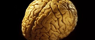
Brain
Before moving on to studying the areas of the head, it is worth saying that the head is considered the most important part of the body for a reason. After all, this is where they are located:
- brain;
- organs of vision;
- hearing organs;
- olfactory organs;
- taste organs;
- nasopharynx;
- language;
- chewing apparatus.
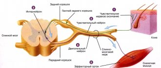
Now we will learn a little more about the brain. What is it and how does it work? This organ is formed from nerve fibers. Neurons (these are brain cells) are able to control the functioning of the entire human body by generating an electrical impulse. In total, twelve pairs of nerves can be observed that control the functioning of organs. Signals sent by the brain reach their destination through the spinal cord.
The brain is kept in fluid all the time, which prevents it from contacting the skull when the head moves. In general, our brain has pretty good protection:
- hard connective tissue;
- soft connective tissue;
- choroid;
- cerebrospinal fluid
The liquid in which our brain “floats” is called cerebrospinal fluid. The pressure of this fluid on the organ is considered to be intracranial pressure.
It is also important that the work of the brain and organs located on the head requires large energy costs. For this reason, we can observe intense blood circulation in this area. This:
- Nutrition: carotid and vertebral arteries.
- Outflow: internal and external jugular veins.
So, at rest, the head consumes about fifteen percent of the body’s total blood volume.
Treatment
Treatment includes:
- relief of symptoms;
- supportive measures.
There are no specific treatments for frontotemporal dementia.
In general, treatment is aimed at:
- symptom control;
- providing support.
For example, if the disorder is compulsive behavior, antipsychotic medications may be used. Speech therapy can help patients with speech disorders.
— Security and support measures.
Creating a safe and supportive environment can be extremely beneficial.
In general, the environment should be bright, cheerful, safe and stable, and should be designed to help the patient navigate. Some stimulation, such as radio or television, is helpful, but overstimulation should be avoided.
Structure and routine help people with frontotemporal dementia stay oriented and give them a sense of safety and stability. Any changes in the environment, routines, or changes in caregivers must be explained to the patient in simple and clear language.
Daily routines, such as washing, eating, or sleeping, help patients with frontotemporal dementia remember them. Having a bedtime routine helps them sleep better.
Scheduled activities at specific times help the patient feel independent and needed by directing attention to enjoyable or rewarding activities. Such activities should include physical and mental activities. Activities should be broken down into smaller chunks or simplified as dementia progresses.
- Caring for caregivers.
Caring for patients with dementia is stressful and demanding, and caregivers can become depressed and extremely tired, often neglecting their own mental and physical health. To help caregivers, the following steps may be helpful:
- Learn how to effectively meet the needs of people with dementia and what can be expected from such patients. Caregivers can obtain this information from nurses, social workers and organizations, as well as from publications and materials on the Internet.
- Seek help when needed: Caregivers can talk to social workers (including those at the local community hospital) about appropriate sources of help, such as day care programs, home visits from nurses, housekeeping assistance during part-time or full-day, as well as assistance with accommodation. Counseling and support groups may also help.
- Self-Care: Caregivers need to remember to take care of themselves. They should not deny themselves communication with friends, as well as favorite hobbies and other activities.
End of life issues
Before a patient with frontotemporal dementia becomes incapacitated, health care decisions must be made and financial and legal matters must be sorted out. These are called advance health directives. The patient must appoint a legally authorized representative to make treatment decisions on behalf of the patient (power of attorney for treatment decisions). The patient should discuss his wishes regarding health care with this person and with his doctor. Such issues are best discussed with all parties involved well in advance, before a decision becomes inevitable.
As frontotemporal dementia worsens, treatment is usually aimed at keeping the patient comfortable rather than trying to prolong life.
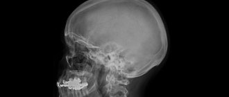
Skull and muscles
The skeleton of the head (skull) has an equally complex structure. Its main function is to protect the brain from mechanical damage and other external influences.
The entire human skull is formed by 23 bones. They are all motionless except for one - the lower jaw. As mentioned earlier, two departments can be distinguished here:
- cerebral;
- facial.
Bones related to the facial section (there are 15 in total) can be:
- paired - upper jaw, palatine bone, lacrimal, inferior nasal concha;
- unpaired - lower jaw, vomer, hyoid.
Paired bones of the medulla:
- parietal;
- temporal
Unpaired:
- occipital;
- frontal;
- wedge-shaped;
- lattice.
The entire brain section consists of a total of eight bones.
The cervical region, to which the skull is attached, allows the head to move. Movement is provided by the muscles of the neck. But on the head itself there are also muscle fibers that are responsible for facial expressions, one exception is the masticatory muscles, which are considered the strongest in this area.
What is the reason?
A headache in the crown area feels like a kind of “helmet”; there is a feeling of pressure especially in the upper part of the skull.
Often this condition is accompanied by a buzzing in the ears and an unpleasant pulsation in the temples. Let's look at the main reasons why the crown of the head may hurt:
- muscle tension;
- head injuries;
- emotional stress;
- migraine;
- alcohol and smoking abuse;
- cervical osteochondrosis;
- cluster pain.
The main causes of pain in the crown area are listed above; now let’s find out why it occurs and who is susceptible to this condition.
Constant stress
With emotional stress, the head muscles are in constant tone, so a person experiences pain that is localized in the center and spreads from top to bottom, creating a feeling of “tightness.” This kind of pain syndrome is usually stable, that is, it does not change its intensity under the influence of stress. But it happens that the pain becomes unbearable and a person is simply forced to take painkillers.
If pain is localized in the parietal part of the head and is accompanied by symptoms such as dizziness and numbness in the limbs, then the cause of this condition may be various psycho-emotional disorders, as well as neuroses.
This often happens to people who have been in a stressful situation for a long time. The body, thus, makes it clear to the person that it is working to the limit and needs rest.
According to statistics, 50% of people who suffer from various nervous system disorders have pain in the top of their heads.
Muscle fatigue
Many people in the modern world lead a sedentary lifestyle. For example, office employees spend a long time in a sitting position, which cannot but affect their well-being and health. Due to muscle tension in the neck and back, the parietal part of the head begins to ache. Such pain can also occur in people who work at a machine in one position, or in avid gardeners.
In addition, the cause of pain can be a constant lack of sleep and an unbalanced diet, as well as mental overload. It is worth noting that women suffer from this pain syndrome much more often than men, so the common complaint of the fair sex “I have a headache” may be true. This is especially true for women who lead a monotonous and sedentary lifestyle.
Migraine
One of the most common causes of pain in the crown area is migraine; it spares no one and occurs in people of any age and gender. It is characterized by the appearance of severe aching pain. Mostly the upper part of the head hurts; the duration of the pain syndrome can vary from an hour to several weeks or even months.
The causes of migraines are usually the following: the release of certain substances into the blood or degenerative changes occurring in the nervous system. This condition is usually accompanied by:
- sharp pain that is throbbing in nature;
- pain, which intensifies after sleep and eating;
- when walking or other physical activity, the pain also intensifies;
- nausea and vomiting.
Migraines can also occur due to excessive drinking and smoking, overeating, stress and excessive exercise.
Cluster pain
As a rule, this kind of pain is localized in a specific part of the head and lasts from several minutes to several hours. Basically, representatives of the stronger sex most often suffer from them, but women also experience them during menopause or PMS. The nature of such pain is usually unstable, the pain either decreases or, on the contrary, intensifies.
Typically this kind of pain is accompanied by the following symptoms:
- redness of the eyes is observed;
- pain increases with physical activity;
- Nausea and vomiting may occur;
- dizziness appears;
- sensitivity to light and noise increases.
Head injuries
The top of the head may hurt after a traumatic brain injury. It turns out that even with a minor injury, pain may appear, which will be accompanied by a decrease in memory, performance and concentration. Sometimes pain can be caused by psychological factors, for example, if the patient is a suspicious person who does not trust doctors and is afraid of various possible complications after an injury.
The pain syndrome can be chronic. Very often it occurs due to a concussion. The patient should immediately consult a doctor if the pain is accompanied by the following symptoms:
- memory loss;
- strong pain;
- temperature increase;
- visual impairment;
- general weakness and deterioration of health.
Pathologies of the cervical spine
If the top of your head constantly hurts, then perhaps the cause is various pathologies of the cervical spine. Osteochondrosis, instability of the cervical vertebrae, pinched blood vessels and nerves - all these reasons can cause headaches.
In this case, in order to get rid of unpleasant symptoms, you must visit a neurologist and, if necessary, a surgeon. If the diagnosis is confirmed, then manual therapy, acupuncture and massage should help. The patient may also be prescribed physical therapy and exercise.
Vascular pain
The upper half of the head very often suffers from pain caused by vegetative-vascular dystonia, high or low blood pressure. Due to excessively increased or, conversely, decreased vascular tone, pressure jumps. Nerve cells are compressed by the walls of blood vessels, which can cause vasospasm. This kind of pain is treated with medication.
In addition, patients must follow a diet, abstain from fatty and fried foods, as well as smoking and alcoholic beverages.
The skin on the crown of the head hurts
In some cases, it is not the head in the crown area that may hurt, but the skin. Painful sensations appear only when touching the skin. This may be due to allergies to various shampoos and hair care products. Sometimes such pain appears due to fungal infections of the scalp. Psoriasis can also be the cause, but in this case flaky plaques will be visible on the scalp. Pain syndrome can be caused by the heaviness of the hair, tight hairstyle or spasm of the blood vessels that nourish the hair follicles.
The people were taken aback! Joints will recover in 3 days! Attach...
Few people know, but this is exactly what heals joints in 7 days!
Head areas
The entire head is conventionally divided into 13 regions. There they also distinguish between paired and unpaired. And so, six of them are classified as unpaired regions.
- The frontal area of the head (attention is focused on it in the next section of the article).
- Parietal (detailed information will be presented to your attention later).
- Occipital (discussed in more detail in a separate section of the article).
- Nasal, which completely matches the contour of our nose.
- Oral, also corresponds to the contour of the mouth.
- The chin, which is separated from the mouth by the geniolabial groove.
Now we move on to listing the seven paired areas. These include:
- The buccal region is separated from the nose and mouth by the nasolabial groove.
- Parotid-masticatory (contours of the parotid gland and muscles responsible for the chewing reflex).
- The temporal region of the head (the contours of the scales of the temporal bone, located below the parietal region).
- Orbital (outline of the eye sockets).
- Infraorbital (below the eye sockets).
- Zygomatic (cheekbone contour).
- Mastoid (this bone can be found behind the auricle, which, as it were, covers it).
Body
It represents the very front part. The body is sloping at the top and front. It distinguishes:
Bottom surface. It has a pharyngeal tubercle, an area where the pharyngeal suture is attached. Two outer lines (edges). They are connected to the pyramids of the temporal element. Slope (upper surface). It is directed into the cranial cavity.
In the lateral part the groove of the petrosal inferior sinus is distinguished.
Frontal region
Now we move on to a detailed examination of the frontal region of the head. The boundaries of the anterior section are the nasofrontal suture, the supraorbital edges, the posterior section is the parietal region, the sides are the temporal region. This section even covers the scalp.
As for the blood supply, it is carried out through the following arteries:
- supratrochlear;
- supraorbital.
They arise from the ophthalmic artery, which is a branch of the carotid artery. A well-developed venous network is observed in this area. All vessels of this network form the following veins:
- supratrochlear;
- supraorbital.
The latter, in turn, partially flow into the angular and then into the facial veins. And the other part goes into the eye.
Now briefly about the innervation in the frontal region. These nerves are branches of the ophthalmic nerve and have names:
- supratrochlear;
- supraorbital.
As you might guess, they pass together with the vessels of the same name. Motor nerves are branches of the facial nerve called temporal.
Clinical manifestations
Three different clinical subtypes of frontotemporal degeneration have been identified.
The most common is the behavioral type of frontotemporal degeneration, found in more than 50% of cases. The basis of the behavioral type of FTD is personality changes and social disinhibition. Unlike Alzheimer's disease, visuospatial orientation is preserved.
A second, less common syndrome is semantic dementia, a disorder of conceptual knowledge. Patients with semantic dementia exhibit behavioral changes and difficulties understanding language, while speech itself remains intact. In addition to speech impairment, patients with prosopagnosia and/or associative agnosia may also be diagnosed with semantic dementia.
The third clinical syndrome is progressive aphasia with lack of fluency of speech, as indicated in the name of the term. characterized by an increasing loss of fluency in spontaneous speech and at least one of the following symptoms: agrammatism, anomia, or phonemic paraphasia.
Behavioral and semantic dementia is more common in men, and progressive aphasia with impaired speech fluency is more common in women.
Parietal region
This area is limited by the contours of the bones of the crown. You can imagine it if you draw projection lines:
- in front - coronal suture;
- posterior - lambdoid suture;
- sides - temporal lines.
Blood supply is facilitated by arterial vessels, which are branches of the parietal branches of the temporal artery. The outflow is the parietal branch of the temporal vein.
Innervation:
- in front - the terminal branches of the supraorbital and frontal nerves;
- sides - auriculo-vesical nerve;
- posterior - occipital nerve.
Occipital region
The occipital region of the head is located below the parietal region, and is limited to the posterior region of the neck. So, the boundaries:
- top and sides - labdoid suture;
- bottom - the line between the tops of the mastoid processes.
Arteries contribute to blood supply:
- occipital;
- posterior ear.
The outflow is the occipital vein, and then the vertebral vein.
Innervation is carried out by the following types of nerves:
- suboccipital (motor);
- greater occipital (sensitive);
- lesser occipital (sensitive).
Nervous system
The article briefly talks about the nervous system of some areas of the human head. From the table you will find out more detailed information. In total, the head contains 12 pairs of nerves, which are responsible for sensations, the secretion of tears and saliva, the innervation of the muscles of the head, and so on.
| Nerve | Brief Explanation |
| Olfactory | Affects the nasal mucosa. |
| Visual | It is represented by a million (approximately) tiny nerve fibers, which are the axons of the neurons of the retina. |
| Oculomotor | Acts as muscles that move the eyeball. |
| Block | Dealt with irritation of the oblique muscle of the eye. |
| Trigeminal | This is the most important nerve located on our head. It innervates:
|
| Abductor | Innervation of the rectus muscle of the eye. |
| Facial | Innervation:
|
| vestibulocochlear | It is a conductor between the receptors of the inner ear and the brain. |
| Glossopharyngeal | Innervates:
|
| Wandering | It has the most extensive area of innervation. Innervates:
|
| Additional | Motor innervation of the pharynx, larynx, sternocleidomastoid and trapezius muscles. |
| Sublingual | Thanks to the presence of this nerve, we can move our tongue. |
Circulatory system
When studying the anatomy of the head, one cannot ignore such a complex but very important topic as the circulatory system. It is she who provides blood circulation to the head, thanks to which a person can live (eat, breathe, drink, communicate, and so on).
The functioning of our head, or rather the brain, requires a lot of energy, which requires a constant flow of blood. It has already been said that even at rest, our brain consumes fifteen percent of the total blood volume and twenty-five percent of the oxygen that we receive when breathing.
Which arteries supply food to our brain? Mainly:
- vertebrates;
- sleepy.
Its outflow from the bones of the skull, muscles, brain, and so on should also occur. This occurs due to the presence of veins:
- internal jugular;
- external jugular.
Arteries
As already mentioned, the vertebral and carotid arteries, which are presented in pairs, supply food to the human head. The carotid artery is the basis of this process. It is divided into 2 branches:
- external (enriches the outer part of the head);
- internal (passes into the cranial cavity itself and branches, providing blood flow to the eyes and other parts of the brain).
Blood flow to the muscles is carried out by the external and internal carotid arteries. About 30% of the brain's nutrition is provided by the vertebral arteries. Basilar provides work:
- cranial nerves;
- inner ear;
- medulla oblongata;
- cervical spinal cord;
- cerebellum.
The blood supply to the brain varies depending on a person's condition. Mental or psychophysiological overload increases this indicator by 50%.
Vienna
When considering the anatomy of the human head, it is difficult to ignore a very important topic - the venous structure of this part of the body. Let's start with what venous sinuses are. These are large veins that collect blood from the following parts:
- skull bones;
- head muscles;
- meninges;
- brain;
- eyeballs;
- inner ear.
You can also find another name for them, namely, venous collectors, which are located between the sheets of the lining of the brain. Leaving the skull, they pass into the jugular vein, which runs next to the carotid artery. You can also distinguish the external jugular vein, which is slightly smaller and located in the subcutaneous tissue. This is where blood collects from:
- eye;
- nose;
- mouth;
- chin
Generally speaking, everything listed above is called superficial formations of the head and face.
Ethmoid bone
The structure of the human skull (the photo with the description shows parts of the ethmoid bone) includes another bone located inside the cranial set of bones. This small bone belongs to the nasal family.
We suggest you familiarize yourself with Why is your head spinning and how can it be dangerous for a person?
It includes in its design a top with a growth called a “cockscomb”, which has “cock’s wings” on the sides and a bottom that is part of the formation of the nose. On different sides of the “cockscomb”, along them, there are numerous openings for nerves passing into the brain.
On the sides of the “cock’s wings” there are flat areas that form parts of the eye sockets. These pieces also contain 1 through passage for vessels. The bottom of the ethmoid bone is filled with many canals, visually resembling a labyrinth.
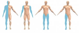
Muscles
To put it very briefly, all the muscles of our head can be divided into several groups:
- chewable;
- facial expressions;
- cranial vault;
- sense organs;
- upper digestive system.
You can guess the functions performed by their names. For example, chewing ones make the process of chewing food possible, but facial ones are responsible for human facial expressions, and so on.
It is very important to know that absolutely all muscles, regardless of their main purpose, are involved in speech.
Symptoms of the disease
With frontotemporal dementia, changes in the human personality occur, not for the better, so dramatic that relatives and friends, colleagues and neighbors simply stop recognizing the person. May appear:
- causeless rudeness, tactlessness, aggression with a complete absence of motives for this and repentance, even a logical explanation for one’s actions subsequently;
- inadequacy;
- theft, petty hooliganism;
- hyperoralism (obsessive smacking, chewing, sucking and the desire to put various objects in the mouth);
- neglect of work responsibilities, passivity, defiant behavior in a team, ignoring social norms;
- indifference to the feelings and problems of other people, lack of empathy, callousness;
- loss of strong-willed qualities, the ability to plan one’s affairs and bring them to completion, an unexpected change of plans, mechanical repetition of any actions;
- mood swings from excitement to apathy, distractibility;
- uncritical attitude towards oneself;
- sloppiness, neglect of personal hygiene, loss of motivation for neatness;
- gluttony, addiction to drinking and sweets;
- increased interest in sex;
- compulsive or repetitive behavior;
- wandering, verbose, incoherent and meaningless speech at the beginning of the disease, later - a decrease in speech activity;
- in later stages - pyramidal signs (pathological reflexes associated with neuronal damage).