Damages and lesions of the cerebral cortex
20.03.2019
Head injury is a complex of organic lesions of the external and internal structures of the head. External tissues include the soft tissues of the face and the bones of the skull. The internal ones are the brain and membranes. Violation of the organic structure leads to general cerebral and specific neurological and mental disorders.
- Classification of head injuries Inflammatory
- Purchased
- Congenital
Classification of head injuries
There is no single classification of head injury, however, from all existing ones, several “backbones” can be distinguished.
The first classification is based on the types of traumatic brain injury:
- Fracture of the cranial bones.
- Concussion. Neurological functions are impaired. Accompanied by complete or partial loss of consciousness, memory impairment, headache, vomiting and nausea.
- Brain contusion. A wound forms in the nervous tissue. The mechanism of injury is the impact of the brain on the opposite wall of the skull. Symptoms are determined by the location of the injury. Typically, trauma is accompanied by impaired consciousness, drowsiness and lethargy, and anxiety.
- Axonal damage. The long processes of nerve cells in the white matter are damaged.
- Compression of the brain. It manifests itself as headache, vomiting, agitation, paralysis of individual limbs or half of the body, epileptic seizures and increased blood pressure.
Damage to the cerebral cortex according to the second classification is based on acute circulatory disorders:
- Ischemic stroke. It develops due to a lack of blood supply as a result of blockage of an artery by a plaque or blood clot. Ischemic stroke is manifested by impaired consciousness, vomiting, acute headache and loss of neurological functions, for example, paralysis of the right side of the face or loss of speech.
- Hemorrhagic stroke. The cortex is damaged due to massive hemorrhage into the brain tissue. The clinical picture is similar.
Inflammatory
Inflammatory diseases of the cerebral cortex:
- Tick-borne encephalitis. Characterized by a stiff neck, high fever, and drowsiness. In later stages, the patient develops hallucinations and delusional disorder. Tick-borne encephalitis is also accompanied by mental agitation, convulsions and weakening of muscle strength in the arms and legs.
- Lethargic encephalitis, or sleeping sickness. The temperature suddenly rises and the throat hurts. Lethargy, drowsiness, and apathy appear. Patients become detached and indifferent. Double vision, speech disorder. Patients sleep during the day and are awake at night. In severe cases, catatonia develops - a waxy stupor: patients freeze in one place for a long time (from an hour to several days). Catatonic stupor is characterized by unnatural postures.
- Measles encephalitis. The temperature rises to 39-40C. Consciousness becomes confused, orientation in space is lost. Damage to the cerebral cortex is accompanied by mental agitation, hallucinations and coma. Cramps often occur in all skeletal muscles. Measles encephalitis is also accompanied by paresis and complete loss of muscle strength.
Purchased
Acquired diseases accompanied by damage to the cortex:
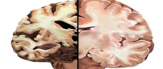
- Alzheimer's disease is a progressive neurodegenerative disease. It is characterized by a decrease in short-term memory capacity, difficulty recalling recent events, loss of long-term memory, speech impairment, poor cognitive abilities and loss of self-care skills.
- Pick's disease. Also a neurodegenerative disease. Accompanied by destruction and atrophy of the cerebral cortex. The disease is characterized by the development of total dementia, impaired attention, and speech disorder. The ability to think logically and build cause-and-effect relationships is lost. Perception is upset. Typically, patients are unaware of their condition, which is called anosognosia.
- Corticobasal degeneration. The disease is characterized by damage and atrophy of the frontoparietal cortex and basal ganglia. The main clinical syndrome is a violation of thinking and memory: the ability for abstract logical thinking decreases and the memory capacity decreases.
Residual organic damage to the central nervous system is a group of congenital syndromes and diseases in children that appear against the background of damage to the cerebral cortex. They are characterized by cerebral insufficiency, slowed intellectual development and impaired neurological functions.
Congenital
Congenital pathology of the cortex is divided into leading syndromes:
- Cerebrasthenic. Such children get tired quickly, they are always lethargic and apathetic. A few minutes after the influx of new information, they become distracted. Attention is scattered, so they remember poorly. Due to decreased appetite, they slowly gain weight.
- Neurosis-like. Characteristics: sleep disturbance, emotional lability, irritability. Somatic symptoms: loss of appetite and stool, excessive sweating, tendency to nausea and vomiting.
- Psychoorganic syndrome. Damage to the cortex in psychoorganic syndrome is accompanied by a decrease in the child’s intellectual abilities, poor memory and difficulty in retaining strong emotions. Reduced attention span: children capture few details in new information.
Didn't find a suitable answer? Find a doctor and ask him a question!
Source: //sortmozg.com/zabolevaniya/povrezhdenie-i-porazhenie-kory-golovnogo-mozga
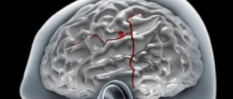
Structure
The performance of important functions by the cerebral cortex is ensured by its anatomical structure. In neurology, the cerebral cortex is represented by many layers that interact with each other, ensuring the full functioning of the system.
The structure of the cerebral hemispheres includes the elements:
- The molecular layer is a collection of chaotically located dendritic formations, the main task of which is to support the work of the associative function.
- The outer layer consists of many nerve cells that differ in shape and concentration.
- The outer layer of the pyramidal type also consists of nerve cells, the shape of which resembles a cone: an impressively sized dendrite emanates from the upper point of the neuron.
- The inner granular layer is represented by a collection of small nerve cells that are localized at a distance from each other. The resulting voids are replaced by a fibrous structure.
- The pyramidal layer contains neurons of medium and large volume, the upper points of which reach the molecular structure.
- The integument is also lined with neurons, the shape of which resembles a spindle, reaching the surface of the white matter.
Regardless of the fact that there are no clear boundaries, each structure performs individual functions, and their combination ensures the regulation of such important processes as the analysis of information and the synthesis of nerve impulses.
Cerebral cortex disorder treatment
Brain dysfunction
Brain dysfunction
The brain, which weighs no more than one and a half kilograms (in an adult), effectively controls all processes in the human body. However, with the slightest violations, serious deviations can occur - in behavior, emotional perception and intellectual development.
Minimal brain dysfunction - this diagnosis is made today to every fifth child, the reason for this is usually a difficult pregnancy or difficult childbirth, as well as prolonged exposure to toxic substances on the body of a pregnant woman.
In addition to perinatal causes, brain dysfunction can also be caused by traumatic brain injuries resulting from a fall, accident or blow, as well as an infectious disease.
Depending on which part of the brain was damaged or deformed, experts distinguish:
- dysfunction of diencephalic structures of the brain (regulate sleep, appetite, thermoregulation, metabolic processes);
- dysfunction of brain stem structures (responsible for basic life functions - breathing, appetite, muscle tone);
- dysfunction of midline brain structures. responsible for the autonomic functions of the nervous system and emotional state.
Minimal brain dysfunction can cause frequent headaches, hyperactivity and hyperexcitability in children, increased nervousness and pain, motor and speech disorders, delayed development, lack of attention and memory, and increased fatigue.
Dysfunction of the cerebral cortex #8211; the reason that the child cannot concentrate and systematically complete even a simple task. Over time, due to the “addition” of other unfavorable factors, minimal dysfunction can provoke the development of epilepsy and various neurotic disorders.
Therefore, if you observe any of the above symptoms in your baby, show him to a specialist. In many European countries, it has become a good tradition to be constantly monitored by an osteopath, who “guides” the child literally from the day of birth.
Dysfunction of brain structures identified at this age is painlessly eliminated or corrected without any consequences for the baby.
The cause of increased fatigue and headaches may be cerebral venous dysfunction. which also requires the participation of a qualified specialist who can identify and eliminate the cause of the violation of venous outflow.
Osteopaths have achieved brilliant results in the treatment of various functional disorders of the brain, whose methods are considered the safest and most effective.
Moscow. st. Lublinskaya. 151, business center Maryino, tel: (495) 545-7310.
When using site materials, an active hyperlink to www.rusosteopathy.com is required. | doctors
What symptoms that make it possible to differentiate damage to the cortical and subcortical structures of the brain can be identified when collecting anamnesis?
Damage to subcortical and cortical structures of the brain can be differentiated based on the following four criteria.
What specific symptoms are observed with lesions of the cerebral cortex?
The most useful symptom. indicating damage to the cortex of the dominant (usually left) hemisphere - aphasia.
Therefore, when collecting an anamnesis, attention should be paid to the entire complex of speech functions, not only the patient’s own speech, but also writing, reading, and understanding spoken speech.
A lesion that involves the left side of the brain but is not associated with speech impairment is unlikely to be cortical.
Damage to the cortex of the non-dominant (usually right) hemisphere is more difficult to recognize and is usually associated with a violation of visuospatial functions.
When the cortex of the non-dominant hemisphere is damaged, ignoring the opposite half of the space or one’s own symptoms is often observed. However, identifying such disorders based on the length of the history is often difficult.
and possibly with physical examination. It should also be kept in mind that epileptic seizures almost always indicate cortical involvement.
How, based on anamnesis data about the type of impaired sensitivity, can one differentiate damage to cortical and subcortical structures?
Most primary sensitivities “reach consciousness” at the level of the thalamus and do not require cortical input for their perception. A patient with severe cortical damage may feel pain, touch, and vibration. Thus, the presence of numbness or decreased sensitivity is more likely to indicate a subcortical lesion.
When the cortex is damaged, more subtle disorders of sensitivity are usually detected, which are associated with a violation of its more complex types: discriminatory sensitivity, sense of localization, graphesthesia. These symptoms may be difficult to determine from history.
Thus, cortical lesions involving motor, sensory, and language areas are usually too superficial to involve the visual subwindows and do not cause visual field defects. In contrast, subcortical lesions often affect the optic fibers, causing vision loss.
Thus, anamnestic evidence of visual field loss is more likely to indicate a subcortical rather than a cortical lesion.
Of course, lesions strictly limited to the occipital cortex cause visual impairment, but usually do not affect motor, sensory and other functions, and therefore, according to the clinical picture, they cannot be confused with a typical subcortical lesion.
- Damage to the spinal cord causes a triad, including symmetrical weakness of the distal muscles, disturbances of pelvic functions, and sensory disturbances of the conduction type (starting from a certain level)
- Damage to the brain stem causes dysfunction of cranial nerves and pathways
- Damage to the cerebellum causes ataxia and action tremor
- Cortical lesions can cause aphasia, epileptic seizures, and partial hemiparesis (involving only the face and arm), while subcortical lesions can cause visual field loss, primary sensory loss, and more widespread hemiparesis (face, arm, and leg).
If the history indicates damage to subcortical or cortical structures, what signs can be found during a neurological examination?
Signs revealed during examination usually correspond to the history,
- 1. Cortical dysfunction: the patient may exhibit aphasia. disturbances of visual-spatial functions, epileptic seizures.
- 2. Motor disturbances: signs of central paresis associated with dysfunction of upper motor neurons in the arm and face indicate damage to the cortex, and similar signs in the arm, face and leg indicate damage to subcortical structures.
- 3. Sensitivity disorders: with a subcortical lesion, the primary types of sensitivity are impaired: pain, tactile, vibration, and with a cortical lesion, the primary types of sensitivity remain relatively intact, but a higher level of sensory processes suffers, which is manifested, for example, by a violation of graphesthesia and stersognoea.
How does brain hypotrophy manifest?
The concept of malnutrition combines disorders associated with insufficient tissue nutrition. When the nutrition of brain tissue is disrupted, its development is delayed and its size is insufficient, as a result of which many problems develop in the body.
Brain hypotrophy can be either congenital or acquired. That is, the causes of this disease are internal and external.
Most often, children under 2 years of age suffer from this disease; in older children and adults, malnutrition is much less common.
Causes of brain hypotrophy
- Chromosome mutation
- Oxygen deficiency during fetal development,
- Unhealthy lifestyle of the mother during pregnancy,
- Mother's age during pregnancy is less than 18 and more than 42 years,
- Infections of a woman during pregnancy.
Hypotrophy of the cerebral cortex can develop due to the following reasons:
- Inadequate child care and inadequate nutrition,
- Acute infectious diseases,
- Brain malnutrition due to drug use and alcohol abuse.
Degrees of malnutrition
Since the brain does not receive enough nutrition, it cannot cope with its work and does not always adequately #171;give commands#187; other organs.
The first degree is characterized by a lag in body weight and height, that is, excessive thinness. At this stage, chronic disturbances in the functioning of the digestive organs are observed. However, psychomotor functions have not yet been impaired; the person is often still quite active.
The second stage of brain hypotrophy is accompanied by a decrease in emotional tone, apathy develops, and interest in life decreases. At the second stage, cortical hypotrophy of the brain may be observed.
In children, it causes a delay in the development of psychomotor functions. The ability to perform various movements and control one’s behavior is not developed in time, and the development of speech skills lags behind.
Externally, the phenomena of malnutrition manifest themselves in the form of dry flaky skin and lethargic behavior. Intercurrent diseases that are caused by impaired thermoregulation (for example, pneumonia) may appear.
At the third stage of malnutrition, the patient may be irritable, and he is practically not interested in what is happening around him. Developmental delay is pronounced, and loss of already acquired skills may also occur. At the third stage, cerebellar hypotrophy may occur.
The processes of malnutrition of the cerebellar tissues gradually increase and are combined with signs of disruption of the functioning of other parts of the brain.
As is known, the cerebellum is responsible for the coordination of movements, therefore, disturbances in its work primarily affect the state of the synergistic and antagonist muscles, that is, the muscles performing motor acts.
As a result, movements become uncoordinated, imprecise, and tremors are often observed.
In adults, damage to the cerebellum and the development of its hypotrophy can be observed during ischemic and hemorrhagic strokes. In addition, alcohol abuse and a viral infection can lead to malnutrition of this part of the brain.
Hypotrophic phenomena of the cerebellum are often accompanied by hydrocephalus and related symptoms. Among them:
- Increased intracranial pressure,
- Headaches, dizziness, tinnitus.
- Various neurological disorders
- Movement disorders
- Decreased visual acuity,
- Epileptic seizures,
- Mental disorders: intellectual and personal,
- Lethargy, apathy,
- Attacks of aggression.
Functions
There are many neurons in the structure of the cerebral cortex: due to the close relationship with the central part of the nervous system, information is perceived, processed, and the formation of reactions, both physiological and mental.
In addition to data analysis, the cerebral cortex performs the following functions:
- ensuring the coordinated functioning of internal organs;
- stabilization of metabolic processes;
- stimulation of the mental function;
- the formation of the relationship between the soft tissues that make up the organ structures;
- control over the functioning of the speech apparatus;
- stimulation of intellectual function;
- improvement of creative abilities;
- stabilization of the psycho-emotional background.
Another important function of the cerebral cortex is the synthesis of electrical signals, the degree of activity of which is determined using amplitude indicators and a person’s well-being.
The anterior part of the cerebral cortex is characterized by low susceptibility to external influences, therefore electrical impulses localized in this area do not manifest themselves in the form of bright reactions. The main functions of the anterior cortex of the cerebral hemispheres are considered to be self-awareness and the formation of abilities: if the anatomical element is damaged, a person loses interest in appearance, work activities and communication with loved ones.
Consequences
Damage to one of the parts of the cerebral cortex leads to the loss of such vital functions as the perception and processing of external information, the transmission of nerve impulses, and the provision of a response to irritating factors.
A malfunction in the functioning of the cerebral hemispheres is an indication for a visit to a neurologist, who will perform an external examination and refer the patient for a comprehensive diagnosis.
Dangerous consequences that develop against the background of untimely diagnosis or poor-quality treatment of damage to the cerebral cortex include atrophy.
This complication not only accelerates the aging process of brain tissue, but also provokes the death of local nerve cells.