When is MR arteriography required?
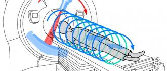
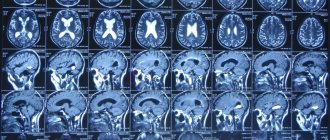
Today, MR arteriography of the vessels of the head is recognized as the most informative and safe diagnostic method. This is due to the fact that MRI allows you to create a 3D model of the intracerebral segments of the main great arteries. Thus, the procedure involves the study of the vessels of the circle of Willis - the carotid arteries and their branches, intracranial segments of the vertebral arteries, the basilar artery, the posterior cerebral arteries, the cerebellar arteries, and the communicating arteries.
Brain arteriography is required in situations if:
- there are signs of vegetative-vascular dystonia;
- the patient complains of tinnitus and spontaneous dizziness;
- the patient suffers from a headache, the cause of which could not be determined;
- vertebrobasilar insufficiency was detected;
- there is a risk of developing a stroke.
Important! Cerebral arteriography is a valuable method of differential diagnosis, both from the point of view of clarifying the diagnosis and in the process of preparing for future surgical intervention. Thanks to the creation of a 3-D model, the neurosurgeon can think through the stages of the operation down to the smallest detail, which reduces the risk of complications.
How is angiography performed?
Indications for angiography using MRI are:
- Traumatic brain injury;
- Vasculitis (inflammation of the vascular walls);
- Vascular atherosclerosis;
- Phlebeurysm;
- Aortic dissection;
- Congenital heart defect;
- Visual and hearing impairments;
- External vascular compression syndrome;
- Frequent headaches;
- Narrowing of arteries in diameter;
- Suspicion of cancer.
Most brain pathologies have a cause-and-effect relationship with the condition of the vessels of the neck and the brain itself. Therefore, the possibilities of angiography are extensive: it can reveal not only the disease, but also the causes that caused it.
A cerebral MR angiogram may be performed:
- For assessment of the arteries of the head and neck before surgery or minimally invasive intervention.
- For more information about the blood supply to the tumor.
- As a replacement for CT angiography when iodinated contrast material cannot be used.
The procedure can be used to diagnose the cause of the following symptoms:
- Severe headaches
- slurred speech
- dizziness,
- double vision,
- weakness or numbness
- loss of coordination or balance.
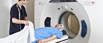
This examination is usually performed on an outpatient basis and takes about one hour. The patient is placed on a movable examination table, and the body is secured using straps and bolsters. If contrast material is used, the doctor or nurse will place an IV to give the solution intravenously.
The study itself usually involves several runs, which can last several minutes. The patient is placed inside the tomograph, and the doctor will supervise the process from an adjacent room. The device can make a lot of noise during operation, so you may need to protect your ears with headphones or earplugs.
What does non-contrast MR arteriography of blood vessels show?
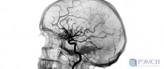
Magnetic resonance imaging of the brain using the vascular bed scanning mode allows us to identify:
- vascular aneurysms;
- vascular atherosclerosis;
- dissection of arterial walls;
- the presence or absence of stenoses and vascular occlusions;
- vascular malformations;
- vascular deformations affecting the passage of blood flow, including tortuosity of various degrees of severity;
- hypoplasia of brain segments, etc.
Vascular arteriography is an indispensable procedure for diagnosing brain diseases. Using magnetic resonance imaging, specialists can see minimal deviations from the norm, which is the key to providing timely medical care.
Preparing for the examination
During the examination, the patient lies on his back on the table. It is important to lie still while the MRI machine is operating. The duration of the procedure is up to an hour and depends on the area being examined. In some cases, it is necessary to administer a contrast agent before the examination.
Usually, before tomography in angiography mode, a radiologist recommends coming for an examination after abstaining from food for 4-6 hours. Then the data obtained as a result of the study will be reliable.
It is better to come to the diagnostic room in loose clothing without metal fittings, because the procedure requires the absence of metal. Otherwise, you will have to change into hospital clothes: a shirt or gown. You also need to remove all jewelry in advance: piercings, earrings, rings, chains and watches.
If the MRI requires the administration of a contrast agent, then you should warn the doctor in advance about diseases such as renal failure and bronchial asthma, as well as about pregnancy, if it is present or only suspected.
6000 4500
| Study | Price |
| MRI of cerebral venous sinuses | |
| 3500 |
**The promotion “Cheaper at night” is valid from 23.30 to 7.00
Arteriography: key advantages of the study
The main advantage of arteriography is that no contrast agent is used during the procedure. With computed tomography, cerebral vessels can only be visualized using contrast. SCT uses iodine-containing drugs, which cannot be used by certain categories of patients, for example, people suffering from renal or liver failure, which makes screening inaccessible for them.
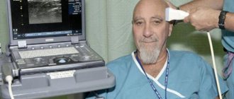
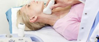
Arteriography has a visible advantage over ultrasound diagnostics. In particular, ultrasound does not allow examination of vessels that are overlapped by the thick bones of the skull.
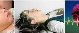
Important! Non-contrast magnetic resonance arteriography is often the only safe way to examine the vascular system. MRI diagnostics is allowed for pregnant women in the 3rd and 4th trimesters of pregnancy and newborns from the first day of life after consulting a doctor.
ARTERIOGRAPHY
ARTERIOGRAPHY
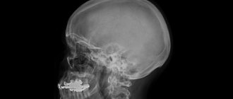
(Greek arteria + -graphö write, depict) - X-ray examination of the arteries by introducing a contrast agent into the lumen of the vessel, followed by radiography. Arteriography is used both for diagnosing lesions of the vessels themselves, and for recognizing diseases of many organs - the brain, lungs, liver, kidneys, uterus, etc. Arteriography of peripheral vessels was first performed by Berberich and Hirsch (J. Berberich, S. Hirsch, 1923) . In the USSR, arteriography for diagnostic purposes was first performed by V.V. Krestovsky (1930), G.P. Kovtunovich (1932).
Arteriography is widely used for arteriovenous aneurysms and fistulas, traumatic arterial ruptures, ligated vessel diseases, occlusive diseases, aneurysms and arterial thrombosis. Arteriography makes it possible to study the anatomical and topographical features of the arteries, their functional state, the speed of blood flow, the localization and extent of the pathological process, the paths of the roundabout circulation, as well as the permeability of the vascular wall. Contraindications to the use of arteriography: severe condition of the patient, acute diseases of the liver, kidneys, pancreas and intolerance to iodine drugs.
Triiodinated contrast agents (hypaque, verografin) used in arteriography contain from 65 to 85% iodine. When introduced into the vascular bed, they are excreted from the body by the kidneys in a short time.
Arteriography can be performed in two ways: open, that is, by surgical exposure of the blood vessel, with a poorly developed vascular network and a difficult to palpate artery, or closed, by percutaneous puncture of the artery: under local anesthesia at the puncture site, 20– 40 ml of 50-70% contrast agent solution.
If arteriography is performed carelessly, a puncture of the opposite wall of the vessel is possible, as a result of which the contrast agent is injected into the paravasal tissue; if painful infiltrates occur, a 0.25% solution of novocaine must be injected into the tissue surrounding the vessel. During puncture arteriography, the needle is connected to the syringe with a rubber tube, which facilitates the technical performance of the study. Arteriography is also performed by percutaneous introduction of a vascular catheter according to Seldinger: the vascular catheter is inserted through the bloodstream or retrogradely. During retrograde arteriography, a mechanical syringe or injector should be used to administer the contrast agent. To prevent the formation of blood clots around a catheter inserted into the vascular bed, it is necessary to infuse a warm isotonic solution of sodium chloride with a small admixture of heparin. In case of an obliterating disease with impaired patency of the main arteries of the pelvis or thigh, it is advisable to perform aortography (see), and in case of unilateral damage to the femoral artery, a vascular catheter can be inserted into the artery of the healthy thigh and with retrograde arteriography, a contrast image of the vessels of the diseased limb can be obtained. This will allow us to determine the level and extent of occlusion of the main vessel, the paths of the circuitous blood flow and the anatomical and functional state of the collaterals.
For arteriography, you can use conventional stationary x-ray diagnostic devices, taking serial images using special devices or single images with a long exposure (from 4 to 6 seconds). Currently, X-ray machines with automatic film feeding are used, as well as electro-optical converters using filming and X-ray television.
Arteriography during surgery becomes important: in addition to clarifying vascular pathology, arteriography on the operating table makes it possible, if necessary, to determine the patency of a vascular graft or anastomosis, that is, to objectively assess the quality of plastic vascular surgery.
The operation is performed in a specially adapted X-ray room or in an operating room, where there is a powerful mobile X-ray machine. A repeat study can be performed after 5-7 days with appropriate indications and joint consultations between a surgeon and a radiologist. Rice.
1, left. Arteriogram of the right thigh with atherosclerosis of the femoral artery.
The tortuosity of the femoral artery is visible. Rice.
2, right. Arteriogram of the area of the right knee joint with obliterating endarteritis: 1 - normal main artery; 2 - narrowed main artery; 3 - obliterated artery
Arteriography makes it possible to differentiate the nature of the pathological process at different stages of the disease. In atherosclerosis of the iliac and femoral arteries, the following is found: narrowing of the lumen of the vessel, jagged contours, filling defects due to atherosclerotic plaques, calcification of the vascular wall, tortuosity of the vessels (Fig. 1), and in the later stages - complete disruption of the patency of the vessel. With obliterating endarteritis, a narrowing of the main artery with clear contours is observed (Fig. 2); when the vessel is occluded, a delimited stump with tortuous collaterals is visible. Arteriography allows us to identify the characteristic features of tissue vascularization in the limb under study in various phases of Raynaud's disease and the effectiveness of therapeutic measures for this disease. Arteriography is important in the differential diagnosis of soft tissue neoplasms of the extremities and pelvis.
Radioisotope arteriography
- a type of radioisotope angiography (see), used to study the condition of the arteries and surrounding tissues. The essence of the study is that when 0.1-0.3 ml of a radioactive drug with a total activity of 2-5 microcuries is introduced into the lumen of the vessel, its dilution in the blood occurs quite slowly, due to which it is possible to obtain quite satisfactory contrasting of the cavities of the heart, aorta and large arteries.
Registration of the passage of a radioactive substance through the arteries is carried out using a scintillation camera (see Scintigraphy) with subsequent filming of the resulting image. Given the need to administer drugs with relatively high activity, radioisotope arteriography uses only isotopes with low radiotoxicity and a short half-life (technetium-99m).
Radioisotope A. is used to diagnose diseases of the heart, aorta, pulmonary artery, kidney arteries, brain, etc.
Bibliography:
Vilyansky M. P. Arteriography in obliterating endarteritis, M., 1959, bibliogr.; Clinical angiography, ed. M.I. Kuzina et al., M., 1973; Krakovsky N.I. and Mazaev P.N. Angiography in vascular surgery of the limbs and neck, p. 21, M., 1964, bibliogr.; Petrovsky B.V. and Milonov O.B. Surgery of peripheral vascular aneurysms, p. 33, M., 1970, bibliogr.; Stepanova G. G. Arteriography data for obliterating diseases of the arteries of the lower extremities, Uzhgorod, 1962, bibliogr.; Tikhonov K. B. Angiography, p. 67, D., 1962, bibliogr.; Angiography, ed. by H.L. Abrams, v. 1-2, L., 1961; Lumpkin M. V. a. O. Arteriography as an aid in the diagnosis and localization of acute arterial injuries, Ann. Surg., v. 147, p. 353, 1958; Maurer HJ Zur arteriographi-schen Diagnostik von Knochensarkomen, Radiol, clin. biol. (Basel), Bd 38, S. 293, 1969; Seldinger SI Catheter replacement of the needle in percutaneous arteriography, Acta radiol. (Stockh.), v. 39, p. 368, 1953, bibliogr.; Vascular roentgenology, ed. by RA Schobinfeer a. FP Ru-zicka, p. 127, NY—L., 1964, bibliogr.
P. N. Mazaev; G. A. Zubovsky (med. rad.).
Arteriography: stages of scanning
MR imaging of the vascular bed is performed according to a standard protocol. Immediately before the procedure, the patient must leave any metal-containing items, jewelry, or electronic gadgets outside the office. Clothing should be comfortable, which will not hinder movement during the procedure and cause inconvenience to the client. If necessary, the nurse can offer a disposable medical gown.
The patient should lie down on a retractable table, after which the assistant will fix the head in a stationary position using special holding devices and put on the head coil. It is important to remember that stillness is the key to obtaining high-quality images. Patients suffering from psychomotor disorders or claustrophobia may be offered sedatives.
To communicate with the doctor, a signal bulb and an intercom are provided, which the patient can use if they feel unwell or have a panic attack.
Important! Arteriography is a painless diagnostic method that does not involve any mechanical impact. During operation of the tomograph, clicks and crackling sounds are heard. Medical earplugs will help cope with discomfort.
Decoding images
Examination of blood vessels reveals the following pathologies:
- Aneurysms (pathological expansion of the walls of blood vessels) and their dissection;
- Congenital heart defects;
- Arterial atherosclerosis;
- Inflammation of the vascular walls (vasculitis);
- Stenosis (pathological narrowing) of blood vessels.
Unlike conventional tomography of the brain, angiography diagnostics can detect hemorrhagic types of strokes. This method, combined with contrast, is also good for detecting tumors. Inside them there is always a densely woven vascular network. When a contrast agent passes through it, the neoplasm stands out in the image as a bright spot with clearly defined edges.
MRI vascular angiography is completely safe for the body, but this type of diagnosis is very informative. It detects the presence of tumors and visualizes pathological structures. All this is necessary to make an accurate diagnosis and prescribe effective treatment for vascular diseases.
"ICLINIC": accurate diagnosis in a short time!
The ultra-modern diagnostic unit is equipped with innovative equipment, thanks to which it was possible to reduce the time spent on diagnostics by 15-20%. The tomographs are equipped with the latest generation “Tim” and “Dot” technologies, which guarantee high image clarity during screening.
Interpretation of tomograms is carried out by expert radiologists who have many years of practical experience. Doctors improve their professional skills in the world's leading clinics, which is why the quality of the services provided is at an extremely high level. If it is difficult to decipher the images, it can be provided.
Still have questions? Ask them to specialists on the ICLINIC website. If necessary, you can leave a request on the portal - managers will call you back at a time convenient for you.
How to prepare for research?
You must wear your own clothing without metal fasteners.
Women should always tell their doctor and technologist if they may be pregnant or breastfeeding.
Metal and electronic objects may interfere with the magnetic field of the MRI machine and may cause burns:
- jewelry, watches, hearing aids.
- pins, hairpins, metal zippers.
- piercing
- mobile phones, digital watches and tracking devices.