Prices Make an appointment
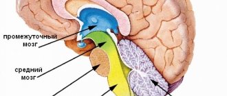
One of the most important structures of the brain is the cerebellum. It is located in the postcranial fossa of the skull, and it is quite difficult to assess its pathology using hardware diagnostics. Diagnostics with ultrasound, computed tomography - do not provide enough information. Magnetic resonance imaging can effectively help in making the correct diagnosis. MRI of the cerebellum and brain is the best choice for obtaining a detailed picture of what is happening and diagnosing diseases.
Cerebellar tumor: symptoms, removal surgery, prognosis and consequences
A cerebellar tumor is a tumor that arises in any part of the cerebellum of the brain. Is benign or malignant. Occupies about 30% of all brain tumors. Pathology occurs at any age.
Some types of neoplasms are more common in children (medulloblastoma, sarcoma), some predominantly in adults (glioblastoma, astrocytoma). According to ICD-10, a malignant neoplasm of the cerebellum of the brain has code C71.
5.
The neoplasm can be primary or secondary in nature. The primary tumor is formed from the cells of the cerebellum and spreads by replacing healthy cells with damaged ones. A secondary tumor is a metastatic type of tissue damage to an organ that is malignant.
From the point of view of the histology of tumors, there are about a hundred types. The most common (75% of cases) among cerebellar tumors is glioma.
Types of cerebellar tumors
There are two types of tumors in the cerebellum: benign and malignant. Benign ones include hemangioblastoma, astrocytoma and some types of cystic formations. They do not have a tendency to transition to a malignant neoplasm and have a favorable prognosis.
Malignant tumors are characterized by rapid growth, require removal and are accompanied by a number of complications. These include sarcoma and medulloblastoma of the cerebellum.
Benign neoplasms
Benign cerebellar tumors include hemangioblastoma and astrocytoma.
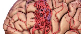
Hemangioblastoma is a neoplasm that arises from the vessels of the brain. The typical age of those suffering from this pathology is 35-65 years. Hemangioblastoma is characterized by multiple complications in the form of concomitant tumors. The pathology has 1 degree of malignancy, which indicates the presence of differentiated cancer cells with slow growth.
Cerebellar hemangioblastoma
Depending on the structure of the tissues, the tumor is divided into the following types:
- Solid hemangioblastoma is tumor cells collected in a soft node, which are surrounded by a capsular membrane and have a dark cherry color.
- Cystic hemangioblastoma is a smooth-walled cyst with a small nodule.
- Mixed hemangioblastoma is a large tumor with small cystic formations inside.
Symptoms of a tumor appear depending on its location and are divided into cerebral, cerebellar and distant. Common symptoms include:
- impaired coordination of movements;
- low activity in the limbs;
- disturbed balance;
- symptoms of hydrocephalus: signs of intoxication of the body, headaches;
- disruptions in brain activity.
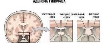
The appearance of hydrocephalus is a common symptom of pathology. This is fluid stagnation in the brain, which provokes an increase in intracranial pressure. Hemangioblastoma also disrupts the functioning of the optic nerves, which significantly impairs vision. As the pathology develops, irritability, neurotic states, blocked ears, and loss of consciousness are observed.
An astrocytoma is a benign tumor that arises in the back of the brain from glial cells. The neoplasm is characterized by clearly defined boundaries and is cystic in nature. The pathology affects the brain areas that are responsible for balance and movement.
As the tumor grows, it begins to compress the brain tissue, which leads to swelling of the organ. Symptoms of the pathology will be expressed:
- headaches in the morning and evening;
- nausea;
- weakness;
- feeling of chronic fatigue;
- disorientation in space;
- numbness in the hands;
- speech disorders.
Astrocytoma can be nodular, diffuse and mixed.
Malignant neoplasms
Malignant neoplasms include sarcoma and medulloblastoma of the cerebellum.
A malignant tumor of the cerebellum of the brain affects not only individual parts, but also the trunk of the organ, which affects the quality and length of life. Cancer cells also affect the ventricle of the brain, which makes it difficult for fluid to leave the ventricular system and causes a severe course of the disease.
Cerebellar medulloblastoma is a type of malignant tumor consisting of embryonic cells. The neoplasm can be localized both in the cerebellum and in other parts of the brain.
The pathology is typical for children 5-10 years old. In adults, the manifestation of the disease is observed at the age of 20-40 years. Moreover, it occurs more often in men than in women.
It is predisposed to the development of metastases not only in the nervous system, but also in the liver, kidneys and bones.
Localization of carcinoma in the cerebellar vermis helps to stop the circulation of spinal cord fluid and poses a threat to the patient’s life.
Depending on the histological structure, there are:
- medulloblastoma (consists of muscle fibers);
- melanotic medulloblastoma (contains cells consisting of melanin);
- lipomatous medulloblastoma (consists of fat cells).
The neoplasm has many clinical manifestations. Symptoms depend on the location of the tumor. Cerebellar medulloblastoma has general cerebral symptoms:
- clouded consciousness;
- increased irritability;
- disorientation;
- personality disorder.
The main manifestation of the pathology is cerebellar ataxia - impaired coordination of muscle movement.
Symptoms of a tumor:
- frequent falls due to poor coordination;
- unsteady gait with legs spread wide apart to maintain balance.
- deterioration of the swallowing reflex;
- frequent headaches.
Another type of malignant tumor is sarcoma. The disease most often affects children aged 1-16 years.
The disease develops rapidly. On average, patients arrive at the clinic for treatment 3 months after the first symptoms appear. At an early stage, the tumor manifests itself as a low-grade fever or an elevated temperature of up to 39 degrees and is accompanied by enlarged lymph nodes. Common symptoms of sarcoma include:
- decreased appetite;
- fast fatiguability;
- drowsiness;
- attacks of headache and vomiting.
Sometimes the manifestation of the disease begins with disorders of the cerebellar functions.
Reasons for appearance
The reasons for the development of pathology are not fully known. Scientists and doctors can only speculate about the factors that provoke the growth of tumor cells. The development of the tumor process can be caused by radioactive radiation and the effect of chemicals on the body.
Additional factors influence the development of pathology in the body:
- Congenital anomalies.
- Separation by gender: men are more often affected by the disease.
- Weakened immunity due to existing cancer.
- Age: Some types of tumors are typical for the young age group, others are more common among middle-aged and elderly people.
Symptoms
Symptoms of the manifestation of pathology consist of general cerebral and focal, as well as distant symptoms. During the development of the disease, all 3 groups of symptoms appear simultaneously and can be of a different nature, depending on the location of the tumor.
General cerebral symptoms
The appearance of cerebral symptoms is associated with increased intracranial pressure. ICP increases due to enlargement of the carcinoma, which leads to protrusion of the cerebellum. The cerebellum, increasing in size, emerges into the 4th ventricle.
This leads to disruption of the outflow of cerebrospinal fluid, which provokes an increase in pressure. The main symptom is increasing headaches, worsening in the morning and evening. They are accompanied by nausea, even vomiting, and severe dizziness.
Weakness and drowsiness, periodic stupor appear.
If carcinoma begins to affect the outflows of cerebrospinal fluid, then the symptoms increase: the patient is forced to keep his head tilted, attacks of nausea and vomiting become more frequent and intensify.
Long-term symptoms
The appearance of long-term symptoms is caused by swelling of the brain tissue caused by a rapidly growing tumor. This causes compression of the nerves in the brain stem. Vomiting becomes more frequent due to sudden movements or changes in position. There is a violation of the sensitivity of one half of the face. Hearing loss, strabismus, and speech problems develop. Epileptic seizures are possible.
Compression of the brain stem provokes a dangerous symptom - respiratory arrest, which can cause death.
Focal symptoms
Focal symptoms are associated with damage to the cerebellar tissue. They will depend on what part of the organ was affected by the neoplasm. There are disturbances associated with coordination of movements.
If the tumor has affected the cerebellar vermis, then the symptoms will manifest as a change in gait. The patient in this case moves like a person in a state of intoxication. Nystagmus appears - involuntary movement of the eyeballs and various speech disorders.
Diagnostics
Pathology examination requires the presence of the following methods:
- blood analysis;
- neuro-ophthalmological examination;
- MRI;
- CT;
- radiography;
- biopsy;
- angiography.
First of all, an examination by a neurologist is necessary to monitor the connection of the neoplasm with reflex disorders and other neuralgic pathologies. As well as an ophthalmologist who will examine the fundus and check visual acuity.
A blood test is necessary to determine tumor markers - laboratory indicators of the progression of a malignant tumor.
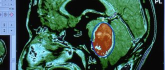
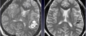
The most informative research methods are radiation. These include computed tomography and magnetic resonance imaging. CT and MRI can determine the size of the tumor, its location and nature.
Using CT, the abdominal cavity, chest, and pelvic organs are additionally examined for the presence of metastases.
X-ray of the skull is one of the available diagnostic methods, since changes in the structure of the bones occur under the influence of a growing tumor.
Angiography is sometimes used, a method for studying the blood supply to the brain.
Treatment
Treatment methods depend on the nature of the malignancy of the tumor and are often complex. Benign formations are completely removed, since they have clearly defined forms, are located on tissue surfaces, do not have a tendency to germinate, and are characterized by a favorable prognosis.
Malignant carcinomas are formed by pathological tissues, can grow deeply into brain tissue, give metastases, and have an unfavorable prognosis. Therefore, in addition to surgical intervention, auxiliary treatment is prescribed in the form of radiation and medications.
The main method of treating tumors is surgery.
Surgery involves performing a craniotomy and removing the affected tissue. It is divided into several types:
- Radical surgery with complete elimination of tumor formation.
- Palliative surgery with partial removal of tumor formation.
Removal of a tumor is impossible only if it has grown into the brain stem, ventricle or neighboring tissues. In such cases, as much of the tumor tissue as possible is removed and an attempt is made to restore cerebrospinal fluid circulation.
Modern methods of tumor removal include:
- A craniotomy allows you to make a hole in the skull, allowing access to the brain tissue.
- Stereotactic radiosurgery using biopsy.
- Endoscopic trephination allows you to perform surgery without opening the skull when removing tumor formations.
After removal of the malignant tumor, the patient is prescribed additional treatment in the form of radiation and chemical therapy, which is aimed at destroying the remaining cancer cells.
Radiation therapy is used both through external irradiation and the introduction of special drugs into brain tissue. For this purpose, equipment is used that allows a capsule with a radioactive substance to be placed into the brain according to coordinates.
With external irradiation, gamma rays are directed to the location of the malignant tumor.
Chemotherapy involves the intravenous administration of drugs that affect cancer cells and selectively destroy them.
Chemotherapy is divided into 3 types:
- Neoadjuvant – prescribed before surgery to destroy tumor cells.
- Adjuvant – prescribed after surgery to eliminate the remaining elements of cancer.
- Targeted – involves the use of drugs whose action is aimed at stopping the growth and destruction of cancer cells.
The course of treatment lasts from 1 to 3 weeks and has a number of side effects.
One of the areas of chemotherapy is targeted therapy. The treatment method consists of administering drugs that do not suppress the division of tumor cells, but stop the reactions provoked by cell growth. This reduces the level of toxins entering the body.
Dietary supplements may also be prescribed to maintain the general condition of the body.
The prognosis for life with pathology depends on the nature of the tumor, the extent of the lesion and its location. Benign tumors have a favorable prognosis provided that the tumor is completely removed.
However, with the rapid growth of the tumor and the lack of timely medical care, the tumor can compress the stem structures that are responsible for the respiratory system.
This is fatal.
The prognosis for malignant tumors cannot be called favorable. Life expectancy can be increased through supportive care. On average, people with cerebellar cancer live from 1 to 5 years.
Source: https://onko.guru/termin/opuhol-mozzhechka.html
Hemangioblastoma
A rather rare type of cancer that affects the blood vessels of the brain. Such neoplasms can be localized in all areas of its spheres. However, they are most often found in the cerebellum, the posterior cranial fossa.
According to their characteristics, hemangioblastomas are benign neoplasms. However, anatomically they are located so close to vital brain structures that the slightest damage to the latter leads to serious dysfunction. The typical location is the pia mater surrounding the brain.
Hemangioblastoma manifests itself as follows:
- Headache.
- Nausea and vomiting.
- Changed gait.
- Double vision.
- Decreased visual acuity.
- Recurrent dizziness.
- Mental, personal changes.
- Feeling of discomfort in the neck area.
- Anorexia.
- Apathy, lethargy.
- Noises in the head.
- Chronic feeling of weakness in the limbs.
- Fainting.
- Speech impairment.
- Eye pain.
The listed symptoms can manifest themselves both sharply and gradually. The worsening severity of the patient’s condition most often indicates new bleeding or increased intracranial pressure. Sometimes the tumor can manifest itself as subarachnoid hemorrhage.
Hemangioblastomas are rarely diagnosed in young patients. They mainly affect people aged 20-40 years. Among men, cancer is diagnosed twice as often.
Cerebellar tumor of the brain: symptoms, causes, diagnosis, treatment and prognosis
A cerebellar tumor is one of the types of neoplasms that can be benign or malignant. Regardless of the histological structure, it poses a threat to life.
Such neoplasms occur in approximately 30% of people with various cerebral tumors. Thanks to histology, more than 100 types have been identified, but in 70% of cases the tumor is understood as a glioma (primary tumor of a pink, grayish-white or dark red node).
The formation of education occurs at any age, but some types are characteristic of a certain type of people.
For example, medulloblastoma occurs in children, and astrocytomas and hemangioblastomas in middle-aged men and women. More often, the disease occurs in Caucasian men. A malignant tumor has ICD-10 code C71.6
Reasons for development
The only proven cause of development is radiation. It is estimated that about 10% of lesions are formed due to genetics or as a result of exposure to oncogenes.
A violation at the genetic level occurs under the influence of:
- toxic substances,
- excessive sun exposure,
- heredity.
The impetus is the use of artificial ingredients in food and electromagnetic fields. The risk of occurrence also increases in those who have reduced immunity (HIV patients).
The mechanisms that play an important role in tumor formation operate in several directions at once. Tissue damage occurs due to pressure from growing tissue. Gradually, the tumor increases in size and disrupts the cells of the brain stem. General cerebral symptoms associated with increased blood pressure develop.
Classification
A tumor is divided into malignant and benign.
The first type includes hemangioblastomas and astocytomas. Sometimes the cells transform into a cyst, represented by a small node. Transformation into a malignant disease occurs in rare cases.
Malignant tumors – without treatment, a guaranteed death outcome and the inability to lead a full life.
Such neoplasms tend to grow rapidly and easily penetrate the tissues of nearby areas.
Cerebellar tumors are divided according to their genesis. The primary form originates from their cerebellar cells and is the result of metaplasia. A secondary tumor implies a metastatic origin. If the first type is benign and malignant, then the second is exclusively malignant.
Symptoms of a tumor of the cerebellum of the brain
Signs are divided into three groups:
- cerebral,
- distant,
- focal.
They all develop simultaneously, but their severity may vary. It depends on the direction of germination, compression of structures located nearby. Sometimes the first signs are general cerebral or distant signs. This is possible due to the special location of the cerebellum between the 4th ventricle and the brain stem.
General cerebral symptoms include:
- Headaches that are felt in the back of the head or in the neck. If intracranial pressure increases, diffuse pain appears.
- Nausea and vomiting. They are not related to eating. Nausea most often appears in the morning and is associated with irritation of specific centers.
- When examined by an ophthalmologist, congestive nerve discs are detected. This symptom appears before all other signs. The veins are probably being compressed.
- Dizziness.
Long-term symptoms occur due to damage to the nerves that exit the brain tissue in the brainstem area. They are characterized by:
- sensory disturbances,
- squint,
- disorders caused by problems with the facial nerve,
- deterioration of hearing, tongue mobility,
Cerebellar (focal) signs are characterized by the appearance of signs depending on the affected area. If the worm is damaged, it is difficult for a person to walk and stand equally. The gait begins to resemble the walking of a drunk. The larger the formation becomes, the more pronounced the symptom becomes in the sitting position.
If a tumor grows in the area of the cerebellar hemispheres, there is a disturbance in the smoothness and accuracy of movements on the side of the body where the cancer is. A person cannot grasp objects, he is unable to bend and straighten his arms. Handwriting and speech are deformed. The latter becomes intermittent and can be divided into syllables. Nystagmus develops (oscillatory movements of the eyeballs).
If the brain tissue is pinched, then another part of it begins to move towards the foramen magnum. When this happens, the risk of losing your own life increases many times over.
Treatment of neoplasm
The main method of treatment is surgical. The question of its use and the scope of actions taken is decided by the neurosurgeon, but often the optimal solution is radical removal of transformed cells.
However, such an operation is not always possible due to tumor growth into anatomical structures. Then the main goal is to remove the maximum possible volume and restore normal liquor circulation.
Radiation therapy is considered a promising method. It does not lead to disruption of the integrity of the tissue, but the beam affects the affected area.
Chemotherapy consists of administering cytostatic drugs that block tumor cells. Helps speed up the healing process:
- radiosurgery,
- immunotherapy,
- gene therapy.
Almost all known methods lead to suppression of activity of normal cells. This leads to the development of side effects.
Prognosis for cerebellar tumor
Treatment results depend on the type of disease. If the tumor is benign, the prognosis is favorable. If compression or death of the structures that are responsible for breathing and heart function occurs, the risk of death increases.
If the tumor was not completely removed, then a repeat operation will be required after a few years. For malignant tumors, patient survival after therapy ranges from 1 to 5 years.
tells the stories of two brain tumor patients:
Source: https://gidmed.com/onkologiya/lokalizatsiya-opuholej/spinnoj-i-golovnoj-mozg/opuhol-mozzhechka.html
Astrocytoma
The name is given according to the origins of the neoplasm - astrocytes located in the cerebellum. This tumor is characterized by slow growth. It rarely spreads to other areas of the brain. But cases of metastasis, even rare ones, still occur.
Symptoms of this type of cerebellar tumor are as follows:
- Morning sickness, morning and night migraines. The manifestation is systematically repeated over several weeks or months.
- Ataxia and dysdiadochokinesia may develop with corresponding damage to the cerebellum. These signs help specialists determine the location of the tumor.
- Nausea, often ending in vomiting.
- Apathy.
- Loss of orientation in space.
- Confused thinking.
- Weakness in the limbs, numbness in the arms and legs.
- Deterioration of visual function. The “picture” appears double or blurred.
- Blurred consciousness.
- Memory problems.
- Difficulty, confused speech.
Cerebellar tumor: symptoms, diagnosis and treatment
A cerebellar tumor is one of the types of brain tumors. A cerebellar tumor can be benign or malignant, with a wide variety of histological structures.
Even if the tumor is benign, due to its special location it can pose a direct threat to the patient’s life due to the possibility of infringement of brain structures with impaired breathing and circulation.
A cerebellar tumor manifests itself as cerebral, distant and focal (cerebellar) symptoms. To diagnose this pathology, it is mandatory to conduct a computed tomography (CT) or magnetic resonance imaging (MRI) of the brain.
Treatment of cerebellar tumors is predominantly surgical. From this article you can learn about the main symptoms, methods of diagnosis and treatment of cerebellar tumors.
Classification and terminology
Among all brain tumors, cerebellar tumors account for about 30%.
Like all tumors of the nervous system, cerebellar tumors can be primary (if their source is nerve cells or membranes of the brain) and secondary (if they are a metastasis of a tumor of another location).
The histological structure of cerebellar tumors is also very diverse (more than 100 types are known). However, the most common are cerebellar gliomas (medulloblastomas and astrocytomas) and cancer metastases.
Cerebellar tumors can have a relatively benign slow growth, located separately from normal brain tissue (as if in a capsule), or they can infiltrate surrounding tissues, which in itself is less favorable.
Symptoms of a cerebellar tumor
All signs of a growing cerebellar tumor can be divided into three groups:
- cerebral (develop due to increased intracranial pressure);
- distant (occur at a distance, that is, not directly next to the tumor);
- focal (actually cerebellar).
In almost all cases, these three groups of symptoms occur simultaneously with each other, just the severity of certain symptoms varies. This is largely determined by the direction of tumor growth and compression of individual adjacent structures.
The special location of the cerebellum in the cranial cavity determines some features of the clinical course of its tumors. A clinical situation is possible when the first signs of a tumor are general cerebral and even distant symptoms.
This is due to the fact that the cerebellum is located above the fourth ventricle and brain stem. Therefore, sometimes the first symptoms of a cerebellar neoplasm are signs of damage to the brain stem and disruption of the outflow of cerebrospinal fluid from the fourth ventricle, and not the cerebellum itself.
And the damage to the cerebellar tissue is compensated for some time, which means it does not manifest itself in anything.
General cerebral symptoms of cerebellar tumor
These include:
- headache. It can be felt in the occipital region and even the neck. It can be periodic or constant with periods of intensification. If intracranial pressure increases, the headache becomes diffuse, accompanied by a feeling of nausea and vomiting;
- nausea and vomiting not associated with food intake. These symptoms are associated with irritation of specific centers in the brain stem. Occurs more often in the morning. Also, these signs may be the result of increased intracranial pressure;
- dizziness;
- congestive optic discs. This change can only be seen during an ophthalmological examination. In the case of a cerebellar tumor (in comparison with brain tumors of other locations), congestive optic discs appear relatively early, even before the cerebellar symptoms directly. Most likely, this is due to fairly rapid compression of important venous outflow tracts in tumors of the cerebellar localization.
CM. SEE ALSO: The human cerebellum and its functions
Long-term symptoms of a cerebellar tumor
In the case of a cerebellar tumor, these symptoms are represented by damage to the cranial nerves (or rather, their compression). The cranial nerves, for the most part, emerge from the thickness of the brain tissue in the region of the brain stem. A growing tumor of the cerebellum puts pressure on the nerve roots, which causes various symptoms to appear. It can be:
- pain and sensory disturbances in one half of the face, difficulty chewing (associated with compression of the trigeminal nerve);
- strabismus (damage to the abducens nerve);
- facial asymmetry (facial nerve damage);
- hearing impairment or a feeling of ringing in the ears (VIII pair of cranial nerves);
- impaired mobility of the tongue and some blurred speech associated with this;
- changes in taste sensitivity.
It should be noted that damage to the nerves of the bulbar group is less common than pairs V-VIII.
In addition to symptoms from the cranial nerves, long-term signs of a cerebellar tumor include the appearance of weakness or changes in sensitivity in one half of the body, epileptic seizures, and increased spastic muscle tone.
Focal symptoms (actually cerebellar)
These manifestations of the tumor process are associated with direct damage to the cerebellar tissue.
The cerebellum consists of several parts: the central one - the vermis and the hemispheres located on the sides of it (left and right). Depending on which part of the cerebellum the tumor is compressing, different symptoms occur.
If the worm is affected, the following symptoms appear: difficulty standing and walking. A person sways when walking and even while standing, stumbles out of the blue and falls.
As the tumor grows, instability occurs even in a sitting position.
If a tumor grows in the area of one of the cerebellar hemispheres, then the smoothness, accuracy and proportionality of movements on the side of the tumor (that is, left or right) are disrupted. A person misses when trying to grab an object; he is unable to perform actions associated with rapid contraction of antagonist muscles (flexors and extensors).
On the affected side, muscle tone decreases. The handwriting changes: the letters become large and uneven, as if zigzag-shaped (this is also due to a violation of the correct contraction of the hand muscles). Speech disturbances are possible: it becomes intermittent, spasmodic, as if chanting, divided into syllables.
Trembling appears in the limbs on the side of the tumor, which intensifies towards the end of the movement performed.
As the tumor grows, the symptoms of damage to the worm and hemispheres gradually mix, the process becomes bilateral.
In addition to the above symptoms, the patient may exhibit nystagmus. These are jerky, oscillatory involuntary movements of the eyeballs, especially when looking to the side.
The proximity of the cerebellar tumor to the fourth ventricle causes disruption of the circulation of cerebrospinal fluid. Internal hydrocephalus develops with headaches, attacks of vomiting and nausea. Overlapping of the openings of the fourth ventricle may be accompanied by Bruns syndrome.
This can occur when there is a sudden change in the position of the head (especially when bending forward), due to which the tumor moves and blocks the openings for the circulation of cerebrospinal fluid. The syndrome is manifested by a sharp headache, uncontrollable vomiting, severe dizziness, temporary loss of vision, and confusion.
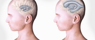
Another dangerous condition that can occur with a cerebellar tumor is infringement of brain tissue. The fact is that a growing tumor occupies part of the space inside the skull, and this space is permanent.
The rest of the brain tissue simply has nowhere to go, and it “moves” in the direction of the nearby openings of the skull (in particular, the foramen magnum). Infringement is also possible in the notch of the tentorium cerebellum (the latter is formed by the dura mater).
Infringement of brain tissue is very dangerous for a person, since at this moment he risks losing his own life.
Medulloblastoma
First of all, let us note the features of the development of this cerebellar tumor in children. Symptoms in younger children are in most cases mild. Are limited to the following:
- Changing habitual behavior.
- Some increase in head circumference.
- Lethargy and apathy.
- Vomit. This syndrome occurs more often in older children than in infants.
When examining a small patient, a specialist may identify an anterior protruding fontanelle, as well as a divergence of the skull bones. Older children often experience statistical ataxia, pathological head tilt, and altered gait. What does this mean? An abnormal tilt of the head indicates both trochlear nerve palsy and growth of the tumor towards the foramen magnum. A potential threat to the patient’s life is the protrusion of the cerebellar tonsils into this hole. This happens due to the same pressure of the tumor on the brain.
Medulloblastoma is characterized by rapid development of the clinical picture. Therefore, specialists can diagnose the disease based on the symptoms that appear in less than two months.
One of the obvious manifestations of this oncological pathology in patients who have left the infancy period will be severe migraines and vomiting in the morning. The symptoms are caused by increased intracranial pressure. As we described above, it is caused by a blockage of the cranial fluids by a rapidly growing tumor.
An examination of the fundus will also indicate increased intracranial pressure - swelling of the optic nerve is visible. This fact is accompanied by patient complaints of blurred vision. However, it will not be too pronounced. In some patients, palsy of the fourth or sixth cranial nerve is additionally detected. There are also complaints of diplopia. It is also caused by pressure from the neoplasm. Some patients with medulloblastoma are diagnosed with speech disorders.
In most cases, the tumor affects the midline structures of the brain. This causes gait disturbance, trunk ataxia, and nystagmus. Sometimes a violation of writing and general clumsiness are visible.
As for adult patients, their medulloblastoma can be characterized by a unilateral manifestation. A common example is dysmetria.
A tumor of the cerebellum of the brain - how dangerous is it for life?
The most common tumor affecting the brain is a cerebellar tumor. It can be either benign or malignant, be primary or be a metastasis of another tumor in the body.
Due to its location, that is, in the cerebellum - the motor center of the brain, the tumor at each stage of the disease can give different symptoms. Thus, experts identify general cerebral, distant and focal (cerebellar) signs of pathology, the strength of which is determined by the size and location of the tumor.
The process of diagnosing a cerebellar tumor is similar to identifying other neurotumors: first, a visit to a neurologist or oncologist is necessary, and only then a brain examination using CT or MRI.
Symptoms of the disease
All cerebellar tumors, symptoms and signs of development, are conventionally divided into three groups:
- General cerebral (occur due to impaired circulation of cerebrospinal fluid).
- Distant (develop against the background of compression of neighboring structures).
- Focal, that is, affecting the functioning of the cerebellum.
They usually appear simultaneously, but their strength may vary depending on the intensity of growth of the neoplasm, its location and impact on neighboring structures.
Due to the location of the “motor center” (it is located above the 4th ventricle behind the brain stem), in each case there is an increase in the symptoms of a cerebellar tumor. But it can vary depending on the intensity of tumor growth and the direction of its growth.
Usually, at first this process is accompanied by a disorder in the functioning of the brain stem and an increase in intracranial pressure, and then by a disruption of the cerebellum, since the brain is able to partially compensate for its functioning.
The appearance of general cerebral signs of pathology is due to the fact that as the size of the cerebellum increases due to tumor growth, it protrudes beyond the cranial fossa, that is, into the 4th ventricle.
This in turn leads to impaired circulation of cerebrospinal fluid and increased ICP.
In a sick person, these processes are signaled by the appearance of a nagging headache in the back of the head, dizziness, nausea and sometimes vomiting.
The appearance of distant manifestations of the development of a cerebellar tumor is explained by displacement of the structures of the reticular formation and includes the following symptoms of pathology:
- impaired sensitivity on one side of the face, difficulty chewing and swallowing food;
- the appearance of strabismus;
- facial asymmetry;
- tinitus, hearing impairment;
- unclear speech;
- change in taste;
- hyper- or hypotonicity of the muscles of one side of the body;
- epileptic seizures.
Focal symptoms of the disease are caused by dysfunction of the structures of the cerebellum itself. These include:
- uncertain and “drunk” gait, impaired stability (if the tumor is located in the cerebellar vermis);
- poor control of the muscles of the limbs, changes in handwriting, tremor (if one of the lobes of the cerebellum is affected, the sign of pathology will be from the side of the tumor);
- speech disorder - it becomes intermittent, the beat becomes complex, and words when pronounced are divided into syllables.
As the tumor moves from one lobe of the cerebellum to another, all of the above symptoms are mixed and can appear simultaneously.
Reasons for the development of pathology
Today, experts cannot accurately identify the etiological factors that provoke the development of a cerebellar tumor. Among the possible reasons, the following are primarily identified:
- Heredity (a factor characteristic of 10% of patients).
- History of radiation exposure.
- Exposure to oncoviruses - herpes, human papilloma, adenoviruses, etc.
- The effect of chemical carcinogenic drugs on the body.
- HIV infection, AIDS.
- Immunosuppressive therapy.
Dysplastic gangliocytoma
Belongs to the category of benign neoplasms. The appearance of gangliocytoma causes abnormal development of the cerebellar cortex. The symptoms of this lesion are as follows:
- Dizziness.
- Migraine.
- Nausea and vomiting.
- Macrocephaly.
Less commonly, patients experience convulsions, subarachnoid hemorrhages, and orthostatic hypotension.
Often manifests itself in patients with diagnosed Cowden syndrome. The pathology is complicated by diseases of the thyroid gland, oral papillomatosis, meningiomas, the formation of polyps in the digestive tract, etc.