Human infection, namely damage to the central nervous system by pork tapeworm, is one of the main causes of epilepsy today. Most often, this disease occurs where people farm and are in constant contact with pigs, and where sanitation and hygiene conditions leave much to be desired. According to recent data, neurocysticercosis affects approximately 50 million people per year. This disease is widespread in developing countries.
What it is?
Neurocysticercosis or cysticercosis of the brain is a disease that is caused by the pork tapeworm (Taenia solium) and affects the central nervous system. Every year, 50,000 people die due to this disease, and the rest remain disabled for life. The most endemic areas for this disease are: Mexico, South America, India, China, African countries, New Guinea.
A person becomes an intermediate host when they ingest pork tapeworm eggs. It enters the body through contaminated water, food and poor hygiene (hand washing). When ingested, cysticerci show a special affinity for the brain, eyes and striated muscles. It is these structures that contain large amounts of glycogen and glucose.
Preventive measures
To prevent cysticercosis of the brain, it is recommended to follow a number of preventive measures:
- It is required to follow generally accepted rules of hygiene. This includes washing your hands before each meal, always using disinfectants or soap. Similar manipulations are performed after visiting the street or toilet;
- you need to visit a doctor to fully examine the condition of the body;
- Do not eat dirty or uncooked food. This is especially true for pork and vegetables that grow in areas where manure is considered the main element of fertilizer;
- Before cooking meat, it must be inspected in detail for the presence of parasite larvae;
- It is allowed to buy food in markets only if the seller has a quality certificate.
Cysticercosis can cause a number of complications, therefore, even before infection, it is necessary to take all possible measures aimed at preventing the penetration of pork tapeworm larvae into the body.
Cysticercosis is considered life-threatening, so self-medication is strictly prohibited.
Causes of the disease
The main cause of the disease is the entry of the parasite into the human body. Initially, Taenia solium enters the body through the gastrointestinal tract and then spreads throughout the body through the bloodstream and is introduced into various tissues and organs.
The adult form of the tapeworm lives in the intestines, and often there may be no symptoms. An infected person releases a huge amount of the pathogen, thereby contaminating household items, especially if he does not adhere to hygiene rules.
After entering the brain, these embryos turn into cysticercus - the larval form of the pathogen. This is a finna up to 13-15 mm in size with liquid inside. The cysticercus shell is thin and not strong, and inside there are suckers and hooks. Cases have been described in medicine when the number of these larvae reached thousands. Then, local inflammation forms in the brain and a fibrous capsule forms around the larvae, which separates the cysticercus from the brain substance.
Most often, the larvae die after a year and a half, but leave behind bubble-like formations, which become calcified over time (cavities with calcium inclusions) and thus the inflammatory process becomes chronic.
Reference! According to data, on average cysticerci live 5-7 years.
In the brain, the larvae settle on the cerebral cortex, in the pia mater, in the basal ganglia, inside the ventricles (free floating or attached to the choroid plexus). In addition to causing local inflammation, they also interfere with the normal circulation of cerebrospinal fluid and compress the brain tissue.
When it enters the meninges, a strong inflammatory reaction is caused in the subarachnoid space and exudates of a dense consistency are formed, which subsequently affect the cranial nerves and the openings of Magendie and Luschka are blocked, which does not allow the cerebrospinal fluid to flow freely and hydrocephalus is formed. All these changes lead to the appearance of epilepsy and various neurological symptoms.
Forecast
Neurocysticercosis is a disease whose prognosis cannot always be predicted, since it is not known how many larvae are in the brain (after all, not all of them can be detected during examination). Late diagnosis, multiple lesions, lack of surgical treatment are unfavorable factors of the course. Sometimes death is possible during an epileptic attack or when the fourth ventricle is blocked with the development of acute occlusive hydrocephalus.
Thus, we can summarize the above: neurocysticercosis is a treacherous disease. Infection occurs unnoticed by humans. The disease may not make itself felt for a long time, sometimes it is completely asymptomatic. The detailed clinical picture does not have specific symptoms, and diagnosis requires a whole range of studies. Treatment is not always effective. In order not to become infected with neurocysticercosis, it is necessary to strictly observe the rules of personal hygiene and thoroughly process foods before eating them.
Symptoms
The clinical picture of neurocysticercosis is very diverse. But the specific symptoms of each patient depend on the following factors:
- number of cysticerci;
- location and attachment;
- what state the larva is in - alive, dead, dying;
- the body's ability to mount an immune response.
The main symptom of neucysticercosis is epilepsy - tonic-clonic or partial seizures. Neurological symptoms are different, but resemble those of a brain tumor. Intracranial hypertension, dementia, focal symptoms, dementia are also detected. The development of encephalitis is facilitated by massive infestation by larvae.
Very often the disease affects the sella turcica and the pituitary gland, which is located on it. And as a result, problems of an ophthalmological and endocrine nature begin.
When the spinal cord is damaged, motor-sensory disorders and pain along the spinal column, reminiscent of radiculitis, are detected.
When the eyes are affected, there is deterioration in vision, loss of visual fields and atrophy of the optic nerves.
Symptoms
Signs of the disease are varied and depend on the affected organ, the level of explicitness of the invasion and the opposing abilities of the body.
Cysticercosis of the brain is expressed in the following symptoms:
- Weak sensitivity;
- Irritability;
- Seizures with clinical manifestations resembling epilepsy;
- Hallucinations;
- Mental agitation, quickly giving way to a state of depression;
- Discoordination of movements;
- Vomiting;
- Cephalgia;
- Dizziness.
Additionally, depending on the area of damage to the brain, the following may also be observed:
- Dysfunction of the cardiac and respiratory system;
- Deterioration or loss of vision and hearing;
- Pain in the occipital region of the head;
- Apoplexy (paralysis) of the cerebral nerves;
- Increased intracranial pressure;
- Pathological changes in the psyche;
- Myalgia in the neck area;
- Gait disturbances, paresis;
- Loss of taste;
- Hypertensive-hydrocephalic syndrome.
When the eyes are invaded, the patient complains of various types of visual impairment, which, without proper treatment, worsen, turning into pathological, often irreversible, forms, up to complete blindness. Helminths are located in all structural parts of the eye (retina, lens, etc.) and its membrane. Pork tapeworm larvae are detected by ophthalmological examination of the eyeball.
During the illness, the patient is constantly bothered by itching and burning like conjunctivitis, increased lacrimation, decreased quality and acuity of visual perception, and redness of the eyes.
Not uncommon consequences of tapeworm seizure of the brain are cases of further parasitic spread to the tissue of the spinal cord. As a result of such an attack, chorionic inflammation, cysts, adhesions, purulent formations, etc. are formed. A person’s subjective well-being also noticeably worsens.
During this period, the patient experiences severe, almost constant pain in the arms and legs, in the abdomen and sternum, and significant disturbances in the functioning of the musculoskeletal system and body sensitivity. A severe form of infection is paralysis.
Parenchymal neurocysticercosis
This form of the disease begins when cysticerci attach to the border of the white and gray matter of the brain. The parenchymal form is characterized by:
- decreased muscle strength in the limbs;
- convulsions - epileptic seizures. And these attacks get worse and become more frequent during the death of the parasite, because they release toxins and the damage increases;
- decreased sensitivity in the limbs;
- loss of visual fields due to damage to the optic chiasm;
- speech disorder;
- with damage to the cerebellum, unsteadiness of gait and dizziness are observed;
- trembling, tremor and involuntary flexion of the arms;
- dementia and loss of mental capacity;
- with massive damage, delusions and hallucinations appear.
Important! It should be remembered that the main symptom is convulsions and epileptic seizures, and that the frequency and duration increases with the start of therapy.
Subarachnoid neurocysticercosis
This form of the disease is characterized by symptoms with meningeal signs, which are not as pronounced as with meningitis. Symptoms include: increased sensitivity to light and sound, nausea and vomiting that does not bring relief, headache and increased intracranial pressure, coma, dementia.
All symptoms depend on the number of larvae and what area of the brain they have affected. The greater their number and the size of the lesion, the more the symptoms worsen, the optic nerves are compressed and visual acuity rapidly deteriorates. Turning your eyes to the side becomes very painful.
The most serious complication is damage to the fourth ventricle of the brain, which often leads to death.
Cysticercosis of the brain
In more than 60% of cases, pork tapeworm larvae enter the brain, less often - other organs. There can be thousands of them, sometimes there are single specimens. The pia maters, superficial parts of the cortex and ventricular cavities are their main localization. After death, the cysticerci become calcified. The parasite's breakdown products support the inflammatory process. The release of antigens can lead to the development of anaphylactic shock.
Rice. 3. The photo shows cysticerci extracted from the human brain (translucent, opalescent, white formations - bubbles).
Symptoms of cysticercosis of the brain
- With a low density of cysticerci and their small size, patients experience symptoms of irritation. Signs of loss of neuronal function are weak or absent. Some patients experience shallow paresis, minor speech impairments and sensitivity disorders.
- Abnormal activity of brain neurons leads to seizures and even epilepsy.
- Increased secretion of cerebrospinal fluid and obstruction of cerebrospinal fluid circulation lead to increased intracranial pressure and cerebral edema. Hydrocephalus and aseptic reactive leptomeningitis develop. The patient develops severe headaches, vomiting, and dizziness. When examining the fundus, congestive optic discs are determined. Brooks syndrome is severe and develops when parasite larvae are localized in the ventricles of the brain. In this case, cysticerci irritate the bottom of the fourth ventricle and prevent the outflow of cerebrospinal fluid from them.
- With cysticercosis, changes in the patient’s psyche from agitation and depression to hallucinations, delirium and dementia are often recorded.
- Cysticerci localized at the base of the brain (usually cysticercus “racemose”) lead to the development of basal meningitis. Headache, vomiting, bradycardia, damage to the optic nerves and palsy of the VI and VII cranial nerves are the main symptoms of the disease.
Rice. 4. In the superficial structures of the brain, white bubbles are visible, inside of which there are larvae (cysticerci).
Course of cysticercosis of the brain
Cysticercosis of the brain occurs in waves. Periods of pronounced deterioration are replaced by light intervals. The disease lasts for many years. There is no complete cure.
Diagnosis of cerebral cysticercosis
Neurocysticercosis does not have pathogonic symptoms, which makes diagnosing the disease extremely difficult. When making a diagnosis, the following signs should be considered:
- symptoms indicating multifocal brain damage;
- predominance of irritation symptoms in the clinical picture;
- signs indicating increased intracranial pressure;
- wave-like course of the disease, when the patient’s serious condition is replaced by periods of well-being.
X-ray examination, computed tomography and magnetic resonance imaging, complete blood count, cerebrospinal fluid analysis and data from serological research methods with cysticercosis antigen will help to correctly diagnose cysticercosis of the brain. In half of the patients, taeniid eggs and pork tapeworm segments are found in the stool, which indicates the presence of the parasite in the patient’s intestines.
Cerebrospinal fluid examination
With cysticercosis, an increased number of lymphocytes and eosinophils is detected in the cerebrospinal fluid, sometimes the protein level increases, and sometimes fragments of the cysticercus and its head are detected. Lumbar puncture in patients should be carried out with great caution, since if the cysticerci are localized in the fourth ventricle, manipulation can lead to the death of the patient. Increased intracranial pressure is indicated by congested optic discs.
General blood analysis
A general blood test for cysticercosis is a mandatory diagnostic procedure. There is often an increased level of eosinophils in the blood.
Serological testing
The complement fixation reaction (CFR) performed with blood serum and cerebrospinal fluid using the cysticercosis antigen has diagnostic value. Immunoblot analysis is more sensitive and highly specific.
Neuroimaging
X-ray examination, CT and MRI can detect decalcified and calcified cysts (locations of larvae), hydrocephalus.
Differential diagnosis
Cysticercosis of the brain should be distinguished from neurosyphilis, brain tumor, epilepsy, meningoencephalitis.
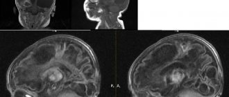
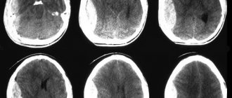
Rice. 4. Computed tomography. Cysticercosis of the brain. The image shows multiple cysts.
Treatment of cerebral cysticercosis
Cysts accidentally discovered during an examination for other diseases do not require treatment.
Anthelmintic treatment
The decision to use antiparasitic therapy is complex. It depends on the stage of the disease, the number and location of cysts with larvae, and specific symptoms.
- Calcified cysts do not require antiparasitic treatment.
- For cysts located inside the cerebral ventricles, treatment with anthelmintic drugs must be approached with caution. Disintegrating cysticerci can cause obstructive hydrocephalus.
- In patients with convulsive syndrome, antiparasitic treatment is carried out only against the background of anticonvulsant treatment.
- For the treatment of neurocysticercosis, the drug of choice is Praziquantel and/or Albendazole. Taking medications for a long time. Treatment is carried out against the background of maintenance therapy with corticosteroids and anticonvulsant therapy. Corticosteroids are necessary to reduce the severity of inflammation that occurs in response to the death of parasites in the brain.
Anthelmintic drugs.
Albendazole is used at a dose of 7.5 mg per 1 kg of weight 2 times a day (15 mg per day per 1 kg of weight) for 15 days. When treating multiple cysts located in the subarachnoid space, treatment is extended to 1 month. Albendazole is prescribed together with Dexamethasone.
Praziquantel is prescribed at a dosage of 15 mg per 1 kg of weight for 15 days.
Sometimes both antiparasitic drugs are used together.
Praziquantel and Albendazole are not used for cysticercosis of the eyes and spinal cord.
Rice. 5. Praziquantel and Albendazole are anthelmintic drugs that are used in the treatment of cysticercosis.
Symptomatic treatment of cysticercosis
Reducing inflammation, preventing seizures, and reducing intracranial pressure are the main areas of symptomatic treatment.
Corticosteroids reduce the severity of inflammation and reduce intracranial pressure. Prednisolone 60 mg or Dexamethasone 6 mg per day are used once.
Patients with convulsive syndrome are prescribed anticonvulsants for treatment or prophylactic purposes.
Surgical treatment of cerebral cysticercosis.
Surgical removal of cysts is resorted to in cases of increased intracranial pressure and hydrocephalus (intraventricular cysticercosis), and with ocular and spinal cysticercosis. Cysts are removed directly or through an endoscopic procedure. When intracranial pressure increases, ventricular shunts are applied.
Disease prognosis
The prognosis of cysticercosis is unfavorable with multiple cysts and localization of cysticerci in the fourth ventricle of the brain. Acute occlusive hydrocephalus and status epilepticus can cause the patient's death. Persistent headaches, frequent attacks of epilepsy and mental changes lead to loss of ability to work.
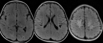
Rice. 6. The photo shows multiple cysts in cysticercosis of the brain.
Intraventricular neurocysticercosis
Like the previous variant of the disease, the intraventricular form is characterized by intracranial hypertension. But this is also accompanied by a sharp attack-like deterioration of the condition. This is directly related to the movement of cysticerci in the ventricles. At one point, they can block the outflow of cerebrospinal fluid, which leads to increased pressure inside the skull.
This variant of neurocysticercosis is characterized by Bruns syndrome. It includes:
- attacks of dizziness;
- fainting;
- vomiting that does not bring relief;
- headache;
- bradycardia;
- disruptions in breathing rhythm;
- increased sweating.
Often it is the fourth ventricle that is affected, which leads to severe hydrocephalus.
Neurocysticercosis of the eye
In addition to severe brain damage, the disease also affects the condition of the eyes and visual functions. Symptoms include:
- drooping eyelid;
- rotation of the eyeball;
- conjunctivitis in the chronic stage;
- movements of the eyeballs become minimal and limited and are accompanied by painful sensations.
Also characteristic is swelling of the orbits and paraorbital areas, loss of visual fields and a feeling of a foreign body in the eye. During therapy, the larva dies and releases a toxin. As a result, optic nerve atrophy or abscess develops.
Diagnosis of the disease
With the development of medicine and the emergence of new diagnostic capabilities, it has become possible to identify the disease in the early stages and with the first appearance of symptoms. The main diagnostic methods include:
- Laboratory blood test. But this method helps to verify the disease only in 50% of clinical cases. From the analysis, eosinophil indicators are important for diagnosis. They indicate helminthic infestation.
- One of the main methods is to determine helminth eggs or their segments in feces. After the parasite is isolated, attention is paid to the scolex, the number of suckers and hooks.
- CSF analysis. In most cases, pleocytosis, increased glucose levels and increased blood pressure are detected.
- Immunological diagnostics. Using the express ELISA method, it is possible to detect antibodies to cysticerci in the blood.
- Serological methods are not very informative and very often give false positive results.
- MRI. Using this imaging method, doctors see foci of inflammation in the spinal cord and brain, in the brain stem and posterior cranial fossa. The use of gadolinium improved the visibility of the cyst walls.
- CT. Using this method, it is possible to identify cystic cavities and the calcifications that form on them. You can also see the focus of inflammation.
- Examination of the fundus. In the anterior chamber or in the vitreous body it is possible to see the cysticerci that float there.
Important ! One of the complications may be chorioretinitis or retinal detachment.
Treatment
Neurocysticercosis can be cured using conservative or surgical methods. Often, drug therapy includes an antiparasitic drug and medications for symptomatic therapy.
Since one of the main symptoms of the disease is seizures, anticonvulsants are prescribed first. Patients whose parenchymal form is in the acute stage are prescribed albendazole. This drug belongs to the group of anthelmintics and is taken for up to 28 days in combination with dexamethasone. But in the case of the development of encephalitis, antiparasitic drugs should not be taken. Since the released toxins will only worsen the patient’s condition. In this case, it is recommended to use corticosteroids.
The larvae are surgically removed from the ventricular cavity. Afterwards, shunts are placed inside them for better outflow of cerebrospinal fluid and to avoid recurrence of intracranial hypertension.
Therapeutic measures
To prescribe effective treatment for neurocysticercosis, it is important not only to evaluate all the symptoms, but also to obtain accurate information about the number of larvae, their viability and location, as well as the resistance of the patient’s immune system to helminths. Treatment is selected individually for each clinical case and is carried out comprehensively:
- medications are prescribed to eliminate the symptoms of the disease;
- anthelmintic therapy is carried out;
- if necessary, lesions are removed surgically;
- ventricular shunts are applied.
Therapy for parenchymal neurocysticercosis
- For a single lesion, treatment is carried out with Albendazole at a dosage of 100 mg per kilogram of weight. The duration of therapy is three days.
- If the degree of infection is moderate, treatment is carried out with Praziquantel in a similar dosage. The duration of therapy is seven days. If side effects occur, steroids are prescribed as a concomitant drug.
- In case of severe infection (when the number of larvae is more than one hundred), Albendazole is prescribed at a dosage of 15 mg per kilogram of weight (this is the daily dosage). The course of therapy is a week. In parallel, treatment with steroids is carried out.
If colloid cysts are found, treatment is carried out as follows:
- if the affected areas are single, treatment with anthelmintic drugs is not carried out;
- for moderate infection, treatment is carried out with Albendazole for one week at a dosage of 15 mg per kilogram of weight;
- in case of severe infection, steroids are prescribed in significant dosages in combination with osmodiuretics.
Therapy for extraparenchymal neurocysticercosis
- If cystic cysts are detected, Albendazole is prescribed at a dosage of 15 mg per kilogram of weight, while the patient takes steroids in parallel. The course of therapy is one month.
- If a patient develops arachnoiditis or angiitis, the doctor prescribes steroids in large dosages. The course of therapy is long and supervised by a specialist. With this diagnosis, there is no need to prescribe special antiparasitic drugs.
- For hydrocephalus, no medications are prescribed; a ventricular shunt is applied.
Therapy for ventricular neurocysticercosis
- Ventricular cysts are removed surgically. As for anthelmintic drugs, they are prescribed at the discretion of the specialist.
- For ependymitis, anthelmintic medications are not prescribed. If the pathology is accompanied by hydrocephalus, a ventricular shunt is required and steroids are prescribed in parallel in high dosages.
What do you need to remember?
- Neurocysticercosis is a serious disease with severe damage to the central nervous system.
- The reason is the pork tapeworm and the eggs of this helminth, which enter the body with poorly processed food, dirty hands and water.
- Main symptoms: convulsions, blurred vision, impaired movement and sensitivity in the limbs, hallucinations, intracranial hypertension.
- The main diagnostic methods include: ELISA, CT, MRI, general blood test.
- Conservative treatment includes anticonvulsant drugs and corticosteroids, and surgical treatment includes placement of an intraventricular shunt.
- The prognosis of the disease depends on the form of the disease and the duration of human infection with pork tapeworm.
Literature
- “Helminth infections of humans”, ed. F.F. Soprunova - M., “Medicine”, 1985
- Zaplotnaya A.A. CNS lesions caused by cestodes: neurocysticercosis / A.A. Zaplotnaya // News of medicine and pharmacy. - 2010. - No. 330. - P. 37-42.
- Ozeretskovskaya N.N., Zalnova N.S., Tumolskaya N.I. “Clinic and treatment of helminth infections” - M., “Medicine”, 1984.
- "Nervous diseases" ed. prof. E.I. Guseva - M., “Medicine”, 1988.
- Neurocysticercosis – a review / Ansari JA // Kathmandu University Medical Journal. - 2003. - Vol. 1, No. 1. - P. 48-55.
- Del Brutto OH, Sotelo J. Neurocysticercosis: an update // Rev. Infect. Dis. - 1988. - Vol. 10. - P. 10751087.
- Grisolia JS, Wiederholt WC CNS cysticercosis // Arch. Neurol. - 1982. - Vol. 39. - P. 540544.
- Garcia HH, Gonzales AE, Evans CAW, Gilman RH Taenia solium cysticercosis // Lancet. - 2003. - Vol. 362. - P. 547–556. Dixon HBF, Lipscomb FM Cysticercosis: an analysis and followup of 450 cases // Med. Res. Counc. Spec. Rep. Ser. - 1961. - Vol. 299. - P. 158.