Epileptic syndrome is a condition in which the convulsive state that usually occurs during epileptic seizures occurs frequently.
A seizure (the main symptom of the syndrome) usually begins suddenly, although the body warns of its occurrence with some symptoms: a day or two before the onset of a seizure, a severe or moderate headache may be observed, appetite disturbance occurs, sleep becomes restless, the person is overly irritable and complains of bad things. well-being. In some cases, the seizure begins with the appearance of an aura in the patient.
In medical practice, auras are divided into:
- motor : in this case, the patient’s seizure begins with the patient freezing in some position or starting to run somewhere;
- sensory : disturbances of the senses (visual, gustatory, olfactory);
- vegetative : discomfort in the heart, heat or cold in the head, urge to urinate, increased peristalsis;
- sensitive : paresthesia of different parts of the body is observed;
- vestibular;
- mental : an unreasonable feeling of fear, joy, euphoria.
The duration of the aura can last only a few seconds, after which the person loses consciousness, emitting a kind of cry, which is a consequence of a spasm of the vocal cords. For the next quarter of an hour, tonic convulsions occur. At this time, the patient’s breathing is delayed, the face becomes bluish-pale, the veins in the neck swell, and cyanosis increases.
For the next 3 minutes, clonic convulsions continue, during which hoarse, noisy breathing is observed. There is a variant of tongue retraction.
Cyanosis slowly recedes, saliva is released in the form of foam (it may turn red as a result of blood entering the saliva due to biting the tongue or the inside of the cheek).
During a seizure, the patient does not respond to external stimuli, dilated pupils do not respond to light, and tendon reflexes do not function. Sometimes the patient can fall asleep without leaving this state.
The entire attack lasts about 4 minutes and after it ends the person feels depressed, drowsy, and weak. The patient does not remember anything about the seizure itself.
Epileptic syndrome, unlike epilepsy, is the result of a previous disease (or accompanies it). Epilepsy is an independent disease, which most often has a congenital character. Also, the syndrome, despite the fact that the symptoms are similar to epilepsy, goes away a little easier.
General information
So, what is the difference between epilepsy and episyndrome? It's simple - epilepsy is an independent disease that occurs as a result of genetic or other reasons. Episyndrome is a symptom or secondary manifestation of any illness or injury and has nothing to do with epilepsy.
Both epilepsy and episyndrome are studied within the framework of neurology and epileptology, and epileptics, in addition, can also undergo treatment in psychiatry, since such patients often develop mental disorders.
The etiology of the epileptic syndrome coincides with a regular epileptic attack, but with the difference that such a seizure occurs once and does not recur.
Establishing diagnosis
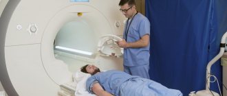
The following methods are used to diagnose epileptic syndrome:
- MRI (magnetic field examination);
- CT (uses x-rays, but the result is more accurate than x-rays);
- electroencephalogram (helps to record seizures and their location).
Article on the topic: Perianal dermatitis: causes and treatment
Using these types of studies, the doctor detects the affected areas of the brain.
Causes
The causes of episyndrome in children and adults are different. Thus, the following factors that contribute to the development of the syndrome are characteristic of children:
- trauma received as a result of childbirth;
- infectious diseases that the mother suffered during pregnancy;
- heredity;
- oxygen starvation of the fetus during pregnancy.
In adults, the reasons may be as follows:
- traumatic brain injuries;
- neoplasms in the brain;
- oxygen starvation (suffocation, drowning, etc.);
- previous infectious diseases of the brain (encephalitis, meningitis, etc.);
- hemorrhages in the brain (this is often influenced by the patient’s age; the older, the higher the risk);
- demyelinating diseases of the nervous system (multiple sclerosis, etc.).
The disease syndrome can also develop in people who have problems drinking alcohol. But an alcoholic may not necessarily experience episyndrome. Often, a person who does not belong to the group of alcohol addicts, but has gone too far with alcohol, may experience an epileptic seizure, diagnosed as a syndrome.
Difference between episyndrome and epilepsy
Epileptic syndrome is a symptomatic anomaly that accompanies a number of diseases. Manifested by loss of consciousness, tonic-clonic convulsions. In frequent cases, it accompanies epilepsy, but is differentiated from the latter by etiology. A seizure can be triggered by:
- mechanical damage to the brain due to trauma;
- insufficient oxygen supply due to poor circulation;
- intracranial edema or swelling;
- chronic diseases affecting the central nervous system (CNS).
Convulsions may be episodic. When the cause is eliminated, the syndrome disappears along with it.
The anomaly cannot be controlled; attacks occur at any moment of the central nervous system reaction to a particular stimulus. They do not entail negative changes in the brain and do not affect intellectual development.
Epilepsy is a common pathology, affecting 0.6% of the population. Usually manifests itself in infancy, less often in adolescence. It is characterized as a chronic neurological disease, in most cases of unknown origin. Pathology is divided as follows:
- Idiopathic (hereditary). There are precedents when electroencephalography of close relatives in a patient’s family indicated the presence of epilepsy, which they did not suspect, and the anomaly did not manifest itself in any way.
- The essential form occurs against the background of brain damage. In either case, a seizure begins as a result of a sudden over-discharging of neurons in the gray matter that spans the entire brain, causing seizures. Attacks occur repeatedly, they can be controlled by warning signs: nausea, headache.
The difference between episyndrome and epilepsy is as follows:
| Syndrome | Neurological disease |
| symptomatic pathology | independent anomaly of chronic course |
| variety of reasons | genetic predisposition, uncertain etiology, rarely essential (acquired) form |
| uncontrolled manifestation | preceded by an aura (a set of signs) |
| episodic seizures | without adequate therapy, worries throughout life |
| does not cause brain damage | occurs against the background of intracranial negative processes |
The onset of epilepsy in 70% of cases occurs in preschool age. Episyndrome (ES) can first appear in both an adult and a child, it depends on the provoking factor. With a chronic neurological disease, psychological deviations are possible: amnesia, psychosis, sudden changes in mood, depression, impaired mental abilities. The listed symptoms are not characteristic of ES.
Symptoms
The main symptoms of the episyndrome are similar to a regular epileptic seizure, the person is in a faint state, and his muscles contract (twitch) uncontrollably, as a result of which the patient convulses.
There are conventionally three phases of a seizure:
- State of fainting.
- Tonic spasms (muscle tension, arching of limbs).
- Clonic phase (convulsive twitching).
This condition may be preceded by an aura (early warning signs of a seizure). Thus, the following types of aura are distinguished:
- sensory (problems with the senses - vision, smell, taste);
- vegetative (fever or cold, heart pain, frequent urination and bowel movements);
- motor (the patient freezes in one position or increases motor activity);
- sensitive (paresthesia in various parts of the body);
- mental (unreasonable feeling of fear or joy, etc.).
Main symptoms
In addition to the classic episyndrome, there are several of its varieties, for example:
Children are characterized by syncope (syncopate) epileptic syndrome, which is expressed in loss of consciousness and tonic-clonic convulsions. The post-syncope phase is characterized by involuntary urination and sometimes defecation.
In addition, the episyndrome includes an abortive seizure - characterized by loss of consciousness and convulsive contractions of the facial muscles, stupor, and loss of motor activity. The most severe variant of the attack is described above. Perhaps an easier course is a short-term loss of consciousness for a few seconds, without muscle contractions. This type of seizure is also called minor. An attack can occur in such a way that the patient does not even notice its presence (for example, the patient was talking with an interlocutor, for a few seconds his consciousness turned off, which the interlocutor noticed, but after the attack has passed the patient continues the conversation as if there was nothing never happened). In such a situation, the patient does not need help.
Clinical picture - how does this happen?
Epileptic syndrome is accompanied by a number of symptoms that are in many ways similar to an epileptic seizure, but are easier to tolerate:
- The seizure comes unexpectedly.
- The duration of the convulsions can be three to four minutes.
- After a seizure, the patient becomes lethargic and drowsy.
- A day or two before the attack, the patient loses his appetite, sleep deteriorates, and there is general weakness in the body.
- During an attack, the patient looks at one point, the facial muscles begin to twitch, and complete loss of consciousness is possible. Immediately after the attack, the patient can communicate freely, will be completely adequate, with absolutely no memory of the attack.
- The twilight state is a manifestation of epileptic syndrome, which is characterized by a disturbance in orientation in place, time, and possibly the appearance of hallucinations. This state can last from several minutes to several hours. After the “return” to a normal state, complete amnesia is observed.
Treatment in children
Only a doctor can prescribe treatment, since only he is able to accurately diagnose and differentiate episyndrome from epilepsy.
First of all, the doctor must determine the cause of the disease, since if it is not eliminated, in most cases a relapse is possible.
So, a child may experience an epileptic seizure due to a high temperature (it needs to be brought down) or due to a lack of certain vitamins (for example, potassium).
As for the treatment of an infant, the difference from an older child is more careful therapy and less intensive treatment.
Since most causes of episyndrome in a newborn are associated with pregnancy and childbirth, not only drug therapy is indicated; in some cases, massage or even surgery will have an effect.
Etiology of the disease
The causes of epileptic seizures are not fully understood. Many studies prove the hereditary nature of the disease. In addition to the hereditary factor, scientists consider the conditions of intrauterine development of the fetus and early postnatal trauma to the child’s brain during childbirth or infection in the natal period with a staphylococcal or streptococcal infection as causes. In 25% of cases, the pathogenesis of the disease could not be determined. In general, the researchers were able to create a classification of the main causes of epilepsy.
Among them:
- brain injury during childbirth or the perinatal period;
- congenital genetic developmental abnormalities;
- head injury;
- cerebral hemorrhages;
- meningococcal, encephalitis, streptococcal infections;
- genetic syndromes;
- tumor processes;
- eclampsia;
- alcohol withdrawal.
Treatment for adults
Epileptic syndrome in adults is treated somewhat differently, since the causes of this disease are different. The principle remains the same; it is possible to cure seizure syndrome only by eliminating the irritating factor, including:
- Infectious agent (meningitis, encephalitis, arachnoiditis).
- Maintenance therapy after a stroke (usually drugs that stimulate brain activity and blood circulation).
- Surgery is necessary if there is a tumor or the need to remove bone fragments after a traumatic brain injury.
Epileptic syndrome can also be treated using standard medications for epileptics (carbamazepine), however, this is justified only if it is impossible to get rid of the disease by other methods.
Treatment
The main goal of treatment is to stop attacks, stop taking medications and improve quality of life. Depending on the results of the examination, either conservative or surgical treatment methods are selected.
Epileptic syndrome can be treated! In the neurology department, specialists treat syndromes.
Our clinic uses the following treatment regimen. At the first stage, the causes of seizures that are subject to treatment are determined (these are various tumors, aneurysms). Then the factors that provoke seizures are excluded (fatigue, mental and physical stress). After this, a thorough diagnosis is carried out and appropriate drug therapy is prescribed.
Status epilepticus
The disease can develop from a normal seizure into a special condition called status epilepticus. This status is characterized by cyclical repetitions of tonic-clonic convulsions, which successively replace each other and do not stop.
The danger of such a manifestation is not only exhaustion of the body, but also an increased risk of injury. It is not difficult to understand that a patient has developed an epistatus; as a rule, the longest seizure lasts no more than five minutes; accordingly, if the seizures do not stop for more than this time, it is necessary to call an ambulance, because it will not be possible to cope with the disease on your own. The doctor will give the patient an injection that will relax all his muscles and stop the seizure.
Post-traumatic epilepsy
Post-traumatic epilepsy (PTE) occurs in most cases some time after a brain injury. The appearance of such a disorder is due to the healing processes of existing damaged structures. After an injury, regeneration processes begin. Functional nerve fibers, neurons, cannot recover. At the site of their death, foci of gliosis form.
Glia are additional cells that normally contribute to the normal course of metabolic processes, the transmission of nerve impulses and perform a number of other functions. The proliferation of glial cells allows one to compensate for damage and restore the conduction of nerve impulses between surviving neurons.
The restoration of nerve endings causes abnormal electrical activity to appear in the affected areas of the brain.
People at particular risk of developing epilepsy should undergo regular examinations by a neurologist after injury. If severe attacks occur, the patient should consult a doctor. First, the specialist conducts a general examination and performs tests to identify neurological disorders. When making a diagnosis, the doctor takes into account the clinical picture, data obtained from EEG and neuroimaging.
Clinical picture
In the process of collecting anamnesis, the specialist tries to clarify with the patient and his relatives the nature of the manifestations of epilepsy. Of particular interest are the frequency and duration of attacks, the time of their occurrence and the characteristics of the patient’s behavior.
An EEG can detect some signs of epilepsy in the periods between seizures. This study makes it possible to find out pathological paroxysms, wave duration and rapid changes in polarity. The data obtained is not always informative, so additional diagnostic methods are required for confirmation.
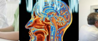
Neuroimaging
MRI is most often used to clarify the nature of residual damage to brain tissue. This study allows us to determine the presence of structural changes. After an attack, MRI helps to exclude bruises and hematomas of the brain.
In addition, this study can identify cysts, fluid accumulation, abscesses, and tumors that could form after injury.
Living with a disease
Is it possible to live with this disease? Yes. The fact that a person has had an epileptic syndrome does not mean that it will relapse. It is likely that the incident is isolated and will not happen again. Nevertheless, it is necessary to adhere to simple recommendations that will allow you not to provoke the disease, including:
- do not stay in a stuffy and hot room for a long time, do not increase body temperature;
- If possible, go on a low-carb diet;
- try to eat less spicy and salty foods;
- give up bad habits (smoking and alcohol), as well as drinking energy drinks;
- stress less.
So, epileptic syndrome is an unpleasant but harmless disease that can occur in any person if certain factors coincide. There is no need to panic about this; you need to consult a doctor and follow his instructions. Do not be ill!
Epileptic syndrome and epilepsy - what are the differences?
These two concepts should be clearly distinguished. The main difference is the origin of these two conditions. SE develops due to some previous brain disease. Epilepsy is a separate disease, the causes of which are unknown.
Epilepsy is accompanied by frequent mental disorders. In sick patients, intelligence decreases, thought processes are disrupted, and mood swings occur frequently. In epileptic syndrome, such manifestations do not occur.
If the underlying brain disease that causes the development of epileptic syndrome is eliminated, all signs of this condition may disappear.
Thus, SE does not mean that the patient suffers from epilepsy.
results
The results of the study showed that SHSPs can be part of the structure of many epileptic syndromes (Table 1).
Table 1. Main characteristics of forms of epilepsy associated with SHSP Note. IFE-PHP is idiopathic focal epilepsy with pseudogeneralized seizures.
Table 1. Main characteristics of forms of epilepsy associated with SHSP (end) Note. IFE-PHP is idiopathic focal epilepsy with pseudogeneralized seizures. Most often, in patients with SHSP, symptomatic focal epilepsy was diagnosed (33.8% of cases), cryptogenic focal epilepsy (23.8%), rolandic epilepsy (12.6%), and FEDSIM-DEPD - 12.3% of cases. Other forms of epilepsy were detected much less frequently.
The onset of epilepsy in patients with SHSP varied over a wide age range from the first month of life to 18 years. The average age of onset was 5.7±4.96 years. Most often, epilepsy associated with SHSP debuted in the following age intervals: from the first month of life to 1 year - 16.8%, from 1 year to 3 years - 25.3% of cases, from 4 to 6 years - 20.8% . As they grew older, the likelihood of developing an epileptic syndrome in patients with SHSP gradually decreased: from 7 to 9 years of age - 15.7%, from 10 to 12 years - 9.3%, from 13 to 15 years - 5.7%, from 16 to 18 years old - 6.4%. It is noteworthy that in most cases (62.9% of patients), the onset of the disease was noted in children of preschool age. The age of onset of various syndromes associated with SHSP could differ significantly (see Table 1).
At the onset of epilepsy, SHSPs were noted in 207 cases, which accounted for 43.9% of patients. SHSP as the only type of paroxysms was observed in 28.3% of cases during the entire period of the disease. In other cases, SHSPs were combined with other types of seizures. Two types of attacks or more were observed in 71.7% of cases, three types or more - in 39.3%.
Along with SHSP, 19 different types of seizures could also be observed in the clinical picture of patients in our group. The following paroxysms were most often observed in epilepsy associated with SHSP: focal automotor - 22% of cases, focal hemiclonic - 20.4%, focal sensory - 20.4%, febrile seizures - 14.6%, focal motor (including versive) - 14.2%, asymmetrical tonic seizures - 8.9%. Other types of seizures were less common: focal atonic (so-called “temporal syncopation”) - 6.8% of cases, atypical absences - 6.2%, autonomic paroxysms - 6.2%, seizures originating from the occipital cortex - 4.7%, focal myoclonus - 3.6%, epileptic spasms - 3.4%, negative myoclonus - 2.9%, myoclonic seizures - 2.5%, focal hyperkinetic - 2.1%, focal adversive and gelastic - 1.3%, atonic - 1.1%, epileptic myoclonus of the eyelids - 0.8% of cases.
It should be noted that for different epileptic syndromes associated with SHSP, the frequency of occurrence of different types of seizures differed (see Table 1).
During continued VEM, various types of pathological activity were detected in most cases (91.3%).
The absence of epileptiform activity was noted only in 8.7% of cases (41 patients). Moreover, all these patients were observed with diagnoses: symptomatic or cryptogenic focal epilepsy. In other groups of epileptic syndromes - idiopathic focal forms of epilepsy and epileptic encephalopathies - epileptiform changes were recorded in 100% of patients during prolonged VEM with sleep included.
Regional/multiregional epileptiform activity was recorded in 91.3% of cases of all forms of epilepsy associated with SHSP.
Interictal diffuse epileptiform activity was detected in 30.1% of cases. Moreover, in 100% of cases, diffuse discharges were detected in epileptic encephalopathies, progressive myoclonus epilepsy and idiopathic focal epilepsy with pseudogeneralized seizures (IFE-PHP syndrome).
The study revealed that 175 (37.2%) patients had DEPD. These graphelements were found in all patients with idiopathic focal forms of epilepsy, pseudo-Lennox syndrome, Landau-Kleffner syndrome, electrical status epilepticus syndrome of slow-wave sleep, cognitive epileptiform disintegration, FEDSIM-DEPD syndrome.
During continued VEM with sleep included, HFSPs were recorded in 39 patients. In all cases, a similar EEG pattern of the attack was revealed - at the beginning of SHSP, the appearance of regional rhythmic epileptiform activity on the EEG is noted. The tonic phase corresponds to the appearance of diffuse rhythmic pointed theta activity in combination with rhythmic fast peak-wave and polypeak-wave complexes. As the tonic phase continues, an increase in the amplitude of the acute-slow wave complexes and their slowdown to 1.5-3.5 Hz are observed, which corresponds to the transition to the clonic phase. Clonic seizures were characterized by the appearance of groups of diffuse polypeak-wave complexes of a low degree of synchronization in combination with rhythmic motor and myographic artifacts. As the attack ends, diffuse polypik-wave discharges become less frequent. The stage of post-ictal confusion corresponds to the appearance of diffuse slowing. In all cases, pronounced myographic and motor artifacts were superimposed on the ictal EEG recording, making it difficult to assess the bioelectrical activity of the brain; in 12 cases, an objective assessment of the EEG pattern of the attack was impossible due to the large number of artifacts.
Analysis of the use of therapy demonstrated that patients were prescribed the following groups of drugs, both in mono- and polytherapy in various combinations: carbamazepine, oxcarbazepine, valproate, levetiracetam (tablets and in the form of an oral solution - Keppra), topiramate, perampanel, lamotrigine, ethosuximide.
Prescription of PET led to the achievement of complete remission in 57.1% of cases of epilepsy associated with SHSP. A reduction in the frequency of attacks by 50% or more on the background of PET was observed in 33.6% of patients. No effect on seizures was noted in 9.3% of cases.
The study showed different effectiveness of PET in the treatment of certain groups of epileptic syndromes associated with SHSP (Table 2).
Table 2. Efficacy of PET in various forms of epilepsy associated with SHSP (%) The highest percentage of remission was observed in idiopathic focal forms of epilepsy (94.7% of cases) and in FEDSIM-DEPD syndrome (77.6%). In the largest group of patients with symptomatic/cryptogenic focal epilepsy, remission was achieved in only 42.0% of cases. In epileptic encephalopathies, remission was observed in 45.2% of patients.
Separately, the effectiveness of PET was analyzed in forms of epilepsy associated with DEPD, which were recorded in patients with idiopathic focal forms of epilepsy, pseudo-Lennox syndrome, Landau-Kleffner syndrome, electrical status epilepticus syndrome of slow-wave sleep, cognitive epileptiform disintegration, FEDSIM-DEPD syndrome (37 .2% of all patients with SHSP). High effectiveness against attacks was obtained, which was significantly higher than in the general group of patients with SHSP. Remission of attacks was observed in 84.6% of cases, a reduction in the frequency of attacks by 50% or more in 14.3%, and no effect on paroxysms in only 1.1% (see Table 2).
Symptoms of epilepsy, main types of seizures
Partial (focal, local) seizures are seizures whose initial, clinical and electrophysiological manifestations indicate the involvement of one or more areas of one cerebral hemisphere in the pathological process.
- Simple partial (focal) seizures occur suddenly, last no more than a minute, and are accompanied by symptoms corresponding to the excitation of certain cortical areas against the background of preserved consciousness. The most typical are tonic or clonic contractions of the muscles of half the face, part of a limb, half of the body, adversion of the head and/or eyes to the side, and sudden stop of speech.
- Complex (complex) partial seizures often begin with gaze fixation, followed by automatism, including repeated steps like stepping on the spot, fussy, swallowing, chewing and other stereotypical movements, snatches of speech that are not related to the circumstances, muttering, and mooing. At the end of automatism, a period of confusion lasting several seconds is noted.
Generalized seizures are seizures, the initial clinical and electrophysiological manifestations of which indicate the involvement of both hemispheres of the brain in the pathological process.
- Absences (simple) seizures, the main manifestation of which is a sudden and sharply interrupted short-term (5-10 sometimes up to 30 seconds with atypical absences) disturbance of consciousness with freezing of the gaze, freezing, sudden interruption of speech and movement (with simple absences) or automatic continuation of what was started actions (for atypical forms). Clonic seizures are an attack of massive clonic muscle contractions, leading to rhythmic flexion-extension movements in the joints seized by convulsions against the background of impaired consciousness. Tonic generalized seizures of the axial type, manifested by short-term tonic tension of the body muscles against the background of impaired consciousness.
- Epileptic myoclonus of the eyelids with or without absences - epileptic: seizures manifested by covering the eyes and frequent (3-6 times per second) rhythmic myoclonus of the eyelids (“fluttering of the eyelids”). They may be accompanied by short absence seizures or occur without loss of consciousness. In most cases, eyelid myoclonus is not accompanied by a disturbance in the level of consciousness. In patients with a documented decrease in the level of consciousness at the time of the attack, absence seizures occur following the phase of myoclonic eyelid twitching and are short-lived. Myoclonus of the eyelids with absence seizures is usually recorded in bright light or during rhythmic photostimulation. In some patients with epileptic myoclonus of the eyelids, one can observe the phenomena of self-induction, that is, self-provocation of attacks by rolling the eyes, performing various manipulations in order to create flickering light.
- Myoclonic seizures are an attack of clonic rapid contractions of individual muscle groups, usually in the facial muscles, with impaired (rarely preserved) consciousness. Myoclonic-astatic seizures are short epileptic generalized seizures that involve a sudden fall. At the beginning of the attack, myoclonus is noted in the axial muscles, which causes lightning-fast bending of the torso, head and neck forward, as well as a sharp raising of the arms up and forward. This is immediately followed by a phase of atonia, resulting in a sudden fall.
- Tonic-clonic seizures are one of the most common manifestations of primary generalized epilepsy. They occur with loss of consciousness, consist of an initial tonic phase, followed by a clonic phase and end with a post-attack impairment of consciousness.
- Atonic seizures are attacks of short-term, sudden loss of muscle tone, especially in the extensor muscles, which leads to a rapid fall, accompanied by impaired consciousness.
Unclassified seizures: rhythmic eye movements, chewing and swimming movements, “salaam” convulsions, flexion and extension seizures (epileptic spasms).
- Epileptic spasms are characterized by short massive contractions of the axial muscles and can be flexor, extensor or mixed. Occurs in children of the first year of life with epileptic encephalopathies (Otahara syndrome, West syndrome). Flexor epileptic spasm consists of a sudden sharp tension of the flexor muscles. There is a nod of the head forward, bending at the torso, and bringing the arms in front of oneself. With an extensor epileptic spasm, on the contrary, predominantly extensors are involved and the kinematics of the attack resembles the movements that occur in a child with the Moro reflex.
- Separately, asymmetric epileptic spasms are distinguished, reminiscent of an asymmetric cervical-tonic reflex: there is a sharp tonic extension and stretching of the limbs on one side and tonic tension of the flexor muscles in the limbs by adduction to the body on the contralateral side, deviation of the head and eyes, usually towards extension. The duration of the attacks is a fraction of a second. However, in most patients, epileptic spasms tend to occur serially, forming groups of 5 to 50 or more in one cluster. Most often, epileptic spasms occur after the child wakes up.
medlibera.ru