Authors: Mukhin K.Yu., Petrukhin A.S.
Part 1:
- Introduction
- Definition, classification of epilepsy
- Idiopathic generalized epilepsy
- Pediatric absence epilepsy.
- Juvenile absence epilepsy.
- Epilepsy with isolated generalized seizures.
- Juvenile myoclonic epilepsy
- Idiopathic partial epilepsy
- Rolandic
- Idiopathic partial epilepsy with occipital paroxysms.
Part 2:
- Cryptogenic generalized epilepsy
- Lennox-Gastaut syndrome.
- Epilepsy with myoclonic-astatic seizures.
- Epilepsy with myoclonic absence seizures
- Symptomatic partial epilepsy
- Symptomatic temporal lobe epilepsy
- Symptomatic frontal lobe epilepsy
- General principles of treatment of epilepsy
Introduction
The rapid development of neuropharmacology in recent decades, the synthesis of new highly effective antiepileptic drugs (AEDs), a radical revision of many principles of treatment of epilepsy, has now made it possible to classify epilepsy as a curable disease. According to generalized data from the leading antiepileptic centers in the world, a pronounced therapeutic effect is achieved in 75-85% of patients suffering from epilepsy. Epilepsy is one of the most pressing problems in pediatric neurology. According to foreign data, the frequency of epilepsy in the pediatric population is 0.5-0.75% of the child population, and febrile seizures up to 5%. There are no exact statistics on this disease in Russia. Firstly, not all doctors are widely familiar with the modern International Classification of Epilepsy. In this regard, the statistics of epilepsy did not include such diagnoses as “epileptiform syndrome”, “convulsive syndrome”, which are not used in any country in the world and, in fact, are the same disease - epilepsy. Secondly, in our country, epileptic people are observed by both neurologists and psychiatrists, which significantly distorts the statistics. In all countries of the world, epilepsy is a neurological disease.
Over the past decade, there has been a very rapid accumulation of new knowledge on the problem of epilepsy. Both experimental and clinical studies have contributed to a better understanding of the underlying pathophysiological mechanisms of the disease. New research methods have significantly changed our ability to diagnose epilepsy and identify etiological factors of the disease. In addition, great advances have been made in the development of medical and surgical treatment approaches.
Diagnosis of the disease
To make a diagnosis, the doctor carries out the following activities:
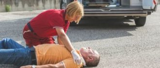
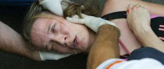
- Listens to the story of a witness who was present during the victim’s seizure. The patient himself with complex partial seizures often does not remember the attack. In simple cases, the patient can talk about how he feels during a seizure.
- A neurological examination is performed. The patient is tested for coordination of movements, performing a finger-nose test, questions are asked to test intelligence, and simple logical problems are solved.
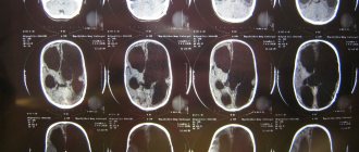
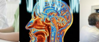
- MRI is necessary for diagnosing epilepsy with congenital structural pathologies and various brain tumors, cystic formations, vascular diseases of the head, multiple sclerosis.
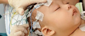
- EEG (electroencephalogram) - determines the location of the focus and the form of epilepsy. In some cases, the examination is carried out several times.
Taking into account all the data that was obtained during the study, as well as the causes and symptoms of partial epilepsy, the doctor builds a treatment strategy for the patient.
Definition, classification of epilepsy
Definition. Epilepsy is a chronic disease of the brain characterized by repeated unprovoked attacks of disturbances in motor, sensory, autonomic, mental or mental functions resulting from excessive neural discharges. The presented definition contains two important provisions. Firstly, epilepsy does not include single seizures, regardless of their clinical manifestations. Only repeated seizures are the basis for a diagnosis of epilepsy. Secondly, epilepsy includes spontaneous, unprovoked seizures (with the exception of reflex forms). By definition, febrile seizures, as well as seizures that occur in acute diseases of the brain (for example, encephalitis, subdural hematoma, acute cerebrovascular accident, etc.) are not epilepsy. Classification. The manifestations of epilepsy are extremely diverse, which from the very beginning of studying the disease made it difficult to create a unified classification. The modern classification of epileptic seizures was adopted by the International League Against Epilepsy in 1981 in Kyoto (Japan). Unlike previous classifications, it takes into account both clinical and neurophysiological (EEG) criteria for most types of epileptic seizures (Table 1). The classification divides all types of epileptic seizures into partial (focal, focal, local, localization-related), generalized and unclassified. Partial seizures are diagnosed when, at the onset of paroxysm, there are clear clinical and electrophysiological criteria for the involvement of certain brain structures. For example, clonic convulsions of one half of the face and arm (faciobrachial seizures) usually indicate the presence of an epileptic focus in the middle-lower parts of the anterior central gyrus; olfactory hallucinations – in the area of the hook; photopsia – in the occipital lobe cortex, etc. If the attack begins as a partial attack, and then involves the entire musculature of the trunk and limbs and signs of involvement of both hemispheres on the EEG, then it should be classified as focal with secondary generalization. The classification clarifies the concept of simple and complex partial seizures. Previously, complex partial seizures were defined as paroxysms with a change in consciousness, and simple ones - without change. According to the Kyoto classification, complex partial seizures should be understood as paroxysms not with a change, but with a complete shutdown of consciousness. Consequently, according to the modern classification, all attacks that occur with phenomena of depersonalization, dream states, cognitive disorders, etc., are not classified as complex, but as simple partial, since the patient’s consciousness during these paroxysms is changed, but not turned off, and the memory of the attacks is preserved . Also, the 1981 classification stipulates that one patient may have several different types of seizures. For example, an attack, starting as a simple partial, can transform into a complex partial, and then into a secondary generalized one. The term “polymorphic seizures” has been removed from the classification, which does not carry any information and is not recommended for use. Thus, the Kyoto classification is at this stage the most complete systematization of epileptic seizures. With the accumulation of clinical experience, the introduction of video-EEG monitoring into practice, the development of neuro-radiological diagnostic methods (CT, MRI, PET), molecular genetics and other sciences, it became obvious that there are a number of special forms of epilepsy, which are characterized by their own clinic (typical types of attacks), course and prognosis. Some of these forms have been known for a long time, such as West syndrome, Lennox-Gastaut syndrome, and Rolandic epilepsy. Others - benign familial neonatal seizures, severe myoclonic (in infancy), juvenile absence epilepsy - have been identified only in recent years. These forms of epilepsy, or according to the International Classification, epileptic syndromes, as a rule, are manifested not by any one type of seizure, but by a combination of them. Epileptic syndromes are defined as separate, independent forms of epilepsy, characterized by a limited age of onset of seizures, the presence of a special type of seizures, specific changes in the EEG (characteristic of a given syndrome), patterns of course and prognosis. For example, one type of seizures - absence seizures - can be part of a number of epileptic syndromes: childhood and adolescent absence epilepsy, juvenile myoclonic epilepsy, with myoclonic absence seizures and others, and the course and prognosis for all of these syndromes are different. A fundamentally new step in the development of epileptology was the creation of a modern classification of “epilepsies, epileptic syndromes and diseases associated with seizures.” This classification was adopted by the International League Against Epilepsy in October 1989 in New Delhi and is currently generally accepted by epileptologists around the world (Table 2). The classification of epileptic syndromes is based on the following principles: 1. Localization principle:
- localization-related (focal, local, partial) forms of epilepsy;
- generalized forms;
- forms that have features of both partial and generalized.
2. Principle of etiology:
- symptomatic,
- cryptogenic,
- idiopathic.
3. Age of onset of attacks:
- newborn shapes,
- infant,
- children's,
- youthful,
- adults.
4. The main type of attacks that determines the clinical picture of the syndrome:
- absence seizures,
- myoclonic absence seizures,
- infantile spasms, etc.
5. Features of the course and forecast:
- benign,
- severe (malignant).
The principles of localization and etiology in classification require clarification. The classification is based on classical ideas about focal and generalized forms of epilepsy. Localization-related forms are determined if the nature of the paroxysms, EEG data and neuroradiological examination confirm the local origin of the attacks. This applies not only to forms with a clearly identified structural defect of the brain (temporal lobe epilepsy), but also to syndromes in which the nature of the seizures and EEG indicate a local onset, but changes on CT are usually absent (rolandic epilepsy, benign occipital epilepsy). It is also possible that there are multifocal forms of epilepsy, in which seizures originate from several foci within one or both hemispheres. In generalized forms of epilepsy, seizures must be generalized from the very beginning, which is confirmed by EEG data (bilateral synchronous spread to both hemispheres). The pathogenesis of generalized forms of epilepsy is still not clear enough. A corticothalamic hypothesis for the occurrence of primary generalization is proposed. In cases where the nature of the attacks and examination data do not allow us to confidently state the local or primary generalized onset of paroxysms, these epileptic syndromes are defined as not amenable to clear classification, that is, having signs of both locality and generalization. The classification divides all epileptic syndromes into symptomatic, idiopathic and cryptogenic. Symptomatic forms mean epileptic syndromes with a known etiology and verified morphological disorders (tumors, scars, gliosis, cysts, dysgenesis, etc.). In idiopathic forms, there are no diseases that could be the cause of epilepsy, and epilepsy is, as it were, an independent disease. Currently, the genetic determination of idiopathic forms of epilepsy has been established. The term “cryptogenic” (hidden) refers to those syndromes whose cause remains hidden and unclear. These syndromes do not meet the criteria for idiopathic forms, but there is no evidence of their symptomatic nature. For example, in the case of a combination of epilepsy with hemiparesis or oligophrenia, the symptomatic nature of the disease is assumed, but CT and MRI studies do not visualize changes in the brain. This case is classified as cryptogenic. It is obvious that with the improvement of the technical capabilities of neuroimaging (for example, PET), most cryptogenic forms will be transferred to the category of symptomatic. Symptoms. The semiology and clinic of various epileptic seizures are described in detail in many manuals on epilepsy. At the same time, the diagnosis of certain forms of epilepsy (epileptic syndromes) has received insufficient attention in the literature. Let us dwell on the diagnostic criteria and principles of treatment of the main epileptic syndromes of childhood and adolescence.
Idiopathic generalized epilepsy
All forms of idiopathic epilepsy are characterized by:
- Genetic predisposition (often a family history of epilepsy).
- Limited age of disease onset.
- No changes in neurological status.
- Normal intelligence of patients.
- Preservation of the basic rhythm on the EEG.
- Absence of structural changes in the brain during neuroimaging.
- The drugs of choice for treatment are valproic acid derivatives.
- Relatively favorable prognosis with therapeutic remission achieved in the vast majority of cases.
Pediatric absence epilepsy.
Absence seizures are a type of generalized nonconvulsive seizures, characterized by a high frequency and short duration of paroxysms with loss of consciousness and the presence of a specific pattern on the EEG - generalized peak-wave activity with a frequency of 3 Hz. Absence seizures are one of the most common types of epileptic seizures in children and adolescents. The onset of absence seizures in childhood absence epilepsy (CAE) is observed in the age range from 2 to 9 years, averaging 5.3 ± 0.3 years. The peak age of manifestation is 4-6 years, with a predominance of girls by gender. Clinically, absence seizures are characterized by a sudden short cessation (or significant decrease in the level) of consciousness with absence or minimal motor phenomena. An aura, as well as post-ictal confusion, are not typical. The duration of absence seizures ranges from 2-3 to 30 seconds, averaging 5-15 seconds. A characteristic feature of absence seizures is their high frequency, reaching tens and hundreds of attacks per day. It is important to divide absence seizures into simple and complex ones. Simple absence seizures are characterized by the cessation of all activity, “freezing”, “freezing” of patients, a fixed “absent” gaze, and a confused, hypomimic facial expression. Simple absence seizures account for only 20% of the clinical picture of DAE. DAE is more characterized by complex absence seizures that occur with a minimal motor component. The following types of complex absences are distinguished: with myoclonic, tonic, atonic, vegetative components, as well as with automatisms and focal phenomena. The most common absence seizures are those with myoclonic and tonic components. Absence seizures with a myoclonic component are observed in 40% of patients with DAE. Manifested by: myoclonus of the eyelids; perioral myoclonus (rhythmic stretching of the lips, like a “goldfish”); perinasal myoclonus (rhythmic twitching of the wings of the nose). During an attack, a number of patients experience a single shudder or several short weak twitches of the muscles of the shoulder girdle and/or arms. Absence seizures with a tonic component are most typical for DAE, observed in 50% of patients. They are manifested by deviation of the head, and sometimes the body, backwards (retropulsive absence seizures), tonic abduction of the eyeballs upward or to the side. Sometimes during an attack there is a slight tonic tension (usually asymmetrical) in the muscles of the upper extremities. Absence seizures with an atonic component are not typical for DAE and are observed only in isolated cases, mainly in atypical forms. They manifest themselves as a sudden loss of muscle tone in the muscles of the arms (loss of objects), neck (passive nod), and legs (atonic-astatic attacks). Atonic absence seizures are more often referred to as atypical absence seizures (peak-wave complex frequency less than 2.5 Hz) within Lennox-Gastaut syndrome. Absence seizures with a vegetative component are observed, on average, in 5% of patients with DAE. They manifest themselves as urinary incontinence during an attack, mydriasis, discoloration of the skin of the face and neck with the appearance of an urticarial rash on the skin of these areas. Absences with a focal component are found in 15% of patients and significantly predominate in patients with tonic absences. During an attack, mild unilateral tension in the muscles of the arm or face appears, sometimes with isolated myoclonic twitches; turning your head and eyes to the side. It should be remembered that the appearance of pronounced focal phenomena during attacks is alarming regarding the presence of partial paroxysms in patients. Automatisms in the structure of absence seizures are observed in 35% of patients suffering from DAE. The most common automatisms that occur are gestures, pharyngo-oral and speech. The status of absence seizures in DAE is noted with a frequency of about 10%. This condition is manifested by a sharp increase in absence seizures, following one after another directly or with a very short interval. Amymia, drooling, and motor retardation (stupor) are observed. The duration of the status ranges from several hours to several days. Generalized convulsive seizures (GSE) are found in 1/3 of patients with DAE. Several months or years pass from the debut of absence seizures to the joining of SHGs. In most cases, SHGs join 1-3 years after the onset of the disease (65% in the group of patients with SHGs), less often - in the range of 4-13 years (35%). Rare generalized tonic-clonic convulsive paroxysms predominate. Factors that provoke an increase in absence seizures include the following: hyperventilation; sleep deprivation; photostimulation; menstruation; intense mental activity or, conversely, a relaxed, passive state. Hyperventilation is the main trigger for absence seizures. Carrying out 3-minute hyperventilation in untreated patients with DAE causes absence seizures in almost 100% of cases; and in patients receiving AEDs, it serves as one of the criteria for the effectiveness of drug therapy. The frequency of detection of epileptic activity in the interictal period with DAE is high and amounts to 75-85%. The main background recording activity is preserved. The most typical EEG pattern is bursts of generalized spike-wave activity. The frequency of peak-wave complexes varies from 2.5 to 4-5 per second. (usually 3 Hz – typical absence seizures). Treatment. Complete therapeutic remission in DAE is achieved in 70-80% of cases and a significant reduction in attacks in the remaining patients. Treatment should always begin with valproic acid preparations (Depakine, Convulex, Apilepsin). Average dosages are 30-50 mg/kg/day Depakine Enterik in 3-4 doses or chrono in 2 doses. Suxilep has also been shown to be highly effective in relieving absence seizures. The average dosage of the drug is 15 mg/kg/day in 2-3 doses. A significant negative aspect of suxilep therapy is the complete lack of effect of the drug on GSP. If absence seizures are resistant to monotherapy with valproate and succinimides, a combination of valproate and suxilep, or valproate and lamotrigine (Lamictal) is prescribed. The average daily dose of Lamictal when combined with valproate is 0.2-5.0 mg/kg/day in 2 divided doses. The use of carbamazepine (finlepsin, tegretol, timonil) is strictly contraindicated in all forms of absence epilepsy due to the high likelihood of increased frequency of attacks.
Juvenile absence epilepsy.
Juvenile absence epilepsy (JAE) is a type of idiopathic generalized epilepsy, which is characterized by the main type of seizures - absence seizures, debuting in the pubertal period with a high probability of the addition of DBS and characteristic EEG changes in the form of generalized peak-wave activity with a frequency of 3 Hz or more. The onset of absence seizures in JAE varies from 9 to 21 years, with an average of 12.5 years. In the vast majority of patients (75%), absence seizures begin in a relatively short time period - 9-13 years. An important feature of JAE is the frequent onset of the disease with GSP - 40% of cases. Absence seizures in patients suffering from JAE are manifested by a short loss of consciousness with freezing and hypomimia. Characterized by a significant predominance of simple absences, that is, attacks without any motor component. The duration of attacks ranges from 2 to 30 seconds, on average 5-7 seconds. At the same time, half of the patients experience very short absence seizures, not exceeding 3 seconds. A characteristic feature of JAE is the relatively low frequency of attacks compared to DAE. In most patients, single absence seizures predominate during the day or 1 attack every 2-3 days. Generalized convulsive seizures are observed in the majority of patients – 75%. In the group of patients with GSP, the disease most often debuts not with absence seizures, but with tonic-clonic convulsive paroxysms. GSPs are characterized by short, infrequent tonic-clonic seizures, usually occurring upon awakening or falling asleep. Unlike DAE, hyperventilation provokes absence seizures in no more than 10% of patients with JAE. HSP in 20% of patients is provoked by sleep deprivation. During an EEG study in the interictal period, results close to normal are found in 25% of patients. The main EEG pattern is generalized peak-wave activity with a frequency of 3 Hz or more (4-5 per second), which is predominantly symmetrical and bilaterally synchronous. Treatment. The effectiveness of treatment for JAE is significantly lower than for DAE. Therapeutic remission is achieved, on average, in 60% of patients, a significant reduction in attacks - 35%, no effect - 5%. Treatment begins with monotherapy with valproic acid. The average dosage is 30-50 mg/kg/day. Due to the extremely high probability of the addition of GSP in JAE, starting treatment with succinimides, as well as using them as monotherapy, is strictly contraindicated. If there is no significant effect from monotherapy with valproate in sufficiently high doses, a combination of valproate with succinimides or lamictal is used. Average doses of suxilep are 20 mg/kg/day; Lamictal – 1-5 mg/kg/day.
Treatment of the disease
When treating symptomatic epilepsy, an integrated approach is used. To do this:
- timely and accurate diagnosis of the disease;
- monotherapy – one effective drug is used;
- experimental way of selecting a medicine;
- the dose of the drug is increased until the symptoms of the disease disappear;
- selection of another medication if there is no effect.
Then stop treating partial epilepsy, signs and symptoms stop appearing for a long period of time. Therapy is carried out on an outpatient or inpatient basis depending on the severity of symptoms. The treatment aims to achieve the following goals:
- prevent new attacks;
- reduce the duration and frequency of seizures;
- reduce side effects from drugs;
- achieve drug withdrawal.
For treatment use:
- nootropics – affect the nerve impulse of the brain;
- anticonvulsants – shorten the duration of an attack;
- psychotropic drugs – neutralize the effects of neurological disorders.
In some cases, long-term use of medications does not produce a positive effect, then surgery is performed. It is shown when:
- tumors;
- cysts;
- abscess;
- hemorrhage;
- aneurysm.
Using surgery, the area connecting the two hemispheres is dissected, cysts and tumors are removed, and sometimes one of the hemispheres is removed. The prognosis of surgical intervention is positive, most patients get rid of the symptoms of focal epilepsy.
Epilepsy with isolated generalized seizures.
Epilepsy with isolated GSP is defined as a syndrome of idiopathic generalized epilepsy, manifested by a single type of seizure - primary generalized tonic-clonic convulsive paroxysms in the absence of an aura and a clear focus on the EEG. The onset of the disease is observed in a very wide age range: from 1 to 30 years with a maximum in the puberty period (average - 13.5 years). Clinically, GSP is manifested by a sudden (without aura) loss of consciousness with the patient falling, convulsions, eyeballs turning, and pupils dilating. First, there is a short tonic phase, which turns into a longer clonic phase, followed by post-ictal stunning. The duration of the GPS ranges from 30 seconds to 10 minutes (average 3 minutes). The frequency of attacks is low - from single per year to 1 time per month, without a tendency to a serial and status course. It is extremely characteristic that most attacks coincide with the period of awakening and, less often, falling asleep. The most significant provoking factor is sleep deprivation and sudden violent awakening. There may be an increase in attacks during the perimenstrual period. An EEG study in the interictal period may be within normal limits in half of the patients. Characterized by generalized peak-wave activity with a frequency of 3 Hz and higher, often with amplitude asymmetry or bifrontal predominance. Different regional patterns may emerge. Treatment. Remission is achieved in 75-80% of patients. Basic drugs are carbamazepine and valproate. In the absence of generalized peak-wave activity on the EEG, treatment begins with carbamazepine, which is more effective than valproate. When generalized convulsive attacks are accompanied by absences or myoclonic paroxysms, or when generalized peak-wave activity appears on the EEG, the basic drug is only valproate. The average dosage of carbamazepine is 15-25 mg/kg/day in 3 divided doses; valproate 20-50 mg/kg/day in 3 divided doses. Reserve drugs include barbiturates (phenobarbital 1.5-3.0 mg/kg/day in 1-2 doses; hexamidine, benzonal), hydantoins (difenin 4-8 mg/kg/day in 2 doses). In rare resistant cases, combinations are possible: carbamazepine + valproate; carbamazepine + barbiturates; valproate + barbiturates; barbiturates + hydantoins. With inadequate treatment, it is possible that absence seizures or myoclonic seizures may be added to GSP with transformation into juvenile myoclonic epilepsy.
What does medicine offer?
Drug treatment consists of prescribing:
- anticonvulsants - Phenobarbital, Difenin, Carbamezepine;
- neurotropic drugs;
- psychoactive and psychotropic drugs.
Other treatments include:
With a correct description of the symptoms of aura and attack, it is easier for the attending physician to identify the type of provoking pathology and prescribe adequate treatment.
This section was created to take care of those who need a qualified specialist, without disturbing the usual rhythm of their own lives.
Juvenile myoclonic epilepsy.
Juvenile myoclonic epilepsy (JME) is one of the forms of idiopathic generalized epilepsy, characterized by onset in adolescence with the occurrence of massive bilateral myoclonic seizures, mainly in the arms, during the period after patients awaken. JME is one of the first forms of epilepsy with a known genetic defect. A two-locus model of inheritance (dominant-recessive) is assumed, with the dominant gene localized on the short arm of chromosome 6. The onset of JME varies from 7 to 21 years with a maximum in the age range of 11-15 years. The disease can begin at an earlier age with absence seizures or DBS, followed by the addition of myoclonic seizures during puberty. Myoclonic seizures are characterized by lightning-fast twitching of various muscle groups; they are often bilateral, symmetrical, single or multiple, varying in amplitude. They are localized mainly in the shoulder girdle and arms, mainly in the extensor muscle groups. During attacks, patients drop objects from their hands or throw them far to the side. In 40% of patients, myoclonic attacks also involve the leg muscles, while the patient feels a sudden blow to the knees and slightly squats or falls (myoclonic-astatic attacks); then immediately gets up. Consciousness is usually preserved during attacks. Myoclonic seizures occur or become more frequent in the morning, after the patient awakens. In 90% of cases they are combined with GSP awakenings and in 40% with absence seizures. The main triggers for attacks are sleep deprivation and sudden violent awakening. Approximately 1/3 of JME patients (usually females) exhibit photosensitivity. Epileptic activity on EEG is detected in 85% of patients in the interictal period. The most typical is generalized fast (from 4 Hz and above) polypeak-wave activity in the form of short bursts. Peak-wave activity of 3 Hz may also occur. Treatment. Along with drug therapy, it is necessary to strictly adhere to sleep and wakefulness; Avoid lack of sleep and photostimulation factors in everyday life. Basic drugs are exclusively derivatives of valproic acid. The average daily dosage is 40-60 mg/kg. If the effectiveness is insufficient, polytherapy is prescribed: depakine + suxilep (for resistant absence seizures); depakine + phenobarbital or hexamidine (for resistant GSP); depakine + lamictal or clonazepam (for resistant myoclonic attacks and severe photosensitivity). Complete drug remission is achieved in 75% of patients, in most cases with valproate monotherapy. However, subsequently, when AEDs are discontinued, relapses are observed in half of the patients. For this form of epilepsy, it is recommended to discontinue AEDs after at least 4 years from the onset of remission.
Rolandic
Benign childhood partial epilepsy with central temporal peaks (rolandic epilepsy).
Rolandic epilepsy (RE) is an idiopathic partial epilepsy of childhood, characterized by predominantly short hemifacial motor nocturnal seizures, often preceded by somatosensory aura and typical EEG changes. The onset of RE varies in the age range from 2 to 14 years. In 85% of cases, attacks begin between the ages of 4 and 10 years, with a maximum at 9 years. Simple partial (motor, sensory, autonomic), complex partial (motor) and secondary generalized seizures are observed. The most typical are simple partial motor and/or sensory paroxysms. The attack begins with a somatosensory aura: a tingling sensation, numbness on one side in the pharynx, tongue, and gums. Then motor phenomena appear: unilateral tonic, clonic or tonic-clonic convulsions of the muscles of the face, lips, tongue, pharynx, larynx; pharyngo-oral attacks, often combined with anarthria and hypersalivation. At the same time, in their sleep, patients make peculiar throat sounds such as “gurgling”, “grunting”, “gargling”. In 20% of patients, seizures may spread from the facial muscles to the homolateral arm (brachiofacial seizures) and involve the leg in approximately 8% of cases. As the disease progresses, attacks may change direction. Secondary generalized seizures are observed in 20-25% of patients with EC. The duration of attacks with RE is short: from a few seconds to 2-3 minutes. The frequency is usually low - on average 2-4 times a year. In the first months after the onset of the disease, attacks may be more frequent, but over time they occur less and less often, even without treatment. RE paroxysms are “rigidly” connected to the sleep-wake rhythm. The most typical attacks are at night, occurring mainly when falling asleep and waking up. Only 15-20% of patients experience attacks both during sleep and while awake. On the EEG in the interictal period, characteristic “Rolandic” peak-wave complexes are detected with high frequency while the basic activity is necessarily preserved. These complexes are slow, diphasic, high-amplitude peaks or sharp waves (150-300 μV), often followed by slow waves, with a total duration of about 30 ms. These complexes resemble the QRS waves of an ECG. Rolandic complexes are usually localized in the central and temporal region; can be observed both unilaterally (usually contralateral to hemifacial seizures) and bilaterally independently. Typical is the instability of EEG patterns, their variability from one recording to another. Treatment. The basic drug is valproate. The average dosage is 20-40 mg/kg/day in 3 divided doses. If ineffective, switch to carbamazepine - 10-20 mg/kg/day in 2-3 doses. Polytherapy is unacceptable! Complete therapeutic remission is achieved in almost 100% of cases. After 14 years of age, attacks disappear (with treatment or spontaneously) in 93% of patients, and after 16 years - in 98%. Given the absolutely favorable prognosis, some authors suggest not prescribing treatment if a diagnosis of EC is established. This point of view is debatable. We recommend the use of AEDs for patients suffering from endometrial cancer. If the doctor has chosen observation tactics without prescribing AEDs, then all the following conditions must be met:
- absolute confidence of the doctor (better than 2 expert epileptologists) in the diagnosis of EC,
- clinical and EEG manifestations of RE are typical,
- both parents are aware of the diagnosis and agree not to give the child AEDs,
- the patient himself agrees not to undergo treatment, knowing that he may experience epileptic seizures for several years.
Features of simple focal seizures
As noted earlier, with simple partial or focal epileptic seizures the patient is conscious. Epilepsy attacks last no more than five minutes. They are characterized by the following symptoms:
- Rhythmic convulsive contractions of muscles with varying strength of manifestation. Spread to the upper and lower extremities, as well as the face.
- Dysfunction of the respiratory system.
- Blueness of lips.
- Profuse salivation.
In addition, attacks are characterized by vegetative symptoms:
- rapid heartbeat;
- heavy sweating;
- feeling of a lump in the throat;
- depression, fear or drowsiness.
Simple attacks are accompanied by sensory reflexes: auditory, taste and visual hallucinations occur, and sudden numbness of body parts occurs.
Idiopathic partial epilepsy with occipital paroxysms.
Idiopathic partial epilepsy with occipital paroxysms - benign occipital epilepsy (BOE) - is a form of idiopathic localization-related epilepsy of childhood, characterized by simple partial seizures with visual disturbances and the presence of specific peak-wave activity in the occipital leads on the EEG. The disease begins at the age of 2-12 years with two peaks of onset - around 3 and 9 years. Simple partial sensory paroxysms with visual disturbances are typical. Characteristic are simple visual hallucinations, photopsia, visual illusions (macro-, micropsia). Transient amaurosis and homonymous quadrant hemianopsia may occur. During an attack, a versive component with forced turning of the eyes and head is often observed. A focus in the occipital lobe often radiates excitation anteriorly (to the temporal and frontal lobe) with the appearance of complex structural hallucinations; switching off consciousness and the occurrence of secondary generalized convulsive attacks. Typically, attacks occur during sleep, especially when patients awaken. Attacks are often accompanied by migraine symptoms: headache and vomiting. The first attacks in young children are the most severe and lasting (up to several hours or even days). The EEG determines the appearance of high-amplitude peak-wave activity in one of the occipital leads or bioccipital leads independently. The morphology of the patterns resembles that of rolandic epilepsy. Characteristic is the disappearance of epiactivity when recording EEG with eyes open. Differential diagnosis. It should be differentiated, first of all, from symptomatic occipital epilepsy, which occurs with structural damage to the occipital lobes. With symptomatic occipital epilepsy, the age of onset of seizures is not limited; complex partial and secondary generalized seizures are more often observed. The morphology of peak-wave complexes on the EEG is different than in idiopathic occipital epilepsy, and it does not change with eye opening. Neuroradiological examination reveals morphological changes in the occipital lobes of the brain. The prognosis is less favorable. Treatment. The drugs of choice are carbamazepine derivatives. The average daily dose is about 20 mg/kg. If ineffective, valproate is used - 30-50 mg/kg/day. Reserve drug phenytoin – 3-7 mg/kg/day. Treatment is carried out only with monotherapy. Resistant cases must be differentiated from symptomatic occipital epilepsy. Complete therapeutic remission is observed in 95% of cases. At the same time, attacks are more difficult to stop and the doses of AEDs are higher than for rolandic epilepsy.
part-1 part-2
Causes
In most cases, partial epilepsy occurs in childhood. The cause of seizures in this case is intrauterine pathologies, heredity or severe genetic diseases. In adults, this type of disease appears much less frequently as a secondary phenomenon.
Diseases and pathological conditions that can cause an attack of partial epilepsy are:
- hematomas and hemorrhages in brain tissue caused by intracranial injuries, hemorrhagic stroke, rupture of an aneurysm;
- acute cerebral circulatory failure caused by ischemic stroke;
- brain tumors, regardless of their nature;
- brain damage caused by meningitis, encephalitis, syphilis;
- abscesses developing in brain tissue;
- endocrine pathologies that cause metabolic disorders in the body and hormonal imbalance.
In some cases, the cause of partial seizures cannot be determined, so the disease is assigned the status of unknown.