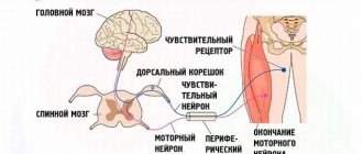
Motor neurons are nerve cells that send electrical signals to muscles that affect the muscle's ability to function.
Motor neuron disease (MND) can occur at any age, but most patients are over 40 years of age when diagnosed. It affects men more than women.
The most common type, amyotrophic lateral sclerosis (ALS), likely affects about 30,000 Americans at any given time, with more than 5,000 diagnoses per year.
Renowned English physicist Stephen Hawking lived with ALS for many decades before his death in March 2020. Guitar virtuoso Jason Becker is another example of someone who has been living with ALS for several years.
Here are some key points about motor neurone diseases. This is discussed in more detail in the main article.
- Motor neuron diseases (MND) are a group of diseases that affect the nerve cells that send messages to the brain.
- There is a gradual weakening of all the muscles of the body, which ultimately affects the ability to breathe.
- Genetic, viral and environmental issues may play a role in the occurrence of IHL.
- There is no cure, but supportive treatment can improve quality of life.
- Life expectancy after diagnosis ranges from 3 years to more than 10 years.
Affected areas
As motor neuron disease gradually progresses, patients experience increasing problems with breathing, swallowing, moving, and communicating with others. The following difficulties may occur:
- Hands, arms . Even when performing daily tasks (washing dishes, turning a tap, pressing keys), increasing difficulty will appear with increasing weakness in the hands and arms.
- Legs . Due to weakening of the legs, it will become increasingly difficult for the patient to move.
- Emotional condition . Sometimes areas that are responsible for emotional reactions can be affected, which is manifested by involuntary laughter or crying of a physiological nature.
- Shoulders, neck . After a certain time, it becomes difficult for the patient to breathe due to weakening neck muscles.
- Breathing . The disease mainly spreads to the respiratory muscles, making breathing difficult.
- Swallowing, speech functions . There may be difficulty eating, drinking and talking.
In some patients, MND causes memory impairment, decreased concentration, difficulty choosing words and learning difficulties. That is, an imperceptible and hidden change in mental functions occurs.
In MND, the intellect remains intact and patients are aware of everything that is happening. For many people, vision, taste, touch and hearing are not affected; the functions of the bladder, intestines, genitals (up to the later stages) and the heart muscle remain functional.
diagnosing
At an early stage, MHD can be difficult to diagnose because the signs and symptoms are common to other diseases, such as multiple sclerosis (MS), nerve inflammation, or Parkinson's disease.
The primary care doctor usually refers the patient to a neurologist, a doctor who specializes in diagnosing and treating diseases and conditions of the nervous system.
The neurologist will begin with a complete history and physical examination of the neurological system.
Other tests may be helpful.
Blood and urine tests: These tests can rule out other conditions and detect any elevated creatinine kinase levels. It is formed when muscles break down and can sometimes be found in the blood of patients with MHD.
MRI brain scan: This cannot detect MHD, but can help rule out other conditions such as stroke, brain tumor, problems with brain circulation, or abnormal brain structure.
Electromyography (EMG) and nerve conduction studies (NCS): These are often performed together. EMG measures the level of electrical activity in the muscles, while NCS measures the speed at which electricity flows through the muscles.
Spinal tap or lumbar puncture: This tests the cerebrospinal fluid, the fluid that surrounds the brain and spinal cord.
Muscle biopsy: If the doctor thinks the patient may have a muscle disorder rather than MHD, a muscle biopsy may be performed.
After testing, the doctor usually monitors the patient for some time before confirming the presence of MHD.
The criteria, known as the El Escorial criteria, can help a doctor check for distinctive neurological signs that can help diagnose ALS.
- muscle contractions, weakness or cramps.
- muscle stiffness or abnormal reflexes
- symptoms spread to new muscle groups
- without other factors explaining the symptoms.
Pathogenesis and pathomorphology of the disease
Motor neuron disease is considered a multifactorial pathology, the development of which occurs from the influence of genetic inheritance with environmental factors.
In 10% of situations, the disease is associated with familial forms, where 25% are characterized by a mutation of the SOD-1 gene. Sometimes sporadic types of pathology are associated with changes in other genes (neurofilaments) as a result of dysfunction or structure, under the influence of which motor neurons begin to degenerate. The mechanism is triggered by the action of provocateurs (age, gender, heavy metals, etc.). The relationship between the occurrence of the disease and infectious agents is unlikely.
Clinically, neurons degenerate when 80% of cellular structures are destroyed, which makes timely therapy of the pathology difficult and requires diagnostic studies during the preclinical stage.
Near the frontal lobes, in the 3rd, 5th layers of the central gyri, the motor nuclei of the 5th, 7th, 9th and 11th cranial nerves in the brain stem, neuronal degeneration and changes in the corticospinal tracts are determined. At various stages of changes in motor neurons, eosinophilic, basophilic inclusions and Bunin bodies are determined.
Axonal spheroids are formed in the proximal sections of axons. Based on the data obtained, disturbances in the movement and degradation of proteins are noted. In muscle structures, denervation atrophy is determined.
Notes
- Disease Ontology release 2019-05-13 — 2019-05-13 — 2020.
- Monarch Disease Ontology release 2018-06-29sonu - 2018-06-29 - 2018.
- Epidemiology of Sporadic ALS. (English) (inaccessible link). Stanford Medicine » School of Medicine. Retrieved October 24, 2015. Archived October 8, 2020.
- Conwit, Robin A.
Preventing familial ALS: A clinical trial may be feasible but is an efficacy trial warranted? (English) // Journal of the Neurological Sciences (English) Russian. : journal. - 2006. - December (vol. 251, no. 1-2). - P. 1-2. — ISSN 0022-510X. - doi:10.1016/j.jns.2006.07.009. - PMID 17070848. - Al-Chalabi, Ammar;
P. Nigel Leigh. Recent advances in amyotrophic lateral sclerosis (English) // Current Opinion in Neurology (English) Russian. - Lippincott Williams & Wilkins (English) Russian., 2000. - August (vol. 13, no. 4). - P. 397-405. — ISSN 1473-6551. - doi:10.1097/00019052-200008000-00006. - PMID 10970056. - “Hawking’s disease” was explained by the appearance of four-stranded DNA // Lenta.ru, 2014-03-07
- Cyrille Garnier, François Devred, Deborah Byrne, Rémy Puppo, Andrei Yu.
Roman. Zinc binding to RNA recognition motif of TDP-43 induces the formation of amyloid-like aggregates (En) // Scientific Reports. — 2017-07-28. — T. 7, issue. 1. - ISSN 2045-2322. - doi:10.1038/s41598-017-07215-7. - Aphrodite Caragounis, Katherine Ann Price, Cynthia PW Soon, Gulay Filiz, Colin L. Masters.
Zinc induces depletion and aggregation of endogenous TDP-43 // Free Radical Biology and Medicine. - T. 48, issue. 9. - pp. 1152-1161. - doi:10.1016/j.freeradbiomed.2010.01.035. - ALSUntangled: research into Lyme disease and the drug Iplex = ALSUntangled Update 1: Investigating a bug (Lyme Disease) and a drug (Iplex) on behalf of people with ALS: [trans. from English] // ALS INFO: [electr. resource]. — 2020. — April 25.
- ALSUntangled Update 1: Investigating a bug (Lyme Disease) and a drug (Iplex) on behalf of people with ALS : [English] : Report / The ALSUntangled Group // Amyotrophic Lateral Sclerosis : journal. - 2010. - Vol. 10, no. 4. - P. 248–250. - doi:10.1080/17482960903208599. - PMID 19701824.
- Khondkarian O. A., Amyotrophic lateral sclerosis, in the book: Multi-volume guide to neurology, ed. S. N. Davidenkova, vol. 3, book. 1, M., 1962
- Amyotrophic lateral sclerosis
- article from the Great Soviet Encyclopedia. - https://www.ninds.nih.gov/disorders/amyotrophiclateralsclerosis/detail_ALS.htm Archived January 4, 2020 on the Wayback Machine “In 90 to 95 percent of all ALS cases, the disease occurs apparently at random with no clearly associated risk factors. … About 5 to 10 percent of all ALS cases are inherited. The familial form of ALS usually results from a pattern of inheritance that requires only one parent to carry the gene responsible for the disease. Mutations in more than a dozen genes have been found to cause familial ALS.”
- Exposure to pesticides and risk of amyotrophic lateral sclerosis: a population-based case-control study.By Bonvicini F, Marcello N, Mandrioli J, Pietrini V, Vinceti M. In Ann Ist Super Sanita. 2010; 46(3):284-7.PMID 20847462
- Pesticide exposure as a risk factor for amyotrophic lateral sclerosis: A meta-analysis of epidemiological studies: Pesticide exposure as a risk factor for ALS. By Malek AM, Barchowsky A, Bowser R, Youk A, Talbott EO. In Environ Res. 2012 Aug; 117:112-9. PMID 22819005
- Are environmental exposures to selenium, heavy metals, and pesticides risk factors for amyotrophic lateral sclerosis?. By Vinceti M, Bottecchi I, Fan A, Finkelstein Y, Mandrioli J. In Rev Environ Health. 2012; 27(1):19-41. PMID 22755265
- Pesticide exposure and amyotrophic lateral sclerosis. By Kamel F, Umbach DM, Bedlack RS, Richards M, Watson M, Alavanja MC, Blair A, Hoppin JA, Schmidt S, Sandler DP. In Neurotoxicology. 2012 Jun; 33(3):457-62. PMID 22521219
- Reflexogenic zones
- article from the Great Soviet Encyclopedia. - Brooks BR, Miller RG, Swash M., Munsat TL
El Escorial revisited: revised criteria for the diagnosis of amyotrophic lateral sclerosis (English) // Amyotroph. Lateral Scler. Other Motor Neuron Disord. : journal. — 2000. — December (vol. 1, no. 5). - P. 293-299. - PMID 11464847. - ↑ 12
https://www.ninds.nih.gov/disorders/amyotrophiclateralsclerosis/detail_ALS.htm Archived January 4, 2020 on the Wayback Machine How is ALS treated? - Aaron Hitler, Leonard Petrucelli.
Solving the mystery of ALS // In the world of science. — 2020. — No. 8/9. — P. 50-56. - Neurology. National leadership. - GEOTAR-Media, 2010. - 2116 p. — 2000 copies. — ISBN 978-5-9704-0665-6.
- Riluzole // PATIENT & CAREGIVER EDUCATION (Russian)
- https://www.ninds.nih.gov/disorders/amyotrophiclateralsclerosis/detail_ALS.htm Archived January 4, 2020 on the Wayback Machine However, the Food and Drug Administration (FDA) approved the first drug treatment for the disease—riluzole (Rilutek )—in 1995. Riluzole is believed to reduce damage to motor neurons by decreasing the release of glutamate. Clinical trials with ALS patients showed that riluzole prolongs survival by several months
- Nagata, Kazuaki
.
Japan recognizes Cyberdyne's robotic suit as medical device, widespread use anticipated (English), The Japan Times Online
(26 November 2015). Retrieved February 10, 2020. - https://mosmedpreparaty.ru/news/4923
- https://www.ab-science.com/file_bdd/content/1490200983_SLAPresentationv03.2017vdef.pdf
- The Bishop of ROSHVE asks the head of the Ministry of Health of the Russian Federation to change the situation with the attitude towards patients with an “unrecognized” illness, NewsRu, August 21, 2013
- Where can you get the right to hope? // “Honest Word” No. 3 (832), 01/23/2013
What causes the death of motor neurons?
According to experts, the main causes of MND are heredity and environmental factors that increase the risk of pathology.
A familial hereditary form has been identified in approximately 5-10% of patients with motor neuron disease. MND was mainly diagnosed (90-95% of cases) in a sporadic form that occurs for unknown reasons.
Epidemiological studies have revealed a possible connection between the effects of shock, mechanical injuries, high sports loads, and harmful substances on the development of the disease.
Currently, studies are being carried out on the mechanisms of damage to intracellular processes, due to which motor neurons and surrounding cells die.
Possible reasons include a strike and malfunction of “editors” in processing RNA, changes in chemical communication connections of the spinal cord, disruption of the transport of metabolic products, nutrients, accumulation of protein aggregates in neurons with disruption of normal functions and adhesion, the appearance of free oxygen radicals, disruption of the energy background cells and lack of nutrients for motor neurons. Also, the death of motor neurons can occur due to being surrounded by glial cells, which can be toxic.
Causes
Motor neurons send signals from the brain to the muscles and bones, and this causes the muscles to move. They are involved in both conscious movements and automatic movements such as swallowing and breathing.
Some MHDs are inherited, while others occur randomly. The exact causes are unclear, but the National Institute of Neurological Disorders and Stroke (NINDS) notes that genetic, toxic, viral and other environmental factors likely play a role.
Main and rare types of MND
Motor neuron disease has several main forms:
- ALS (amyotrophic lateral sclerosis) develops in 80% of people with motor neuron disease, causing mainly weakness, cramps and wasting in the muscles, stiffness of the upper and lower extremities;
- PBP (progressive bulbar palsy) affects about 10-25% of people, spreads to the neurons of the spinal cord and brain, and is manifested by difficulties in chewing, swallowing, and slurred speech;
- muscular progressive atrophy (8%) - a rare type of MND, affects motor neurons in the lower extremities, developing weakness, muscle atrophy, fasciculations and weight loss;
- PLS (lateral primary sclerosis) (2%) primarily affects motor neurons in the upper extremities, developing muscle spasticity, unsteady walking, and difficulty speaking.
Symptoms of motor neuron diseases
Below is a brief description of the symptoms of some of the most common forms of motor neurone disease.
Amyotrophic lateral sclerosis (ALS), also called Lou Gehrig's disease or classical motor neuron disease, is a progressive, ultimately fatal disease that disrupts signaling in all muscles of the body. Many doctors use the terms motor neuron disease and ALS interchangeably. The disease is caused by disorders of both upper and lower motor neurons. The first symptoms are usually noticed in the arms and legs or in the muscles responsible for swallowing. About 75 percent of people with classic ALS develop weakness of the bulbar muscles (the muscles that control speech, swallowing and chewing). Muscle weakness and atrophy occur on both sides of the body. Patients lose strength and the ability to move their arms and legs and hold their body upright. Other symptoms include muscle spasticity, spasms, and muscle cramps. Speech may become slurred or guttural. When the muscles of the diaphragm and chest wall do not function properly, patients lose the ability to breathe without mechanical support. Although the disease generally does not impair a person's intellectual abilities or personality, some recent scientific research suggests that some people with ALS may develop cognitive (mental) impairment. Most people with amyotrophic lateral sclerosis die from respiratory failure, usually within 3 to 5 years of the onset of symptoms. However, about 10 percent of patients survive for 10 years or more.
Progressive bulbar palsy , also called progressive bulbar atrophy, affects the lower motor neurons responsible for activities such as swallowing, speaking, chewing, and others. Symptoms include weakness of the glossopharyngeal, jaw and facial muscles, progressive loss of speech function, and atrophy of the tongue muscles. Limb weakness in the disease with signs of motor neuron damage is almost always obvious, but less noticeable. People are at increased risk of choking and aspiration pneumonia caused by the passage of fluids and food through the lower respiratory tract and lungs. Affected individuals have emotional outbursts of laughing or crying (called emotional lability). Stroke and myasthenia gravis may have certain symptoms similar to those of progressive bulbar palsy and should be excluded when diagnosing this disease. In about 25 percent of people with ALS, early symptoms begin with associated bulbar abnormalities. Many clinicians believe that progressive bulbar palsy by itself, without signs of pathology in the extremities (arms or legs), is extremely rare.
Pseudobulbar palsy , which has many symptoms similar to progressive bulbar palsy, is characterized by degeneration of the upper motor neurons that transmit signals to the lower motor neurons in the brain stem. Patients develop progressive loss of the ability to speak, chew, and swallow, as well as progressive weakness of the facial muscles. Patients may develop voice disturbances and an increased gag reflex. The tongue may become immobile and lose the ability to protrude from the mouth.
Primary lateral sclerosis (PLS) damages the upper motor neurons of the arms, legs, and face. This occurs when specific nerve cells in the motor areas of the cerebral cortex (the thin layer of cells covering the brain that is responsible for most high-level brain functions) gradually degenerate, causing movements to be slow. The disease often affects the legs first, then the torso, arms, and finally the bulbar muscles. Speech may become slow and difficult. When nerve cells are damaged, leg and arm movements become clumsy, slow and weak, and spasticity occurs, resulting in the inability to walk or perform tasks that require fine hand coordination. Balance problems can lead to falls. Speech may become slow and slurred. Patients typically experience pseudobulbar affect and an overactive response. Primary lateral sclerosis is more common in men than women, and the onset of the disease usually occurs between 40 and 60 years of age. The cause of the disease is unknown. Symptoms progress gradually over many years, resulting in progressive stiffness (spasticity) and clumsiness in the affected muscles. PLS is sometimes considered a form of amyotrophic lateral sclerosis, but the main difference is the sparing of lower motor neurons, a slow rate of disease progression, and a normal life expectancy. Primary lateral sclerosis may be mistaken for spastic paraplegia, an inherited upper motor neuron disorder that causes spasms in the legs and usually begins in adolescence. Most neurologists follow the clinical course of the affected person for at least 3-4 years before making a diagnosis. This disease is not fatal, but can affect quality of life.
Progressive muscular atrophy is characterized by slow but progressive degeneration of exclusively lower motor neurons. The disease largely affects men, with onset earlier than other motor neuron diseases. Weakness usually begins in the arms and then spreads to the lower body, where it can be more severe. Other symptoms may include muscle wasting, clumsy arm movements, and muscle cramps. The muscles responsible for breathing may be damaged. Exposure to cold may worsen symptoms. The disease develops together with ALS in many cases.
Spinal muscular atrophy (SMA) is an inherited disease affecting the lower motor neurons. This is an autosomal recessive disease caused by disturbances in the SMN1 gene (the gene responsible for the production of a protein that is important for the functioning of motor neurons (SMN protein)). In SMA, insufficient levels of SMN protein lead to degeneration of lower motor neurons, causing weakness and wasting of skeletal muscles. Weakness is often more severe in the muscles of the arms and legs, as well as the muscles of the trunk. Spinal muscular atrophy in children is divided into three types based on age of onset, severity, and progression of symptoms. All three types are caused by defects in the SMN1 gene.
Post-polio syndrome (PPS) is a condition that can affect polio survivors, which can occur for decades after they have recovered from polio. Poliomyelitis is an acute viral disease that destroys motor neurons. Many people affected by the disease early in life recover, only to experience new symptoms many decades later. After acute polio, the surviving motor neurons are responsible for more of the muscles they control. Post-polio syndrome and post-polio muscle atrophy are thought to occur when surviving motor neurons are lost through aging or injury/disease. Many scientists believe that SPP is a hidden symptom of muscle weakness previously affected by polio, rather than a new motor neuron disease. Symptoms include fatigue, slowly progressive muscle weakness, muscle atrophy, cold intolerance, and muscle and joint pain. These symptoms most often occur among the muscle groups affected by the initial disease and may consist of problems breathing, swallowing, or sleeping. Other symptoms of SSP may be caused by skeletal deformities, such as long-standing scoliosis, which have led to chronic changes in the biomechanics of the joints and spine. Symptoms are more common in older people and those most affected by earlier illness. Some people have only minor symptoms, while others develop muscle wasting, which can be misdiagnosed as ALS. SSP usually does not threaten the patient’s life. According to medical statistics, 25 to 50 percent of those who have had paralytic poliomyelitis usually develop post-polio syndrome.
Symptoms and clinical picture of MND depending on the form
A whole group of diseases affect motor neurons. Neurons include two groups of cells necessary for voluntary movements.
The first group includes motor neurons located in the precentral gyrus, and the second group includes neurons located in the spinal cord and brain stem.
Depending on the location of the dead motor neuron, the form of the disease and its symptoms will depend.
With spastic paraplegia, the motor neuron of the first group is affected, with spinal muscular atrophy, infantile paralysis - the second group, and with amyotrophic lateral sclerosis, both groups of neurons.
Forms of MND and their manifestations:
- Spastic paraplegia is defined by increased spastic tone of the legs with gait disturbance. It is caused by genetic mutations.
- Spinal muscular atrophy by weakness and reduction of muscles, lack of activation or innervation by nerve fibers. Depending on the time of onset of the first symptoms and the nature of the distribution of muscle weakness, spinal muscular atrophy is classified.
- A more common pathology of the musculoskeletal system is ALS , the incidence of which increases with age. As a result, the necrosis of nerve cells progresses, weakness and muscle atrophy increase.
- Cerebral palsy , polio is a viral disease of lower neurons.
Diagnosis of motor neuron disease
There are no specific tests to diagnose most motor neuron diseases, although genetic testing for SMA genes currently exists. Symptoms may vary among patients and in the early stages of the disease may be similar to those of other diseases, making diagnosis difficult. The physical examination is followed by a thorough neurological examination. Neurological tests will evaluate motor and sensory skills, nervous system functioning, hearing and speech, vision, coordination and balance, mental status, and changes in mood or behavior.
Tests to rule out other diseases or measure the extent of muscle damage may include the following:
Electromyography (EMG) is used to diagnose lower motor neuron disorders, as well as muscle and peripheral nerve disorders. In an EMG, a doctor inserts a thin needle-shaped electrode attached to a recording instrument into the muscle to evaluate electrical activity during voluntary contraction and at rest. Electrical activity in the muscle is caused by lower motor neurons. When motor neurons are lost, characteristic abnormal electrical signals occur in the muscle. Testing usually takes about an hour or more, depending on the number of muscles and nerves being tested.
EMG is usually performed in combination with nerve conduction velocity testing. Nerve conduction studies measure the speed at which impulses are transmitted in nerves from small electrodes attached to the skin, as well as their strength. A small pulse of electricity (similar to a shock from static electricity) stimulates the nerve that controls a specific muscle. The second set of electrodes transmits a response electrical signal to the recording device. Nerve conduction studies help differentiate lower motor neuron disease from peripheral neuropathy and can detect abnormalities in sensory nerves.
Laboratory tests of blood, urine, and other substances can help rule out muscle diseases and other disorders that may have symptoms similar to those of motor neurone disease. For example, analysis of the cerebrospinal fluid, which surrounds the brain and spinal cord, can detect infections or inflammation that can also cause muscle stiffness. Blood tests may be ordered to measure levels of the protein creatine kinase (necessary for chemical reactions that produce energy for muscle contractions); high levels allow the diagnosis of muscle diseases such as muscular dystrophy.
Magnetic resonance imaging (MRI) uses a powerful magnetic field to produce detailed images of tissue, organs, bones, nerves and other structures of the body. MRI is often used to rule out diseases that affect the head, neck and spinal cord. MRI images can help diagnose brain and spinal cord tumors, eye diseases, inflammation, infection and vascular disorders that can lead to stroke. MRI can also detect and monitor inflammatory diseases such as multiple sclerosis. Magnetic resonance spectroscopy is a type of MRI scan that measures levels of chemicals in the brain and can be used to assess the integrity of upper motor neurons.
A biopsy of muscles or nerves can confirm the presence of neurological disorders, in particular impaired nerve regeneration. A small sample of muscle or nerve tissue is taken under local anesthetic and examined under a microscope. The sample can be removed surgically through an incision in the skin or by biopsy, in which a thin, hollow needle is inserted through the skin into the muscle. A small amount of muscle tissue remains in the hollow needle when it is removed from the body. Although this test can provide valuable information about the extent of damage, it is an invasive procedure and many experts believe that a biopsy is not always necessary for diagnosis.
Transcranial magnetic stimulation was first developed as a diagnostic tool to study areas of the brain associated with motor activity. It is also used as a treatment for certain diseases. This non-invasive procedure creates a magnetic pulse inside the brain that causes movement in an area of the body. Electrodes attached to various areas of the body collect and record electrical activity in the muscles. Measures of evoked activity can diagnose motor neuron dysfunction in motor neuron disease or monitor disease progression.
Differential diagnosis
The diagnosis of MND is confirmed by electroneuromyography, which reveals a generalized denervation process. To identify spontaneity, duration of motor units, fasciculations, fibrillations, a three-level needle EMG is performed.
Stimulation electroneuromyography makes it possible to detect slowing of motor fibers and pyramidal insufficiency.
Transcranial magnetic stimulation reveals a decrease in excitability in central motor neurons. MRI makes it possible to exclude other pathologies with a similar clinical picture (hernia compression or tumor).
Treatment options
Motor neuron disease is incurable, and the goal of therapy is to slow down the development of the process and reduce the severity of symptoms, since there are no methods for a complete cure. The effectiveness of Rilutek, a presynaptic inhibitor, has been proven.
As with ALS, antioxidant agents may be used. Experts also prescribe creatine, carnitine, and vitamins. Another drug is Riluzole, but the drug is not officially available to patients in the Russian Federation. Like Rilutek, the drug reduces the amount of glutamate released during the transmission of nerve impulses.
Polytherapy includes the elimination of pain from muscle spasms, for which Carbamazepine is prescribed at a dose of about 100-200 mg several times a day. With increased muscle tone, you need to take muscle relaxants (Sirdalud, Baclofen).
Respiratory support is a major factor influencing quality of life and prognosis. With the progression of the pathology, weakening and atrophy of the muscles of the diaphragm and other muscle groups is observed; non-invasive ventilation of the lungs is required.
This normalizes sleep, reduces fatigue and shortness of breath. A device with a special mask is used.
If swallowing functions are impaired, then feeding is carried out with a nasogastric tube or gastrostomy tube. Special nutrition is prescribed to make up for the lack of calories.
It is difficult to talk about the prognosis for MND. In rare cases, people live for decades. The person may die within a few months. But on average, life expectancy is 2-5 years from the onset of detection of the disease. There are no specific preventive measures.
Treatment[ | ]
Patients with ALS require supportive care to relieve symptoms[20].
Gradually, patients' respiratory muscles begin to weaken, respiratory failure develops, and the use of equipment to facilitate breathing during sleep (IPPV or BIPAP) becomes necessary. Then, after complete failure of the respiratory muscles, 24-hour use of a ventilator is required.[20]
Research is underway on a treatment method that uses blocking the genes that cause this disease[21].
Slow progression[ | ]
Riluzole (Rilutek) is the only drug that significantly slows the progression of ALS[22][23]. Available since 1995. It inhibits the release of glutamate, thereby reducing damage to motor neurons. It prolongs the life of patients by an average of a month and slightly delays the moment when the patient will need artificial ventilation[24].
HAL therapy, a new robotic treatment method, has been officially approved for use in ALS rehabilitation in Europe and Japan[25].
In 2020, a new drug, Radicava (Edaravone), was approved for the treatment of ALS for the first time in 22 years[26].
Clinical studies are also being conducted on the use of the drug Masitinib[27].
In Russia[ | ]
In Moscow there are:
- Charitable foundation to help people with ALS and other neuromuscular diseases “Live Now”
- Charitable foundation for helping patients with ALS G. N. Levitsky. https://www.alsportal.ru
- assistance service for patients with ALS at the ANO "Hospital of St. Alexis".
At the same time, in Russia, many ALS patients do not receive adequate medical care[28]. For example, until 2011, ALS was not even included in the list of rare diseases, and the only drug that slows the progression of the disease, Riluzole, was not registered[29].
ALS - motor neurons die completely
During ALS, evenly expressed symptoms of central and peripheral paralysis are combined, the predominance of some symptoms over others (segmental-nuclear or pyramidal variants). In the later stages of the disease, indicators of peripheral paralysis predominate.
During ALS, damage to motor neurons in the anterior horn of the spinal cord begins. As the pathology progresses, bulbar disorders appear mainly in men.
There are thoracic, cervical, diffuse and lumbar onsets. The classic variant is the cervical one, the first symptoms of which are characterized by weakness and atrophy of the hand, impaired small movements, and the presence of fascicular twitching of the affected muscles.
Stephen Hawking is the most famous ALS patient
Subsequently, weakness and atrophy of the muscles of the shoulder girdle appear; patients find it difficult to dress and raise their arms above the horizontal plane. First, one side of the body is affected, then symptoms appear on the other arm with the development of upper mixed paraparesis.
Weakness and stiffness of the lower extremities develop; patients find it difficult to walk long distances or navigate stairs.
The extensor muscles are damaged with the manifestation of mixed lower paraparesis. In the later stages, pseudobulbar and bulbar syndromes and the appearance of dysphagia appear.
The first symptoms of chest onset will be fasciculations, muscle atrophy in the abdomen, back and weakness. It becomes difficult for patients to stand and bend over. At the next stages, mixed unilateral hemiparesis appears with the involvement of the muscles of the opposite part of the body. At the initial stage, abdominal reflexes disappear. As a result of respiratory disorders, death occurs.
Diffuse onset is manifested by signs of peripheral motor neuron dysfunction with pronounced fatigue and respiratory disorders. Body weight decreases until dysphagia appears, and death occurs as a result of respiratory failure.
Usually, damage to the masticatory and facial muscles occurs later; it is difficult for the patient to stick out his tongue, pull his lips into a tube, or puff out his cheeks.
Complications of ALS include paralysis, paresis of the neck muscles and limbs, impaired swallowing, respiratory failure, contractures of the limbs, aspiration pneumonia, urosepsis, depression, painful muscle spasms, and cachexia. With the progression of motor disorders, the patient dies.
Risk factors
These are some of the risk factors associated with IHL.
Heredity: In the United States of America (USA), about 1 out of every 10 cases of ALS is inherited. SMA is also known to be an inherited condition.
Age: After 40 years of age, the risk increases significantly, although it is still very small. ALS most likely occurs between ages 55 and 75.
Sex Men are more likely to develop MHD.
Some experts link military experience to a higher likelihood of developing this disease.
Studies have shown that professional football players are more likely to die from ALS, Alzheimer's disease and other neurodegenerative diseases than other people. This implies a possible association with repetitive head trauma and neurological diseases.


