What is Gaffa disease?
According to the Handbook of Diseases of Freshwater, Marine and Aquarium Fish, Gaffa disease (Yuksovsky disease, Sartlan disease) is an acute disease of fish characterized by shrinkage of the stomach and intestines and respiratory depression. Sick fish lose weight, their intestines and stomach decrease in volume and resemble thin threads. The disease got its name from the location of the outbreaks where the toxic fish were caught.
Questions and Answers What is rotavirus?
In the Encyclopedic Dictionary of Medical Terms, the disease is designated as food intoxication resulting from eating certain types of fish caught in stagnant water in early spring.
In the bulletin of the All-Russian Scientific Center of the Siberian Branch of the Russian Academy of Medical Sciences No. 3 (67) for 2009, the disease is defined as a chronic nutritional toxicosis of five species of fish, cats, humans, and some other animals of unknown etiology, manifested by symptoms of damage to the skeletal muscles, nervous system and kidneys.
The disease was first discovered back in the 1920s among fishermen of the Frisch Gaff Strait in the Baltic Sea. Since then, the disease has been rare, and its names have varied depending on the location of the new outbreak. In the 1930s, the disease was called Yuksovskaya due to several recorded cases on Lake Yuksovskoye in the Leningrad region, and in the late 1940s and mid-1980s - Sartlanskaya (after outbreaks of the disease on Lake Sartlan in the Novosibirsk region).
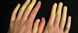
The etiology is not fully understood. It is believed that the source of the disease is toxic fish, in particular perch, pike, carp, and crucian carp. Fish become toxic by feeding on plankton, as well as contaminated toxic substances washed out of the ground.
Blue-green algae toxins are also a likely cause of the disease. When studying the conditions for the occurrence of the disease among fish, it was noted that in the studied reservoir, mass death of fish was observed simultaneously with the development of blue-green algae. Algae, entering the body of fish, not only made them toxic, but also, accumulating in organs and tissues, caused the death of the fish themselves.
Trigeminal neuralgia - Fothergill's disease
Pathogenesis. The primary link in the pathogenesis of this disease is, as a rule, damage to the peripheral segment of the nerve - a branch or root. The latter is confirmed by the well-known fact that turning off the affected branch, for example, alcoholism, leads to the cessation of painful attacks until the nerve regenerates.
Under the influence of the compression factor and prolonged subthreshold stimulation from the periphery, an algogenic system is believed to arise in the brain, which is stable, highly excitable and responds to any afferent sendings with paroxysmal-type excitation.
Clinic and diagnostics. The disease usually develops after the age of 50 years. The maxillary or mandibular nerve (the second or third branch of the trigeminal nerve) is most often affected. Often, at first, the pain is local in nature, projected into the area of a particular tooth or gum, which is why patients turn to dentists. Soon the area of pain increases, usually capturing the zone of innervation of the corresponding branch. The peculiarity of the pain is the short duration of the paroxysm, which usually lasts for a few seconds. Despite this, the unbearable nature of the pain forces trigeminal neuralgia to be classified as extremely severe suffering. In addition, it should be added that during the period of exacerbation, painful attacks are provoked by any irritants - talking, chewing, eating, facial movements. Patients cannot wash or shave, have difficulty eating only liquid food, and often explain themselves in writing. The pain is compared to the passage of an electric current, painful twitching, lumbago (“as if a red-hot rod had been stuck in the face”). It is characteristic that during painful paroxysms, patients do not scream or rush about, but, stunned by terrible pain, freeze, afraid to move. In more rare cases, patients rub their cheek or press on their temple in order to relieve an attack (antagonist gesture).
It has long been noted that with trigeminal neuralgia, attacks of pain are often accompanied by spasms of the facial muscles - a painful tic (tic douloureux). Muscle spasms can be partial, involving the orbicularis oculi muscle, buccal muscle, or take on a hemifacial character.
Attacks of pain are usually accompanied by autonomic disorders - nasal congestion or discharge of liquid secretions, lacrimation, facial hyperemia. Paroxysms rarely occur at night. If the first cardinal sign of trigeminal neuralgia is its characteristic pain attacks, then the second can rightfully be considered the so-called trigger zones - areas of the skin or mucous membrane of various sizes that are hyperexcitable. Irritation of these areas - touch, cold wind, displacement of the skin - usually causes a painful attack. Trigger zones appear during the period of exacerbation of the disease and disappear during the period of remission. There is a tendency towards the location of trigger zones in the medial parts of the face. Trigger zones are also located on the oral mucosa, and in some patients - only in this area. Often the trigger zone is a tooth.
During an objective examination of patients, in addition to the trigger zones, there is often pain when pressing at the exit point of the corresponding branch of the trigeminal nerve on the face, and sometimes areas of hyperesthesia in the zone of innervation of the affected branch. As the disease progresses, certain changes are often revealed in the clinical picture: in pauses between attacks, a feeling of dull pain or a burning sensation may persist, hyperesthesia is replaced by hypoesthesia and even anesthesia. However, the cardinal signs of neuralgia - its characteristic pain paroxysms and trigger zones - remain. It should be assumed that there is a single disease that has different causes, but a single pathogenesis.
The diagnosis is based on identifying the paroxysmal nature of pain, the characteristics of pain attacks, the presence of trigger zones on the skin of the face or in the oral cavity, provocation of attacks by eating, washing, shaving, etc. In some cases, a false impression may be created about the unusual duration of pain attacks over several hours. Meanwhile, status neuralgicus is a series of painful attacks following each other. However, even in this situation, a thorough interview allows us to establish the true nature of the attacks.
In rare cases, neuralgia of individual branches of the trigeminal nerve occurs—the lingual nerve, the superior alveolar nerves, and the inferior alveolar nerve. All of them are a kind of partial form of trigeminal neuralgia, manifested by characteristic pain attacks and trigger zones with limited localization. So, with neuralgia of the lingual nerve, attacks of pain occupy the anterior 1/3 of the corresponding half of the tongue and the trigger zones are also located here. With neuralgia of the upper alveolar nerves, attacks of pain are localized in the area of the teeth and part of the upper jaw, and with neuralgia of the lower alveolar nerve - in the area of the teeth of the lower jaw; trigger zones are usually found in the area of the corresponding gum, a particular tooth.
Neuralgia of the lingual nerve should be distinguished from neuralgia of the glossopharyngeal nerve, in which pain and trigger zones are localized mainly in the area of the root of the tongue, tonsil, palatine arch, as well as from glossalgia. In the latter case, a surprisingly large number of patients complain of pain, burning sensation, sensation of hair on the tongue, and dry mouth. There are no pain attacks or trigger zones. All sensations go away while eating. The disease is a classic form of psychogenic algia. The diagnosis of idiopathic trigeminal neuralgia requires the exclusion of all other causes of nerve compression and facial pain. Essentially, toothache, sinusitis, and glaucoma are secondary forms of trigeminal neuralgia. Occasionally, the latter can be observed in multiple sclerosis and tumors of the brain stem, which must be taken into account, especially in young patients and in the presence of additional focal symptoms.
Treatment. In 1962, carbamazepine entered the practice of treating trigeminal neuralgia, which opened up real prospects for successful treatment in at least 70% of cases of this severe suffering. Other anticonvulsants are also widely used: difenin, suxilep, clonazepam, sodium valproate. The dose of the drug is selected individually. The daily dose of carbamazepine (finlepsin) is usually 400-1200 mg/day. After achieving a therapeutic effect (cessation of pain attacks), the dosage of the drug is reduced to the minimum at which the therapeutic effect is maintained, and this dose of the drug is used by the patient for a long time (maintenance therapy). When carrying out maintenance therapy over time, the effect decreases in a number of patients; drugs can be used either alone or (if insufficiently effective) in combination.
In some cases, baclofen is effective, which enhances presynaptic inhibition in the neurons of the nucleus of the spinal tract of the trigeminal nerve, as well as pimozide (orap).
As in the treatment of other pain syndromes, the use of antidepressants, as well as transcutaneous electrical stimulation of the affected branch, may be useful. The low effectiveness of conservative treatment for trigeminal neuralgia in the era before the use of antiepileptic drugs led to the development of a variety of surgical treatment methods, many of which are currently of only historical interest. Surgery is used both on the peripheral branches of the trigeminal nerve and on intracranial structures.
“Chemical cutting” of the nerve by injecting 80% alcohol with novocaine into its exit site on the face is now almost abandoned, since it brings only a temporary effect. With the regeneration of the nerve, pain is restored, but a productive process occurs in the nerve, which leads to a decrease in the effectiveness of both repeated blockades and the use of antiepileptic drugs.
The discovered possibility of compression of the trigeminal nerve root in the posterior cranial fossa by an abnormally located vessel or an aneurysm opened a new page in neurosurgical treatment. Similar neurovascular conflicts have been noted with facial hemispasm, vestibular ataxia, spastic torticollis, and glossopharyngeal neuralgia.
Symptoms and diagnosis of pathology
Grisel's disease is often diagnosed in weakened children aged 6 to 11 years (especially girls). Parents often notice that after sleep the child holds his head in an unnatural position: it is tilted to one side and turned to the other, the patient complains of severe pain in the neck. He is able to both bend and straighten his neck, albeit with slight discomfort. But rotational movements are almost impossible and very painful. More precisely, the head moves in the direction of the turn without any problems, but movement in the opposite direction is significantly limited.
If you palpate the neck, you can easily detect unnaturally dense muscles on the side where the head is turned. On the side of the head tilt, on the contrary, the muscles are relaxed. Sometimes you can feel the protruding process of the second vertebra. In the area of the posterior surface of the pharynx, you can also see an elevation, which depends on the level of displacement of the atlas.
The first stage of diagnosis is collecting a detailed medical history. Then an X-ray examination is performed. On an x-ray, one can easily distinguish the forward displacement of the atlas and its rotation around the vertical axis.
What causes Grisel's disease?
The cause of the disease is often inflammation of the lymph nodes in the pharynx. Sometimes this can be a general inflammatory process in the nasopharynx, as well as purulent arthritis , which develops in the atlanto-epistropheal joint. This condition is usually accompanied by a sharp increase in body temperature.
After the acute period of the disease has passed, torticollis persists. Grisel explained this phenomenon by the fact that the atlas is displaced due to contracture of the muscles that are responsible for the movements of the skull. As a result of inflammation, these muscles begin to shorten, thereby provoking subluxation of the atlas and a forced tilt of the head. Doctors note that a negative factor may be weakness of the ligamentous apparatus.