Brain cells are so sensitive to a lack of oxygen that even a slight disruption of blood flow leads to serious neurological pathologies. Ultrasound examination of blood vessels makes it possible to solve the problem of cerebrovascular diseases at an early stage. Doctors say that this is the most reliable diagnosis of these diseases. An ultrasound scan showing the vessels of the head and neck provides a unique opportunity to see a two-dimensional image of the circulatory system of the area under study, and it can be done at a price of 1,000 to 12,000 rubles.
What is Doppler Doppler Doppler of the Head and Neck Vessels?
Ultrasound diagnostics is an instrumental research method. Ultrasound waves penetrate the body tissues and are reflected from them, which is recorded by a special sensor. The data is processed by a computer, then displayed on a monitor, after which the image of the internal environments is examined by a doctor. An additional function of ultrasound diagnostics is Dopplerography. With its help, you can evaluate the speed and nature of blood flow in the veins and arteries. If the blood flow moves in the direction of the sensor, the computer colors it red; if it moves in the opposite direction, it turns blue.
Ultrasound of the great vessels: essence and goals
The main factor that has a negative impact on the treatment of vascular diseases is untimely contact with a neurologist and late diagnosis of the disease. The circulatory system is closed in a cycle, so a violation of the blood supply to tissues in one area inevitably causes problems in another area. Main indications for duplex scanning of the great arteries:
- problems with vision, coordination, understanding, or speech production;
- headache and frequent dizziness;
- fainting or attacks of impaired consciousness;
- unsteadiness of gait;
- numbness of the limbs.
Duplex scanning of the main arteries of the head is also carried out if any pathologies of the vascular system are detected during other studies. Diagnostics are necessary so that the neurologist can assess the effect of the disease on the brain and prescribe adequate treatment.
The neurology clinic at the Yusupov Hospital employs neurologists with extensive experience who treat patients even with serious illnesses that other specialists have refused to treat. An individual approach, modern treatment methods, high-precision and effective equipment allow us to promptly identify problems and develop ways to eliminate them.
MAG ultrasound is a combined type study that combines classic ultrasound with the use of the Doppler effect. This will allow you to check the condition of the blood vessels, as well as the movement of blood through them in the brain.
An ultrasound type angiogram is performed using sound waves that penetrate deep into the human body. In this case, the patient does not feel anything. Waves that the human ear cannot hear are emitted and then picked up by a specially designed sensor. When information is reflected from the walls, the computer converts it into a two-dimensional image in black and white.
The objectives of conducting an examination of main arteries:
- detection of a congenital anomaly in the circle of arteries that are responsible for supplying blood to the brain
- determination of stenotic processes and angiospastic type conditions, as well as occlusion (all these processes affect the access of oxygen to tissues)
- identifying the effect of chronic diseases on blood circulation in the brain
- study of pathologies of blood vessels in the circle of Willis
- determination of vascular tone and their reactivity
- aneurysm detection
- regulation and quality control of therapeutic measures aimed at improving blood circulation in the brain
After all the data is received, the doctor will be able to draw conclusions about the need for treatment from another specialist, prescribing additional examinations or treatment procedures. Because the data is available immediately, it is easy to determine the impact of the disease and its treatment on blood flow.
Indications for use of the procedure
The main medical indication for performing TCD or USDG of cerebral vessels is deformation, narrowing (stenosis) or blockage (occlusion) of the extracranial (extracranial) vertebral (vertebral) or carotid arteries and intracranial (intracranial) middle, posterior, and forebrain arteries. In clinical practice, the study is prescribed for:
- early detection of intracranial vascular lesions;
- clarification of the degree of blood flow disturbance after traumatic brain injury;
- detection of vascular stenosis after infectious diseases;
- selection of optimal therapy for migraine to clarify the vasospasm factor;
- assessment of hemodynamics in the brain after organ transplantation;
- identifying the causes of poor blood circulation in the brain due to curvature of the spine, cervical osteochondrosis, compression of the vertebral arteries;
- monitoring the state of cerebral blood flow during surgical operations;
- detection of microembolism in patients with transient circulatory disorders.
An ultrasound of the vessels of the neck and head is prescribed by a doctor at the slightest suspicion of a change in cerebral circulation. The study is also widely used for the prevention of cerebrovascular lesions in people suffering from atherosclerosis or other vascular diseases of the brain. The technique helps prevent the development of strokes and evaluate the tactics of complex treatment of patients.
Uzdg btsa: what is it, indications, implementation, interpretation of results
Ultrasound Dopplerography of brachiocephalic arteries and vessels, or Doppler ultrasound of the BCA and BCS - what is it? This is an ultrasound diagnostic method that allows you to assess the condition of the vessels of the neck and the nature of blood flow in them.
With its help, you can assess existing disorders of the blood supply to the brain and determine the likelihood of developing such disorders in the future.
Doppler ultrasound is still used today, but it can be called outdated compared to other research methods - duplex and triplex scanning of the brachiocephalic arteries.
The essence of the study
The brachiocephalic arteries are the vessels that are responsible for supplying blood to the brain, tissues of the head, shoulder girdle and arms. They are separated from the aorta at shoulder level. The research method is based on the use of the Doppler effect, which is named after the Austrian scientist Christian Doppler who discovered it.
Its essence lies in the fact that ultrasonic waves from moving objects are reflected with a changed frequency: if the object moves towards the sensor, the frequency increases, when moving away from the sensor it decreases.
The main advantages of Dopplerography of neck vessels:
- Dopplerography is a non-invasive and therefore painless method;
- it does not require much time - the examination takes only a few minutes;
- it is an inexpensive study;
- At the same time, Doppler ultrasound is informative and will help the doctor make the correct diagnosis.
Ultrasound of the BCA allows you to determine:
- condition of the vascular walls;
- vascular damage even in the early stages;
- presence and severity of atherosclerosis;
- presence and severity of stenosis;
- speed and nature of blood flow.
Doppler ultrasound also shows the anatomical features of the vessels of the neck - for example, how strongly they twist around the spine.
With all these advantages, keep in mind that Doppler ultrasound is a more primitive method compared to duplex and triplex vascular scanning. They are more informative, but require more sophisticated equipment and are more expensive.
Doppler ultrasound cannot show the cause of blood flow disturbance, which is located outside the vessels, that is, if blood circulation is not obstructed due to the formation of atherosclerotic plaques or blood clots.
A duplex or triplex scan will provide more information that will help your doctor choose the right treatment. The information content is further enhanced by the use of color mapping method during ultrasound diagnostics.
Rules for preparing for the procedure and procedure
To prepare for the study, you need to adjust your diet. The day before the study, it is necessary to exclude foods that affect vascular tone:
- drinks containing caffeine (tea, coffee, energy drinks);
- alcohol;
- salty foods.
Two hours before the procedure, refrain from taking hot baths and smoking, as well as staying in smoky rooms: in the first case, arteries and veins expand, in the second, they contract.
If you do not adhere to these recommendations before the study, the reliability of the results will significantly decrease and you will not be able to find out the real condition of the vascular walls.
In addition, the results of BCA ultrasound are distorted by drugs that change vascular tone and affect the nature of blood flow . You should consult with your doctor about which medications need to be stopped before testing.
As noted above, Doppler ultrasound takes very little time . An experienced ultrasound specialist will only need a few minutes to perform all the necessary manipulations.
To carry out the procedure, you must undress to the waist. During the examination, the patient lies on the couch, periodically changing position - on his back, on his side, on his stomach - so that the diagnostic specialist can examine all the vessels that need to be checked.
Also, during ultrasound diagnostics of neck vessels, additional functional tests can be performed:
- holding your breath;
- changing the horizontal position of the body to a vertical one;
- taking certain medications (most often nitroglycerin).
Typically, such additional tests are prescribed in the presence of genetic abnormalities in the structure of the vascular system.
Principles for deciphering results and normal indicators
A diagnostic specialist can prepare a conclusion immediately after the procedure is completed . When interpreting the results, the data obtained during the study is compared with normal indicators of the condition of the arterial walls and the nature of blood flow in each of them.
The conclusion states:
- condition of the walls of blood vessels;
- description of pathological changes, if any;
- nature of blood flow.
Normally, there are no narrowings or atherosclerotic plaques in the vessels that would impede the movement of blood; the blood flow has no turbulence. In this case, the blood moves freely through the arteries and veins , providing the brain, tissues of the head and arms with sufficient oxygen and nutrients.
At the same time, in a young body, the vessels are elastic, which protects against the development of a stroke, but with age they lose their ability to stretch.
The ultrasound specialist’s conclusion must be shown to the attending physician , who will compare the data obtained with the clinical picture, advise you on all issues and, if necessary, prescribe treatment.
Deviations from the norm and possible diagnoses
With changes in blood vessels and the nature of blood flow, the following concepts may appear in the conclusion:
- stenosis is a pathological narrowing of the lumen of a vessel, as a result of which blood cannot pass freely through them;
- aneurysm - a protrusion of a vessel wall as a result of its thinning, in which the wall of an artery or vein expands more than twice (learn about carotid artery aneurysm);
- atherosclerosis - the formation of cholesterol plaques on the walls of blood vessels, which impede blood flow, resulting in an increased risk;
- occlusion - disruption of the patency of a vessel due to damage to its wall;
- turbulent flow - when blood moves through a vessel, turbulence is formed.
When is the examination performed on children?
Dopplerography of the vessels of the brain and neck is prescribed in pediatric practice. This research method helps to correctly diagnose a child and conduct a course of correct therapy for long-term headaches. If a newborn has perinatal pathology, then assessing the condition of the vessels of the head and neck provides an excellent opportunity to prevent serious disorders that over time can lead to disability.
When performing an ultrasound or TCD, a person does not experience radiation exposure, so the method is ideal for examining children of any age. Indications for Doppler ultrasound in young patients:
- suspected injury to the cervical vertebrae;
- residual (residual) effects of perinatal encephalopathy;
- psycho-emotional disinhibition;
- delayed speech development;
- asthenic syndrome (lethargy, high fatigue);
- poor memory, inattentiveness.
The essence of Dopplerography
This is a highly informative technique with which many brain disorders are diagnosed. Ultrasound Dopplerography (or Doppler ultrasound) combines ultrasound diagnostics and studies based on the Doppler effect.
In a technically simplified way, Dopplerography can be described as follows: reflected high-frequency sound waves acting on blood vessels visualize the structure of arteries and veins, and Dopplerography of the vessels of the head shows the movement of red blood cells in real time.
Using a computer, the two images obtained provide an accurate picture of the movement of blood. The device, thanks to color coding, fully provides clarity of blood flow and the nature of existing pathologies. The study uses several modes, depending on the area being studied:
- Scanning of the great arteries, studying the blood flow in the vessels.
- Duplex venous and arterial scanning, which gives the doctor a two-dimensional color sketch, which records the condition of the vessels in the skull, neck, and both lobes of the brain.
- Triplex scanning, complementing the first two studies.
In order to comprehensively display the state of the cerebral vessels, main arteries and veins that provide blood flow, these modes are used simultaneously.
Ultrasound scanning of the vessels of the head and neck reliably displays the clinical picture on a computer monitor by means of an emitted ultrasonic wave that penetrates the vessels and is reflected from the red blood cells, depending on the direction and speed with which they move. Special sensors catch reflected waves and convert them into an electrical signal that forms a dynamic sketch.
If there is spasm, narrowing of the lumens, blood clots, then the flow of blood changes. This is immediately recorded on the screen. To study arteries and veins located in the brain, the sensor is installed on the bones of the skull, which have a minimum thickness.
Many patients are interested in how triplex ultrasound examination of cerebral vessels is performed, what it is, and why it is done. With conventional Doppler sonography, only vascular patency is assessed, but with duplex scanning, the structure of blood vessels, the intensity and speed of blood flow can be determined.
Triplex ultrasound examination examines the structure of blood vessels, the dynamics of blood movement and provides a full-scale assessment of vascular patency in color format.
Contraindications
Dopplerography is a painless procedure. The study does not violate the integrity of tissues and does not have a negative effect on the body, so there are no contraindications to its implementation. Difficulties can arise only in one case - if the patient for some reason cannot take the supine position required for an ultrasound scan session. A relative contraindication is:
- presence of a wound in the sensor installation area;
- pronounced subcutaneous fat layer;
- location of the vessel under the bone;
- heart rhythm disturbance.
When is transcranial Doppler ultrasound of the brain necessary?
Duplex scanning, as vascular ultrasound is also called, is prescribed by a doctor if a cerebrovascular accident is suspected. The following symptoms indicate this pathology:
- bursting pain in the head;
- dizziness, especially when changing body position and throwing back the head;
- periodic darkening and flickering of spots before the eyes;
- fainting;
- progressive impairment of memory, attention, thinking;
- noise in ears;
- paroxysmal numbness, weakness in the limbs.
Some systemic diseases occur with vascular damage and poor circulation. Therefore, to assess their progression and the effectiveness of treatment, the doctor prescribes an ultrasound examination of the blood vessels. It is shown when:
- atherosclerosis;
- diabetes mellitus;
- after a cerebral stroke;
- increased levels of cholesterol in the blood (hypercholesterolemia);
- cervical osteochondrosis;
- systemic vasculitis;
- heart defects;
- neurocirculatory dystonia;
- obesity;
- long history of tobacco smoking;
- arterial hypertension;
- head and neck injuries;
- coronary heart disease.
All people over 55 years of age should undergo vascular ultrasound once a year if their immediate family members have had a heart attack, stroke, coronary heart disease, atherosclerosis or hypertension. This indicates a hereditary predisposition and a high risk of developing such conditions.
Contraindications
The study does not violate the integrity of tissues, is painless and does not have a negative effect on the body. Therefore, there are no absolute contraindications to its implementation.
Difficulties arise only if a person for some reason cannot take the position necessary for research.
What does ultrasound examination of the vessels of the head and neck show?
The technique provides the specialist with extensive information about the blood supply to the brain based on the following data:
- venous outflow passing from the cranial cavity;
- the speed of venous blood flow through the arteries that supply the brain;
- the degree of development of the reserve (collateral) vascular network;
- kinks, tortuosity, or other vascular abnormalities;
- violation of vascular patency, the degree of its severity.
In atherosclerosis, the location of atherosclerotic plaques and the presence of a blood clot are identified. In hypertension, a decrease in elasticity, thickening of arterial walls, and spasm of the cerebral arteries are determined. If blood flow from the brain is disrupted, dilated veins with reduced blood flow may be found. If a change in the direction of blood flow is visible, this indicates the occurrence of various obstacles in its path: the formation of an aneurysm, dissection of the arterial wall.
Articles on the topic
- Omnik - instructions and mechanism of action, contraindications, side effects, dosage regimen and analogues
- Nausea with pressure - treatment with medications and folk remedies
- Cytomegalovirus in children - types of infection, first signs, interpretation of tests and drugs for therapy
Indications and contraindications
A certain type of procedure provides for specific indications for it. Thus, Dopplerography of the vessels of the neck of the brain is prescribed for migraines, dizziness, nausea and vomiting, loss of consciousness, sharp deterioration of vision, and tinnitus. Indications for conducting a study to assess venous blood flow through the jugular veins may include diagnosing:
- osteochondrosis;
- stroke;
- ischemic disease;
- vegetative-vascular dystonia;
- diabetes mellitus;
- hypertension, etc.
The vessels of the legs are examined for varicose veins, swelling of the extremities, excess body weight, vascular network, burning of the legs.
Ultrasound Dopplerography of certain arteries (iliac, renal, etc.) or veins is indicated in the case of specific manifestations provoked by a violation of their patency, blood flow, or the formation of an aneurysm. The same procedure is prescribed if there is a risk of compression of the veins by tissues or the formation of blood clots.
Ultrasound scanning of pregnant women is performed if the patient has diabetes mellitus, kidney and genitourinary system diseases, congenital pathologies, or vascular diseases. In addition, a risky situation may be the reason for examination: preeclampsia or recent trauma to the abdominal cavity - vascular ultrasound in this case helps to determine the degree of risk for pregnancy.
Most experts will say that there are no restrictions on the procedure. This statement is true: the procedure is truly absolutely painless and safe for health.
However, before undergoing an ultrasound scan, especially if you have to work with the vessels of the neck and head, you need to consult a neurologist.
Limiting factors for performing this type of ultrasound are acute inflammatory disease and skin ailments. In this case, it is recommended to engage in therapy for the current illness and only then proceed to the diagnostic procedure.
Preparation
No special preparation is required to perform an ultrasound scan of the neck and head. Before examining the structure of cerebral vessels, you need to notify your doctor about all medications you are taking, since there are medications that can affect the results of the procedure. The session is performed in a supine position, with a low pillow placed under the head. Before starting the study, the patient is asked to relax and breathe calmly. The procedure is carried out according to general methodological principles.
Before starting an ultrasound scan of the neck, the doctor palpates the carotid artery to determine the mobility, location of the vessel, and the strength of its pulsation. In the process of ultrasound scanning, simple techniques are used to study the functions of the external and main arteries: 8-10 branches are pinched with a finger, then a test is performed with tilts and turns of the head. Then the doctor studies the speed of blood flow. Next, a transcranial study is performed, which evaluates the tortuosity of the internal branches of the vertebral and carotid arteries, vascular tone, and blood flow along its entire length.
How to make a diagnosis
Now that we have found out what Doppler sonography is and why it is needed, we can briefly dwell on how Doppler ultrasound is done.
The patient does not require any special preparation for the procedure. A few hours before the examination, you need to stop smoking, because nicotine affects the condition of the arteries, which can distort the diagnostic results. You also do not need to take a hot bath, visit a solarium, sauna. The day before an ultrasound scan, you should refrain from consuming foods, drinks, or medications that dilate or constrict blood vessels or thin the blood.
At the appointed time, the patient comes to the ultrasound room and takes a horizontal or sitting position on the couch. The doctor applies a conductive gel to the area of the body being examined and uses a sensor to diagnose the blood vessels. It is not painful and takes 20-60 minutes.
First, the examination is done in black and white B-mode. The doctor sees on the monitor a two-dimensional image of the vessels and their anomalies. At this stage, narrowing or dilation of blood vessels, the presence of blood clots, plaques, and aneurysms can be diagnosed. An assessment was also made of the degree of patency of arteries and veins, their sizes, diameter and wall thickness, and localization.
Next, the procedure is carried out in ultrasound mode. If color mapping is done, at this stage the specialist sees a color image of the blood flow on the monitor. In this case, the color of venous blood is blue, and arterial blood is red. The doctor evaluates and analyzes the nature of blood movement in the cavities of the arteries and veins, its consistency, and the degree of filling of the vessels; uniformity and speed of blood flow.
After this, an examination protocol is filled out. Based on its results, conclusions are drawn about the presence, absence or threat of pathology or disease.
Vascular ultrasound is not the only option for studying the state of blood flow in the cervical and cephalic vessels. It is a comprehensive ultrasound-type method, which is focused on the most complete diagnosis of cerebral vessels. Ultrasound in the neck and head area is:
- USDG
- Triplex scanning
- Duplex study
You need to choose a technique depending on the purpose of the study, pathology and some other nuances. A duplex type of study will help check not only the blood flow, but also the walls of the blood vessels. The echolocation principle is used. As a result, a two-dimensional image will be obtained. The gap in them can be studied from different angles.
The triplex type of research is auxiliary to the duplex type. A color image is obtained showing blood circulation. Using this graph you can check the direction, speed and other indicators.
Both methods are considered visualization, because their combination helps to accurately identify the course of blood vessels, their structure, diameter, the presence of wall problems, various anomalies and atherosclerotic plaques.
Ultrasound scanning of the BCS is a fairly informative and accurate method that allows you to identify abnormalities in the neck and head. Ultrasound scanning of the BCA is the same as Ultrasound scanning of the BCA. In general, the BCS and BCA are arteries of the brachiocephalic type (trunk). The method is used to study the large arteries that branch from the aorta and carry blood to the brain. There are three arteries of the brachiocephalic type: the carotid, under the clavicle and in the spine.
To conduct the study, the person is placed on his back on a soft couch. A cushion is placed under the neck, the head lies without a pillow. The doctor applies a special gel to the sensor and skin - this is necessary for the passage of ultrasonic waves into the internal environment of the body.
The vessels of the neck are examined by pressing the sensor to its lateral surface. At this moment you cannot move your head or talk. During the procedure, the doctor will press the sensor several times to assess the elasticity of the blood vessels.
The vessels of the head are examined through the thinnest areas of the cranial bones: the orbit, temporal bone, occipital bone and foramen magnum. The sensor is placed on the closed eye, above the auricle and behind it. After this, the patient is seated and the back of the head and the place where the neck connects to the head are examined.
In this way, the doctor examines all the vessels that bring blood to the brain and carry it back to the heart.
The procedure takes about half an hour and does not cause any discomfort. During this procedure, the diagnostician may ask you to hold your breath, breathe frequently, and turn your head. This is necessary for the best image accuracy and assessment of the functional state of the vessels.
On the day of the test, you should not take medications that affect blood pressure. It is advisable to refrain from drinking strong coffee, nicotine and alcohol - all these substances change the state of the vascular bed and can distort the results of the study.
How to do an ultrasound of cerebral vessels
Scanning the blood vessels of the head and neck can be done in 30-50 minutes. In order for the device to show accurate data, you need to remove air between the skin and the transducer (sensor). To do this, a thin layer of a special gel is applied to the place of its attachment, which must be thoroughly washed off after the session. Doppler ultrasound begins with the vessels of the neck. The doctor applies the sensor to the desired areas and slowly moves it along the blood flow. Then the specialist moves on to examining the vessels of the head.
For this, the uniform rules of ultrasound diagnostics are applied: data is recorded through the temples, which act as windows for better conduction of the ultrasound signal. The sensor picks up the ultrasound that is reflected from the vein or artery and then sends it to the monitor. The resulting picture does not resemble the usual image of a vessel. During an ultrasound scan of the neck and head, sometimes there is a need to conduct various functional tests. To do this, the doctor asks the patient to press the vessels with a sensor or fingers and breathe deeply.
Why is an ultrasound examination of the BCA and BCS prescribed and how is it carried out?
Currently, there are a large number of non-invasive methods for diagnosing vascular lesions.
Using ultrasound waves, you can obtain reliable information about the condition of the walls of blood vessels, the presence of atherosclerotic plaques in them and other lesions, and the intensity of blood circulation. The BCA and BCS ultrasound method allows us to assess the condition of the cervical arteries that supply blood to the brain tissues.
This is the main and most effective way to prevent stroke.
Uzdg btsa and btss: how is it deciphered and what kind of examination is it?
Ultrasound Dopplerography of the BCA or BCS stands for ultrasound Dopplerography of the bracheocephalic arteries (BCA) or vessels (BCS). The bracheocephalic arteries are blood supply systems that supply blood from the aorta to the brain tissue through the neck. They are localized in the cervical spine. Their study is carried out using a special sensor that emits ultrasonic waves.
The method is based on the famous Doppler effect, which is often used in medicine for diagnostic purposes. Ultrasonic waves are reflected from moving substances and, depending on the intensity of movement of the masses, produce images of varying densities on the monitor screen (the range of densities is expressed by the intensity of the color: from white to black).
Using this examination method, a specialist can identify:
- the presence of atherosclerotic plaques;
- blood circulation intensity;
- the nature of damage to the vascular walls (if there are pathologies);
- presence of artery stenosis;
- the presence of aneurysms on the walls of blood vessels (these are local thinning of the epithelium, as a result of which it “swells” and disrupts the uniform flow of blood, it begins to move in “swirls”, and excess cholesterol settles in hollow places).
The main purpose of the examination is to identify vascular lesions in the neck and head in the early stages in order to provide the necessary treatment in a timely manner and prevent a cerebral stroke. This condition is dangerous due to spontaneous death. And it begins, as a rule, with the usual disruption of adequate blood supply due to pathologies of the vascular bed of the neck.
Among all the available methods for examining the walls of blood vessels, ultrasound doppler ultrasound is the most accessible. Firstly, it is relatively cheap, the average price is from 500 to 5000 rubles. Secondly, almost every clinic has the necessary equipment to carry out the procedure.
Other methods: duplex and triplex examination of vascular walls are more expensive, equipment for their implementation is extremely rare, but at the same time they have greater reliability and are preferable for making a diagnosis.
What are the bracheocephalic vessels?
All vessels of the bracheocephalic arteries have a similar name. Simply put, these are the vessels of the neck through which blood enters the skull and through which it is then discharged back in the form of venous blood.
The bracheocephalic vessels range from the brachial girdle (where the bracheocephalic arteries separate from the aorta) to the base of the neck. It is in this area of the body that the described examination is carried out.
In what cases is a study prescribed?
Doppler ultrasound is prescribed in the following cases:
- atherosclerosis;
- osteochondrosis of the cervical spine;
- cerebrovascular diseases;
- migraine.
In the absence of the above diagnoses, the technique can be prescribed by a doctor if the following symptoms are present, when there is a possibility of circulatory pathologies in the cervical spine:
- Frequent headaches.
- Neck pain.
- Darkening in the eyes with a sudden change in body position in space.
- "Floaters" before the eyes.
- Noise in ears.
Based on the research data, the doctor finds out the cause of the described symptoms and prescribes treatment. In most cases, they are associated with impaired blood supply to the brain.
Contraindications
The only contraindication for the study is deep neck wounds. In other cases, TCD is permitted, since the method is non-invasive and absolutely safe. It is allowed even for women during pregnancy and breastfeeding. Childhood is also not a contraindication.
Preparation for the procedure
You should begin preparing for the procedure 2 days before the expected date of the examination. The patient is required to follow a diet. The consumption of drinks and foods that affect vascular tone is prohibited. These include:
- coffee;
- strong tea;
- alcohol;
- energy;
- excessively spicy and salty dishes;
- smoking.
You should also discuss any medications you are currently taking with your doctor. They can also cause blood vessels to narrow or widen. This fact distorts the examination data and may become an obstacle to making a reliable diagnosis.
The procedure for conducting the examination and the principle of operation of the device
It is advisable to arrive for the procedure 30 minutes before the scheduled time to rest and calm down. Walking and physical activity affect vascular tone and distort the examination results.
The procedure itself is carried out in a specially designated room. The doctor invites the patient to undress to the waist and lie down on the couch. A special gel is applied to the skin of the neck to facilitate the sliding of the device.
The doctor moves the sensor of the device over the neck area in different places. Be prepared to be asked to repeatedly roll over onto your stomach or side. This is necessary for a comprehensive assessment of the condition of the cervical arteries.
The average duration of the procedure is 3-5 minutes. During this time, the doctor will have time to collect all the necessary data to assess the state of blood supply in the neck and make a diagnosis.
The device is absolutely safe; it emits only ultrasonic waves, which are reflected from the internal media of the body and provide data about them on a special monitor. The operating principle of the equipment was described in more detail above.
Interpretation of diagnostic results in adults
The results of this study are assessed according to the following indicators:
- Thickness and elasticity of vascular walls . Normally, the thickness of the artery wall should be within 1 centimeter. If any deviations or local thickenings are present, the doctor assumes the presence of atherosclerotic plaques.
- Blood flow speed . The maximum, which corresponds to the systolic phase of the heart, and the minimum, corresponding to diastole, are assessed. The indicator is estimated as the ratio of these two speeds. There is no single norm for an adult. It is identified by a doctor individually based on medical history: the presence of concomitant diseases, abnormalities in the heart, gender and age.
Consequences of deviations
When blood flow in the cervical region is disrupted, the brain suffers. He does not receive enough oxygen and nutrients, which immediately results in headaches and impaired short-term memory. The most dangerous consequence is a complete lack of blood supply to any part of the brain. This condition is called a stroke.
Cost of ultrasound examination of BCA and BCS
The cost of the procedure depends on the condition of the equipment, the prestige of the clinic in which it will be performed and the specialist making the diagnosis based on the study. The minimum price for ultrasound examination is 500 rubles. Maximum – up to 5000 (prices in Moscow average 2500 rubles).
It is important to understand that the latest equipment allows you to obtain more reliable results and make an accurate diagnosis.
Where can I do an ultrasound examination of the BCA?
To undergo this examination, you need to contact your local physician. He will tell you which clinic in your city has it available. If desired, you can contact private clinics. There, the procedure is carried out without a queue by individual registration.
In Moscow
- Clinic "Dobromed" M. River Station, st. Lyapidevsky, 14. Telephone. Price: 2600 rubles.
- Clinic "Doctor.ru". M. Polezhaevskaya, st. Sorge, 2. Phone: 8(495)195-02-54. Price from 2500 rub.
In St. Petersburg
- Clinic "Lab Story". Moskovsky district, st. Basseynaya, 45. Phone: 88123183488. Price from 2176 rub.
- Clinic "Doctor Sun". St. Marata, 78. Phone: 88124071800. Price – 220 rubles.
Conclusion
To confirm the diagnosis of atherosclerosis, a thorough examination of the vessel walls is necessary. Only in case of their visible damage should treatment begin. Doppler ultrasound is an accessible and relatively inexpensive technique. Its data is highly reliable and allows the doctor to objectively assess the patient’s circulatory condition.
Source: https://holest.ru/lechenie/uzdg-bca-i-bcs-chto-za-obsledovanie/
Decoding of ultrasound examination
The results, which show normal patency of the vessels of the head and neck, are as follows:
- the carotid artery (CA) on the left arises from the aorta, and on the right from the brachiocephalic vessel;
- the internal branch of the common carotid artery (CCA) has no other branches until the entrance to the skull;
- the spectral wave in the CCA shows that the speed of diastolic blood flow is the same in the external and internal branches;
- many additional branches branch off from the external branch of the CCA;
- the waveform in the external branch is triphasic, the blood flow velocity in it during diastole is less than in the CCA;
- the waveform in the internal branch is monophasic, the blood flow velocity during diastole is greater than in the CCA;
- the vascular wall has a thickness of no more than 0.12 cm.
Possible disorders and diagnoses
If an ultrasound scan reveals results that are abnormal, this indicates the following diseases:
- Stenosing atherosclerosis. Atherosclerotic plaques are observed. Their features may indicate the ability to embolize. In the early stage of the disease, increased intima-media thickness can be seen on the image.
- Non-stenotic atherosclerosis. The results of the study show an uneven change in echogenicity in large arteries, a narrowing of the lumen of blood vessels by 20%.
- Temporal arteritis. The pathology is expressed by uniform diffuse thickening of the vascular walls and decreased echogenicity. If the disease is advanced, then atherosclerotic lesions are also present.
- Vascular malformations. The patient has an abnormal vascular network of completely different sizes. The veins extending from the affected area are hypertrophied and have signs of lipotic infiltrates and calcification. The consequence of vascular malformation is the so-called steal syndrome and cerebrovascular accident.
- Hypoplasia of the vertebral arteries. Pathology is a decrease in the diameter of blood vessels to 2 or less millimeters. The disease is often accompanied by the entry of the cervical vertebrae into the canal of the transverse processes.
What is ultrasound of the BCA
The main vessels that are responsible for saturating the brain and soft tissues of the head with blood are the brachiocephalic vessels. This category includes the carotid, vertebral, subclavian artery, as well as their connection, forming the brachiocephalic trunk.
The main pathology of this system is atherosclerosis, which directly leads to ischemic stroke. In order to find out how high or low the conductivity of the arteries supplying the brain is and to prevent complications in time, the patient is prescribed a BCA ultrasound procedure (ultrasound examination of the brachiocephalic arteries).
What it is?
Ultrasound of the brachiocephalic arteries is an accessible and informative method for examining the vascular system that supplies blood to the brain. This procedure consists of examining the arterial vascular wall and analyzing changes in its diameter. Mainly, the IMM (intimate media complex) of the carotid artery is assessed. The intima of blood vessels is the part that lines the walls of blood vessels from the inside.
The thicker this layer, the narrower the lumen of the vessel becomes. Consequently, blood supply to the brain is reduced. For a more detailed study, a variation of the method is used - ultrasound.
This is an ultrasound technique using the so-called Doppler effect.
Ultrasound is based on the reflection of an echo signal from a static organ, that is, only the arteries themselves are visualized, without the ability to assess the rate of blood flow to the brain.
Dopplerography is based on recording the difference between the ultrasound pulse going into the vessel and the response echo signal, which is reflected directly from the bloodstream, more precisely, red blood cells (erythrocytes).
This allows you to determine the speed of blood movement in the vessels. Three large arteries (carotid, vertebral and subclavian), as well as smaller vessels, are examined.
The indicators are displayed on the monitor in the form of a graph.
Ultimately, the doctor receives the following data:
- degree of vascular patency;
- the state of the intimate-media complex of the carotid artery and the state of the walls of neighboring arteries;
- the presence of stenosis (narrowing) of the arteries and its degree;
- blood circulation rate (this includes the intensity of the blood supply to the brain and the outflow of blood through the veins to the heart muscle);
- the degree of damage to the vessel by atherosclerosis;
- anatomical features (congenital or acquired).
Based on the final results, the medical specialist makes a diagnosis and chooses the correct treatment tactics.
Ultrasound of the brachiocephalic vessels is performed primarily to identify possible pathologies, as well as for patients with previously diagnosed hormonal and clinical disorders that pose a risk of developing vascular and heart diseases (metabolic syndrome). The procedure is mandatory in the post-infarction and post-stroke period.
Transient ischemic attacks are precursors of stroke
Other indications for this procedure are:
- hypertension stage II (moderate) and stage III (severe);
- diabetes mellitus II, III, IV degrees;
- diagnosed atherosclerosis;
- history of cerebral vascular surgery;
- genetic predisposition.
An examination must be performed if the following symptoms occur:
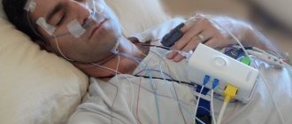
Doppler ultrasound of blood vessels
- memory impairment;
- frequent headaches and dizziness;
- repeated episodes of short-term loss of consciousness;
- inability to concentrate;
- pulsations in the cervical and head areas;
- failure in visual perception of the picture;
- persistently elevated cholesterol levels;
- loss of sensory (sensitivity);
- sudden disorder of speech function;
- temporary disorder of cerebral circulation (transient ischemic attack);
- idiopathic tinnitus.
This symptomatology indicates a violation of the blood supply to the brain. In addition, medical experts strongly advise people with nicotine addiction to undergo a BCA ultrasound, since smoking puts the vascular system at significant risk.
The procedure for ultrasound examination of brachiocephalic vessels has no contraindications or age limits and can be performed on women during the perinatal period and children.
Some limitations of the procedure are due to the individual characteristics of the patient: the presence of a surgical scar on the neck, voluminous calcium deposits in the walls of the BCA, the specific size of the patient’s neck.
Preliminary preparation
No special preparation is provided, but fulfilling certain conditions will help make the procedure easier and get more accurate results.
Firstly, on the day of the examination you need to stop smoking, drinking coffee and strong tea. It is advisable to limit salty foods in your diet. You should not drink alcohol for three days.
Because such eating behavior can have a negative impact on vascular tone and the filling of blood vessels.
Secondly, you must notify your doctor if you are taking any medications.
A day before the procedure, it would be better to stop using medications that affect concentration, nootropic and psychostimulants.
Such medications affect the state of the vascular system and can distort the actual results. Doctors also advise taking a walk in the fresh air immediately before the test.
Price
You can do an ultrasound scan in almost all clinics that are equipped with ultrasound machines. You can undergo the procedure free of charge with a referral from your attending physician. However, the disadvantage of such a study is a long queue. Sometimes you have to wait for a free ultrasound examination for several weeks; moreover, the patient will not always be able to choose a convenient time for the examination. The procedure is also carried out during examination or treatment in some hospitals (cardiological, neurological and others).
Private clinics quickly perform ultrasound examinations at an affordable price and at any convenient time. The cost depends on the level of the medical institution and the degree of qualification of the diagnostician. Average price for Doppler ultrasound of head and neck vessels in the Moscow region:
| Study title | Session time | Price in rubles |
| Duplex (double) brain scan | 30-45 minutes | 2500-3500 |
| Triplex (triple) brain scan | 40-60 minutes | 4000-5000 |
| Doppler ultrasound of extracranial vessels | 30 minutes | 950-12100 |
| TCD of the arteries of the head and neck | 30 minutes | 920 — 10000 |
The essence of the study
Special vessels, the brachiocephalic arteries (BCA), are responsible for the blood supply to the brain, tissues of the head, arms and shoulder girdle. They are determined from the aorta at the level of the shoulders. The condition of these vessels can be examined and their function assessed using Doppler ultrasound (USDG). What kind of examination is this, ultrasound examination of the BCA (BCS)? This method is based on the ability of blood particles to reflect ultrasonic waves emitted by the sensor. The ultrasound machine, in turn, picks up these reflections and graphically depicts the blood flow in the arteries.
The Doppler ultrasound method is non-invasive. It is safe and painless, therefore it has no contraindications or age restrictions for patients.
How is it carried out?
Diagnostics is carried out according to the following algorithm:
- The patient removes shoes and clothing covering the lower limbs. If there is jewelry in this area, you also need to get rid of it during the procedure. They can affect the operation of the sensor, causing the results to be distorted.
- After this, the patient lies down on the couch, taking the following position: legs are spaced shoulder-width apart, while the feet should be facing outward. To examine the vessels located under the knee, he will need to roll over on his stomach or stand up.
- The doctor applies a gel intended for ultrasound on both legs of the patient. The composition ensures maximum contact between the equipment and the skin. By moving the device's sensor, the specialist examines the required area. During the examination, the doctor may apply pressure with a finger on the vessels. This is necessary to obtain the most complete picture of their condition. A breath test is also performed. Its essence is as follows: the doctor asks you to take a deep breath and, holding the air, strain a little. By installing the sensor at a certain point, the specialist observes constantly changing images on the equipment monitor. During the process, the patient will hear unusual sounds from the speaker of the ultrasound machine - they indicate that blood flow parameters are currently being recorded. The data is automatically uploaded into an electronic database. If any abnormalities are detected during the study, the doctor takes pictures of them on thermal paper.
- Upon completion of the examination, the patient wipes off the gel with a napkin and gets dressed.
After this, the doctor must issue a conclusion, which should reflect the following information:
- general analysis of the condition of each vessel located in the lower extremities;
- assessment of the interaction of respiration and blood circulation;
- Is the venous collector easily compressed by the ultrasound device sensor;
- the thickness of the walls of blood vessels, whether there are atherosclerotic plaques on them;
- assessment of valve functioning;
- the presence of blood clots in the vessels;
- determination of the localization of perforating veins.
If a blood clot is detected during the diagnostic process, it is also described according to the following points:
- blood clot size;
- how much does it block the vessel;
- the clot is attached to the wall or fluctuates in the lumen with the blood flow;
- thrombus density;
- whether it moves under the influence of the equipment sensor.
The duration of the procedure, including filling out the protocol, is usually 20-30 minutes.
Next, the patient is sent for an appointment with a phlebologist or vascular surgeon. Only specialists are involved in decoding; independent interpretation of the results can lead to false conclusions. At the appointment, the doctor draws up the most appropriate treatment regimen. When analyzing the course of existing diseases, he compares the results obtained with previous ones. As practice shows, treatment of varicose veins in the legs and other pathologies occurs faster if the condition of the patient’s blood vessels is assessed using Doppler ultrasound.
What does the study clarify?
After ultrasound with Doppler, the specialist is able to determine a number of factors.
Vessel patency
It is important to assess how freely blood flows through the vessel, whether there are any blood clots or blood clots in the lumens of the veins and arteries.
To what extent does the course of the vessel correspond to the normal trajectory?
The existing direction of blood flow during scanning is compared with the norm along the trajectory of the anatomical course of the vessel. An increase in the tortuosity of blood vessels leads to disruption and slowing of blood flow.
Diameter and location of the vessel lumen
Vasomotor function is a change in the diameter of the lumen of a vessel under the influence of certain factors. This function is necessary to regulate blood pressure in blood vessels, heat exchange, and metabolism. Pathological narrowing of the lumen of the vessel leads to increased pressure in the spasmodic area, and local blood supply is disrupted.
Length of visibility of the changed lumen
When identifying pathological changes in the lumen of the vessels of the neck and head, it is important to identify the area of localization of the pathology, as well as the length of visibility of the changed lumen. This will allow you to accurately diagnose and prescribe treatment.
What is an ultrasound scan of the brachiocephalic arteries (BCA): how it is performed and what it shows, interpretation
Doppler ultrasound of the brachiocephalic arteries (BCAUSDG) is a method for diagnosing pathologies of neck vessels and disorders of blood flow in them. The study helps to determine in the early stages insufficient blood supply to the brain and associated pathologies. Ultrasound scanning is widespread and affordable.
What is ultrasound examination of BCS
Brachiocephalic vessels participate in the blood supply to the head, neck, shoulder girdle, and upper extremities. These include the brachiocephalic trunk, common carotid, subclavian, and vertebral arteries.
Extracranial vessels of the head and neck are subject to examination. Using ultra-high-frequency waves, the condition of the vascular wall and blood flow in the arteries are studied. In the presence of vascular atherosclerosis, the size of the plaques and their composition are determined.
In addition, ultrasound examination of the BCA allows us to identify:
- vasculitis (inflammatory vascular diseases) of various etiologies;
- stenosis, its severity;
- pathological formations that interfere with normal blood supply (benign and malignant tumors).
Dynamic assessment of blood flow is necessary to calculate the risk of ischemic strokes and other vascular accidents. In the presence of significant obstruction, hemodynamics are disrupted, which contributes to the appearance of the “steal” symptom (decreased blood supply to the brain). This provokes neurological pathologies.
Preparation rules
A few days before the study, you need to start preparing for an ultrasound scan. It is necessary to avoid foods that affect vascular tone.
These include:
- coffee;
- alcohol;
- tea and foods rich in caffeine and tannin;
- energy;
- salt;
- pepper.
2–3 days before BCA ultrasound, it is worth limiting smoking. Any medications taken by a person are studied by a doctor. If possible, medications that affect blood vessels are discontinued before the procedure. These measures are necessary for reliable research and accurate verification of the diagnosis.
How is ultrasound examination of the brachiocephalic arteries performed?
Ultrasound of the BCA is considered a non-invasive procedure. This technique is simple and allows you to quickly obtain accurate and reliable information about the condition of the vessels and the blood flow in them.
On average, ultrasound examination of the BCA lasts no more than 10 minutes. The subject is positioned on the couch on his back with his neck and chest exposed. During the examination, he changes the position of the body to more accurately visualize all vascular structures.
A conductive gel is applied to the neck and upper chest area, then an ultrasound probe is inserted. The diagnostician sequentially examines all parts of the neck, recording the necessary indicators.
During the ultrasound examination of the BCA, if necessary, the following functional tests are performed:
- breath-hold test while inhaling;
- changes in body position (from horizontal to vertical);
- pharmacological loads (vasodilators - nitroglycerin - are more often used).
After performing an ultrasound of the BCA, the functionalist doctor fills out a research protocol, which will later be deciphered by a specialized specialist.
Watch a methodological video of how an ultrasound scan of the neck vessels is performed:
Decoding the research results
After performing an ultrasound examination of the BCA, the data are carefully analyzed by a neurologist or therapist. Having completed deciphering the results, the doctor makes a conclusion, based on the results of which a diagnosis will be made.
Parameters that need to be assessed using ultrasound examination of the BCA:
- Condition of the vascular wall.
- The nature of pathological changes (if any).
- Features of blood flow.
The data is analyzed and compared with age norms. Based on this, the doctor makes a conclusion about the nature of the pathology.
Normally, large vessels have:
- free clearance;
- structured vascular wall;
- clear contours;
- adequate blood flow rate.
Pathologies detected by ultrasound of the BCA can be of different nature. Combined disorders (atherosclerosis and vessel aneurysm) are often encountered. Some pathological processes are not a consequence of the disease, but the result of age-related involution. Such results should not be regarded as normal, but these features must be taken into account when making a diagnosis.
- The most common finding on ultrasound examination of brachiocephalic vessels is atherosclerosis. In the early stages, it can manifest itself only as a slight thickening of the vascular wall. As the disease progresses, linear or round hyperechoic (lighter than normal tissues) formations are revealed on the vascular wall.
- Stenosis (narrowing of the lumen) and occlusion (complete closure of the vessel) can be a consequence of chronic inflammatory processes in this area. On Doppler Doppler Doppler (USDG), BCS are manifested by thickening of the vascular wall and a decrease in the arterial cavity. When occlusion occurs, the lumen of the vessel is completely closed. Often with such changes, collaterals (additional vessels) appear. Video of internal carotid artery stenosis:
- Various deformities of arteries and veins are in most cases congenital. Doppler ultrasound of the BCA shows abnormally short or tortuous vessels.
- Aneurysms are sac-like protrusions of the vascular wall. On ultrasound, the aneurysmal sac appears as a thin-walled formation. In this place the vascular wall is most vulnerable. Therefore, any provoking factor can lead to rupture of the aneurysm and hemorrhage.
- Vasculitis, including Takayasu's disease, may not produce a typical ultrasound picture. Inflammatory changes that occur in the arteries are not specific. Examination may reveal wall degeneration (thinning), rupture, or inflammation (increase in vessel thickness).
- In case of tumor diseases, ultrasound reveals a neoplasm that compresses the vessel or grows into it. The appearance of the tumor depends on the stage, histological structure and degree of malignancy.
It is impossible to carry out differential diagnosis based only on ultrasound data of the BCA. If a violation is detected, it is necessary to undergo a full range of examinations.
How much does the procedure cost?
The price of the study varies between 1200–3200 rubles. The cost depends on the region, clinic, equipment and qualifications of the specialist.
Cost of the study:
| City | Price in rubles |
| Moscow | 1600–3100 |
| Saint Petersburg | 1200–2200 |
| Kazan | 1500–1800 |
| Novosibirsk | 1200–2400 |
If left untreated, vascular diseases lead to irreversible changes in the brain. Timely diagnosis of pathologies is the only way to avoid complications.
Source: https://uziman.ru/golova-i-gorlo/bca-uzdg-bcs