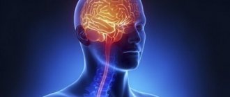
The plexus of nerve fibers in the brain connecting the right and left hemispheres is the corpus callosum, consisting of more than two hundred million nerve fibers. The corpus callosum is the largest structure that connects the hemispheres. Once formed, the corpus callosum grows further in length and width. The intersection of fibers and their penetration from one hemisphere to the other begins at twelve weeks.
With congenital partial or complete absence, they speak of agenesis of the corpus callosum .
In the case of aplasia (agenesis) and hypoplasia of the corpus callosum , the main commissure formed by the commissural fibers is either partially or completely absent and the third ventricle remains open. With agenesis, there are columns of the fornix and transparent septa, and in the case of hypoplasia, only the posterior commissure is absent, and the corpus callosum is shortened. Defects in the corpus callosum generally accompany other disorders in the brain, although they can also occur in isolation.
These types of defects begin to appear approximately in the second week after conception.
The frequency of their occurrence is one in two to three thousand.
Pathogenesis and etiology of the disorder
As mentioned above, the development of agenesis can be triggered by heredity, but most often the reasons for its occurrence cannot be determined.
This pathology has two clinical syndromes. In the first case, the patient’s intellectual abilities and motor activity are preserved, and the disease manifests itself as disturbances in the processes of transmitting impulses from the left hemisphere to the right and vice versa. For example, a patient who is right-handed cannot determine which object is in his left hand, because this requires the transfer of information from the right hemisphere to the left, where the speech zone is located.
In the second case, together with agenesis of the corpus callosum, the patient also has other malformations of the brain, including disturbances in the processes of neuron migration or hydrocephalus. In such cases, patients suffer from severe convulsive seizures and also lag behind in mental development.
All we can do is hope and pray
Although agenesis of the corpus callosum is not an extremely rare disease, it has been poorly studied.
Today, doctors do not have sufficient knowledge about the causes of its occurrence in each specific case; only factors that can serve as an impetus for the development of this pathology have been identified.
Also, no effective methods for treating this condition have been found and it is carried out only according to the symptoms of those disorders that were caused by agenesis. In this case, all measures are aimed at the effect, but do not in any way affect the cause.
From this we can conclude that there are no effective measures to prevent agenesis of the corpus callosum.
- Tingling all over the body like needles
- The whole body aches but there is no fever
Anatomy of the corpus callosum
The commissure is oblong, 2–4 centimeters wide, its length depends on age and gender, but does not exceed 10 centimeters. Consists of several departments.
The splenium of the corpus callosum is a thickening in the posterior part. The middle section is the trunk. The main part is 2/3 of the entire length of the solder.
The front one is a knee that ends in a thin beak.
The structure of the MT is such that it lies deep in the longitudinal sulcus and connects only the hemispheres, without affecting the rest of the brain.
Diagnostics[ | ]
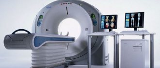
Diagnosis of the disorder is carried out in utero or after birth. Diagnostic methods: ultrasound, MRI, CT.
The main diagnostic criteria to suspect the presence of AMT:
1. Radial pattern of grooves on the medial surface of the brain. The grooves on the medial surface of the brain hemispheres extend from the roof of the third ventricle. The cingulate gyrus is not formed
2. Parallelism of the bodies of the lateral ventricles
3. The distance between the bodies of the lateral ventricles is increased
4. Abnormal expansion of the posterior horns of the lateral ventricles
5. No transparent partition
6. High location of the third ventricle
Intrauterine diagnosis of AMT[ | ]
Department of Radiation Diagnostics of St. Petersburg State Medical Academy named after. Mechnikov carried out MRI diagnostics of AMT in utero. From 2004 to 2009, 270 fetal MRIs with various pathologies were performed, and 35 cases of AMT were identified. 35 cases of AMT were identified. In 29 cases, AMT was complete (83%), in 2 - partial (5.7%), in 4 - atypical (11.4%). The corpus callosum was not identified in 33 cases (94%); increase in the distance between the bodies of the lateral ventricles and parallelism of the lateral ventricles - 28 (80%), deformation of the anterior horns of the lateral ventricles - 30 (86%); dilatation of the lateral ventricles - 28 (80%); colpocephaly (abnormal expansion of the occipital horns of the brain) - 27 (77%); deformation of the grooves of the medial surface of the hemispheres (radial pattern of grooves) - 17 (49%). In 12 cases (35%) AMT was isolated, in 23 fetuses (65%) it was combined with other brain anomalies, in 7 cases the anomalies were multiple.[3]
Predisposing factors
In its normal state, the corpus callosum is a dense plexus of nerve fibers designed to unite the right hemisphere of the brain with the left and ensure the exchange of information between them. This structure is formed from 10 to 20 weeks of pregnancy, the corpus callosum is formed at 6 weeks.
Agenesis can manifest itself in varying degrees of severity: absence, partial or incorrect formation, as well as underdevelopment of the corpus callosum.
In most cases, the cause of this disorder cannot be determined, but there are a number of factors that contribute to the occurrence of such a pathology. Predisposing factors include:
- process of spontaneous mutation;
- hereditary causes;
- chromosome rearrangement;
- the effect of toxins due to taking medications during pregnancy;
- insufficient provision of the fetus with nutrients during intrauterine development;
- viral infections or injuries suffered by the mother during pregnancy;
- disruption of metabolic processes in the mother's body;
- alcoholism during pregnancy.
Identifying the causes of such pathologies is difficult; it is only possible to identify the factors that can provoke their development.
Agenesis of the corpus callosum: what is the danger of the disease?
Among the anomalies of brain development, one of the most common diseases is congenital structural pathology, agenesis of the corpus callosum, in which there is a violation of associative connections between the two hemispheres.
Agenesis of the corpus callosum: causes
A disease associated with abnormal development of the brain, congenital, quite rare, manifested in the complete or partial absence of a structural formation connecting the hemispheres of the brain, is called adhesion of the corpus callosum.
The corpus callosum connects the cerebral hemispheres
Normally, the corpus callosum, or major commissure, is represented by a dense junction of nerve fibers that unites the cerebral hemispheres, left and right, and ensures coordination between them.
The formation of this structure from a morphological point of view corresponds to the period of 10-20 weeks of pregnancy; the beginning of differentiation of the tissue of the corpus callosum is attributed to the middle of the sixth week.
This pathology can have varying degrees of structural manifestation and be expressed in the form of a total absence, partial (hypogenesis) or incorrect (dysgenesis) formation, underdevelopment (hypoplasia) of the corpus callosum.
Instead of the normal structure, which looks like a wide, flat strip, the large commissure takes on the appearance of shortened septa or transparent pillars of the arch.
As a rule, it is difficult to accurately determine the cause of a congenital structural malformation of the brain.
Predisposing factors
- Heredity (family cases of manifestation, with autosomal or X-linked inheritance)
- Spontaneous mutations
- Chromosome rearrangement
- Intrauterine infections (usually viral) or trauma
- Exposure to toxic substances, teratogenic effect of drugs during uterine development
- Fatal fetal alcohol syndrome (due to maternal alcoholism during pregnancy)
- Nutrient deficiency in the fetus
- Metabolic disorders in the mother
To date, it is not possible to unambiguously name the cause of agenesis of the corpus callosum; we can only identify the factors causing its occurrence.
Read: Keratosis follicularis
You can get acquainted with the structure of the brain by watching the proposed video.
Symptoms and diagnosis
The clinical manifestations of the disease vary; depending on this, it can be detected in a severe form in childhood, usually up to two years, or in adults, asymptomatically and sometimes completely by accident.
Children whose disease, for one reason or another, was not diagnosed in the prenatal period, appear healthy at birth, and their development is normal until they reach three months.
It is at this stage that the first symptoms of the disease appear, usually in the form of so-called infantile spasms, a type of epileptic seizures.
Symptoms
- Interruption of the formation and further development of the structure of the corpus callosum in the first stages
- Development of porencephaly, a defect in the mantle of the brain
- Hydrocephalus – lack of ability to track with the eyes, later – lack of voluntary movements
- Atrophic phenomena of nerves, visual and auditory
- Microencephaly
- The appearance of neoplasms and cysts in the hemispheres
- Polymicrogyria (underdeveloped gyri)
- Premature, early sexual development
- Manifestation of spina bifida syndrome
- Manifestation of Aicardi syndrome
- Development of lipomas
- Pathology of the gastrointestinal tract, tumor formation
- Slowing and then inhibition of psychomotor development
- Manifestations of varying degrees of retardation, mental and physical
- Coordination problems
- Developmental delays, abnormalities of various organs
- Low muscle tone
- Skeletal abnormalities
In patients with preservation of intelligence and motor functions, the manifestation of the anomaly is a disruption in the exchange of information between the hemispheres, for example, in the difficulties experienced by a right-handed person when asked to name an object in his left hand.
The disease is diagnosed by scanning the brain
Diagnosis of the disease can be detected by performing a brain scan procedure.
Carrying out prenatal diagnosis of such an anomaly as agenesis of the corpus callosum is associated with great difficulties. Most often, the anomaly is diagnosed during the second or third trimester of pregnancy.
Read: Red urine after beets
The main method used is echography; MRI and ultrasound procedures are also used.
Carrying out an echography procedure in the prenatal period does not allow diagnosing all cases of the disease, including due to the characteristics of fetal presentation.
In addition, with partial agenesis, detecting the defect is even more difficult.
Diagnosis of the disease is complicated by the fact that agenesis is often combined with a number of developmental defects and various genetic symptoms.
To obtain a complete picture of the examination, in case of suspected anomaly, it is necessary to perform karyotyping, as well as a thorough ultrasound analysis and diagnostics using magnetic resonance imaging.
The combination of various modern examination methods makes it possible to more reliably diagnose cases of agenesis of the corpus callosum in the prenatal period.
Agenesis of the corpus callosum: treatment and prognosis
To date, there are no effective treatments for the disease.
Treatment is aimed at minimizing the symptoms of the disease
Therapeutic measures consist of treating serious symptoms, reducing them to a minimum manifestation.
Functions of the organ
For a long time after its discovery, scientists could not understand what specific functions the corpus callosum performs. It was initially assumed that it contained a focus of epileptiform activity.
For patients with epilepsy, surgeons cut it open to eliminate seizures. The seizures passed, but people's behavior changed. Thus, a patient with a dissected corpus callosum hugged his wife with his right hand and pushed her away from him with his left.
And only experiments on animals, and subsequently with the participation of a volunteer, made it clear:
- The obvious function is to physically connect the right and left hemispheres, since the corpus callosum is located deep in the longitudinal fissure between the hemispheres. Nerve fibers connect both symmetrical and asymmetrical sections of different hemispheres, as well as different sections of the same hemisphere.
- Information communication between the hemispheres. Neuronal fibers (white matter) fan out in all directions, transmitting information received by different lobes of the hemispheres.
- Coordination of the hemispheres. MT ensures not only the exchange of received information between the left and right hemispheres, it is also responsible for analyzing this information and responding adequately.
Anatomy and functions
The corpus callosum is covered on top by a small layer of cerebral gray matter, which explains, accordingly, the gray covering on it. Upon visual examination, 3 main sections can be distinguished:
- trunk (or midbrain);
- knee (part of the brain located in the front);
- beak or splenium of the corpus callosum (posterior section).
The brightness of the large commissure (when viewed on photographs or in section) is provided by fibers that are located radially and are located in each of the hemispheres.
The middle section, when viewed, looks like a bulge, which is also the longest part of the entire brain. The posterior section is visually visible as a thickening relative to other sections and zones, which is freely located above neighboring areas of the brain. The gray matter is represented by stripes and is located on top.
Functions provided by the corpus callosum:
- transfer of information (impulses) important for the functioning of the body from one hemisphere to the other;
- formation of the main characteristics that define personality and its characteristics;
- basic (basic, defining) skills and the possibility of their application throughout a person’s life;
- work on the formation of the emotional and personal sphere.
The main and practically the only function of the corpus callosum is the transmission of information from one hemisphere to the other and ensuring normal human life due to the synchronization of their work. Therefore, it is an important part of the brain. Studies have shown that rupture of the corpus callosum leaves both hemispheres working and does not lead to death. However, they work in a separate mode, which affects human behavior.
Manifestations and signs of anomaly
Agenesis of the corpus callosum of the brain manifests itself in different ways, depending on the degree of the disorder; the main symptoms in the presence of this anomaly are:
- processes of nerve atrophy in the organs of hearing and vision;
- cysts and neoplasms in the part of the brain where the hemispheres connect;
- microencephaly;
- prone to seizures;
- presence of facial dysmorphism;
- the occurrence of defects in the development of the visual organs;
- porencephaly;
- pathological changes in the fundus;
- delays in psychomotor development;
- schizencephaly;
- presence of lipomas;
- disturbances in the development of the gastrointestinal tract and the presence of formations;
- early puberty and so on.
In addition to the above, the disease can manifest itself with Aicardi syndrome. This genetic disease is extremely rare and is characterized by abnormal development of the brain and visual organs. Agenesis also causes changes in bones and skin lesions.
In dry but important remainder
Thus, the corpus callosum of the brain, despite its tiny size, has a great influence on a person’s life. It allows the formation of personality, is responsible for the emergence of habits, conscious actions, the ability to communicate and distinguish between objects.
That is why it is extremely important to take care of your health during pregnancy, since the main metabolic disorders are formed during this period.
We should not forget that the corpus callosum forms the intellect and makes a person a personality. Despite all attempts to study this structure, scientists have not yet been able to uncover all its secrets, and therefore very few methods for treating disorders, if any, have been developed.
The main ones are drug therapy and a special set of exercises - exercise therapy, which allows you to maintain optimal indicators of physical development. Measures to eliminate the symptoms of disorders should be taken immediately, otherwise the desired improvements may not occur.
Corpus callosum in men and women
Scientists have not yet reached a unanimous opinion on the relationship between the size of the corpus callosum and the functions it performs. At first, American scientists argued that women have a wider MT than men, and this supposedly explains the peculiarities of women’s intuition.
A few years later, French scientists refuted this theory and stated that the male corpus callosum, in relation to the size of the brain, is larger than that of women, and the brain itself is larger.
But they were able to derive one pattern: the different density of neural fibers between the segments of the hemispheres responsible for speech is a distinctive feature of the weaker half of humanity.
In women, both hemispheres are involved in the work at once, while the brain of men uses one hemisphere to a greater extent than the other.
Dysplasia and hypoplasia - incorrect or underdevelopment of MT
Hypoplasia is a serious, but fortunately quite rare diagnosis. In essence, this, like agenesis, is a violation of the intrauterine development of brain tissue. If during agenesis the corpus callosum of the brain is completely absent, then with hopoplasia it is underdeveloped. Of course, treating this disease with modern medicine is impossible.
Therapy involves a set of measures that minimize deviations in the patient’s development. Neuropsychologists recommend that patients regularly perform a specially designed set of physical exercises that help restore connections between the hemispheres, as well as information wave therapy.
Hypoplasia, or, as this disease is also called, microcephaly, is a complex pathology, during the course and development of which there is a significant decrease (the value is taken based on normal indicators) in the volume of the brain and, accordingly, the corpus callosum as well.
In most cases, along with the diagnosis of hypoplasia, other disorders are observed, including improper development of the present parts of the corpus callosum (dysplasia or dysgenesis), insufficient development of the spinal cord, underdevelopment of the limbs and a number of internal organs.
The main cause of developmental disorders and reduction in size (or complete absence) of the corpus callosum is one or another congenital pathology. Factors causing such changes:
- Presence of bad habits in a pregnant woman (smoking, taking drugs or alcohol);
- intoxication;
- exposure to radiation (ionizing);
- consequences of complex and serious diseases - rubella (acquired in adulthood or especially during pregnancy), influenza, toxoplasmosis.
Particular attention should be paid in the early stages (1-2 trimesters) of pregnancy - during the formation of brain structures.
Symptoms of the disease:
- decrease in brain volume relative to normal indicators (main symptom);
- changes in the usual structure of the brain convolutions and some structures (the convolutions are flat);
- insufficient development of the temporal and frontal lobes of the human brain for normal functioning;
- reduction in the size of the pyramids - elements of the medulla oblongata (pyramidal syndrome develops);
- disorders and malfunctions identified in the functioning of the cerebellum;
- dysfunction of the brain stem (part of the brain);
- in most cases there is an intellectual impairment;
- physical development disorders;
- neurological disorders and characteristic disorders;
- pathology of the optic thalamus.
With hypoplasia, the skull is smaller than a person should normally have.
Despite the development of modern medicine, there is no high-quality and effective treatment for such a disorder. It is possible to reduce symptoms to a minimum.
It is important to remember that this anomaly leads to a decrease in life expectancy. The main measure of influence is taking medications.
If appropriate measures are not taken at the early stage of development and formation of the anomaly, then the majority of patients in the future (already in childhood and adolescence) will experience various problems in the field of neurology.
Also, many patients with hypoplasia have moderate and severe intellectual impairment, developmental delay, both mental and physical.
Thus, according to various medical studies, in at least 68-71% of cases diagnosed with hypoplasia of the corpus callosum, there is a consequence such as mental retardation. In addition, the disorder leads to the emergence of more serious mental disorders, such as schizophrenia.
Dysgenesis of the corpus callosum can cause changes in the muscle system and in the skeleton as a whole. It is a common cause of scoliosis.
There are delays in the mental and psycho-emotional development of children and adolescents. Intellectual impairment, neurological problems, and developmental delays are also observed, so patients in most cases require constant supervision and intensive treatment.
If measures are taken to eliminate the manifestations, children can learn the necessary skills, including mastering a simple school curriculum.
Establishing diagnosis
However, echography does not make it possible to detect the disease in all cases, and if the agenesis of the corpus callosum is partial, then its detection is even more difficult.
Difficulties in diagnosing the disorder arise because this pathology is often associated with a number of other disorders and genetic symptoms. In order to conduct a more detailed examination of the patient, specialists resort to karyotyping, ultrasound analysis and MRI.
Using a combination of examination techniques, it is possible to obtain a complete picture of the disease.
Support groups[ | ]
In the United States, the National Organization of Disorders of the Corpus Callosum has been organized, which supports individuals with developmental disorders of the corpus callosum and their relatives. With the support of the organization, annual thematic conferences are held with the participation of representatives of the scientific medical community involved in the study of pathologies of the corpus callosum.[5]
Support groups have also been organized on social networks and instant messengers.
Malformations of the corpus callosum and their consequences
The first trimester is the most important stage of pregnancy. It is during this period that all the main organs are formed, and it is at this time that they are most vulnerable. The neural tube forms towards the end of the trimester, at 12-13 weeks.
The expectant mother undergoes an ultrasound, and it shows whether the newborn has any abnormalities in the structure of the brain, including the corpus callosum, because they will lead to mental retardation.
Therefore, it is very important to monitor your health, undergo all examinations on time and take vitamin complexes during pregnancy.
The main malformations of the corpus callosum:
- Dysgenesis
- Agenesis
- Hypoplasia
All these defects entail a number of disturbances in the functioning of other organs. But most importantly, they are the cause of mental retardation.
Parents of children with these defects talk about fairly favorable prognoses if there are no concomitant diseases of the central nervous system.
Such children, of course, are doomed to take medications for life: nootropics that improve the nutrition of brain tissue, neuroleptics that correct behavior, hormonal and anticonvulsant drugs.
Such children are observed by neurologists and neurosurgeons. Rehabilitation is carried out by psychologists, defectologists, speech therapists, neuropsychologists, and psychiatrists.
The 30% chance of a favorable outcome that doctors give for such defects depends on the hard everyday work of the parents. It is necessary to deal with both physical and speech, psychomotor, and mental development.
The child will not always want to study. He can get tired quickly, become moody, absent-minded, even aggressive.
Agenesis
Agenesis of the corpus callosum in the fetus is the complete or partial absence of the main commissural commissure between the hemispheres. It can be either an independent disease or part of other congenital defects, for example, Aicardi syndrome.
Reasons for the absence of the corpus callosum:
- Genetic anomaly. A chromosomal mutation can be hereditary (not necessarily in the next generations, it is quite possible that the precedent was in the 5th-6th generation or earlier). An accurate diagnosis can be made by a geneticist after an examination.
- Intrauterine infection. Three decades ago, the term TORCH appeared, which unites the most common infections dangerous to the fetus - toxoplasmosis, mycoplasmosis, syphilis, rubella, cytomegalovirus and herpes. Unfortunately, they lead to serious developmental defects, miscarriage and death of newborns.
Partial agenesis of the corpus callosum is not so dangerous. The functions of the missing areas are taken over by the neighboring segments, as well as the anterior and posterior commissures. However, it also requires constant monitoring of the baby’s condition and drug treatment.
Usually the deviation is visible on ultrasound, but sometimes the disease goes unnoticed. In the first few months of life, the problem becomes obvious. Symptoms of agenesis of the corpus callosum:
- Functions of vision and hearing are impaired. The baby hears poorly or does not perceive sounds at all, and does not focus his gaze on an object. This is noticeable already at 1.5-2 months.
- The size of the head, and therefore the brain, is smaller than normal. During a routine consultation, a neurologist measures the circumference of the child’s head with a centimeter.
- Disturbances in the structure of the bones of the facial part of the skull are especially pronounced in the area of the eyes and nose.
- Various forms of epilepsy - convulsions, freezing, loss of consciousness.
- Delayed mental, psychomotor and speech development.
Parents can notice these symptoms themselves and consult a doctor. There are several signs that can only be detected through examinations:
- tumors, neoplasms, cysts - both at the site of the corpus callosum and in other organs, most often in the stomach and intestines;
- adhesions and characteristic spots on the fundus;
- disturbances in the electroencephalogram can appear only during functional tests, although sometimes the EEG in such children represents a variant of the norm.
It represents disturbances in the development of tissues of both a separate area of the corpus callosum and the entire organ. It is caused by the same reasons as agenesis - chromosomal abnormalities, intrauterine infections and poor lifestyle of the expectant mother.
Smoking, drinking alcohol and drugs lead to improper formation of the fetal neural tube.
MT dysgenesis is not life-threatening, but problems are inevitable. First of all, such children have problems with oral and written speech, perception of light signals and reaction to external stimuli.
Hypoplasia of the corpus callosum of the brain is a general underdevelopment of the brain. Hypoplasia, like other developmental anomalies of the corpus callosum, can be an independent disease or be associated with other developmental defects.
Main features:
- disproportionate skull. As a rule, this is noticeable, but deviations can sometimes be insignificant;
- the structure of the convolutions is changed - they become flat;
- muscular dystonia. The baby's movements are uncertain, infants do not hold their heads well and do not lean on their arms. If the disease was overlooked in the first months of life, then at an older age awkwardness and clumsiness are noted. Children often fall and drop objects;
- disturbances in the functioning of the nervous system. Sometimes it is very difficult to predict the reaction of a sick baby to a particular stimulus;
- developmental delay. The baby begins to walk later than others and has difficulty mastering speech.
Usually, abnormalities in the structure of the brain are visible on ultrasound during pregnancy. But poor quality equipment, low qualifications of the doctor, and the specific position of the fetus during the examination can make it difficult to make a diagnosis in the early stages.
Up to two years of age, children with hypoplasia of the corpus callosum can develop on par with their peers, and only after this age do symptoms begin to appear.
There are several ways to define both hypoplasia and dysplasia:
- Magnetic resonance imaging - allows you to see organic lesions of the brain and all its parts.
- Electroencephalogram - shows how the patient reacts to certain stimuli and records epiactivity.
- Neurosonography - ultrasound of the brain through the fontanel. Possible only in the first 1.5 years, then the connective tissue is replaced by bone.
Agenesis of the corpus callosum: causes and signs of the disease
One of the congenital pathologies - agenesis of the corpus callosum of the brain - occurs as a result of genetic causes and external adverse effects during pregnancy.
Anomalies occur in a small number of patients, and only in 20% of cases is it accompanied by serious disorders that interfere with normal life. The corpus callosum is a small node consisting of nerves that connect both lobes of the brain organ. This element of the main organ of the nervous system is flat and wide. With agenesis, there is no scar tissue, but the exact causes of this pathological process have not yet been established.
Causes of pathology
Abnormal brain formation is always a congenital pathology; it appears quite rarely . From a morphological point of view, the failure occurs at approximately 10-20 weeks of pregnancy, when connective tissue begins to form.
The strength of severity and manifestation may differ: adhesions are either completely absent, or partial agenesis of the corpus callosum is detected. Factors that contribute to the development of this condition:
- changes in chromosomes, including hereditary ones;
- spontaneous mutations that have no explainable cause;
- intrauterine infection as a result of viral infection of the mother;
- the effect of narcotic substances and drugs;
- alcohol syndrome in the fetus due to maternal abuse;
- metabolic disorders in a woman or deficiency of nutrients in the embryo.
The disease is subject to intrauterine diagnosis, but the accuracy of determining agenesis of the corpus callosum is low.
Clinical symptoms
In childhood, agenesis of the corpus callosum is detected; it manifests itself only in the case of a severe form, since it is accompanied by severe symptoms. In adults, agenesis of the corpus callosum is found incidentally, mainly during examination for other diseases. Often this pathology is mild and does not cause any difficulties in life.
In some infants, the disease is detected by 3-4 months of age, when developmental problems begin.
Agenesis of the corpus callosum is manifested by such first signs as: mild epileptic seizures, seizures and infantile spasms. Other symptoms, some of which can be detected during intrauterine examination:
- change in the process of formation of the corpus callosum;
- hydrocephalus or microencephaly;
- porencephaly, diseases of the mantle of the brain;
- atrophy of the auditory or optic nerves;
- syndrome of changes in spinal tissue, Aicardia;
- cysts and other neoplasms in the hemispheres;
- poorly formed convolutions;
- the appearance of lipomas;
- early sexual development in older age;
- abnormalities of internal organs;
- tumor processes often occur in the intestines and disturbances in the functioning of the gastrointestinal tract for no apparent reason;
- slow psychomotor development;
- somatic problems;
- poor coordination, weak muscles, skeletal abnormalities.
Agenesis of the corpus callosum in the fetus does not show any external signs.
Diagnostics
Ultrasound is most often used for detection. A specialist with sufficient experience and knowledge can accurately determine whether a child has agenesis of the corpus callosum. Additional techniques are used if the anomaly is partial and mild.
Additionally, patients are prescribed MRI and CT scan of the brain. Sometimes accompanying blood tests are required. It is very difficult to confuse agenesis with other diseases, but to be on the safe side, some experts advise seeking advice from 2 or more doctors to get a more complete picture of opinions.
Is the pathology curable - video
Modern medicine cannot cure agenesis, but there are methods for correcting the disease. They are selected separately for each clinical case. The doctor takes into account the child’s general well-being, as well as the severity of agenesis of the corpus callosum.
Special medications are used to relieve symptoms. However, doctors are sure: most methods are not able to permanently relieve symptoms. Potent drugs are used for therapy:
- Phenobarbital . Reduces the number of attacks during infantile spasms.
- Benzidiazepines . Psychoactive products that inhibit psychomotor reactions and reduce the number of seizures.
- Corticosteroids . Necessary to combat epileptic seizures.
- Neuroleptics . Used to treat mental disorders.
- Nootropics . Used to correct brain functions.
- Diazepam . Corrects behavioral disorders.
- Neuropeptides . Improves communication between nerve endings.
In addition to medications, surgery is sometimes used to stimulate the vagus nerve. To do this, an electrical pulse generator is introduced.
Such an operation is possible only if agenesis of the corpus callosum threatens acute pathologies of important organs.
Surgery is also prescribed if other methods of therapy do not produce any results.
The generator is installed in the subclavian area. After the stimulator is installed, the child should visit the doctor every few months.
The advantage of installing a stimulator for agenesis of the corpus callosum is a significant reduction in the number of attacks.
Patients tolerate any symptoms better, but the device may not be effective in some situations. Sometimes pathology causes scoliosis, in which case exercise therapy and physiotherapy are prescribed for treatment. Less commonly, spinal surgery is required to improve the condition of bone tissue.
Prognosis for the disease
The development of partial agenesis of the corpus callosum in rare cases leads to pathologies that shorten the patient’s life. This form of the disease occurs in 80% of cases and is not accompanied by severe symptoms. Neurological correction, exercise therapy and physiotherapy are occasionally required.
However, pronounced forms and average degrees of pathology provoke various disorders. In this case, it is impossible to talk about a favorable prognosis. Most often, the main disorders are problems with intelligence, mental development, and severe neurological pathologies . Drug and folk therapy in this case does not give any effect.
Despite the favorable outcomes that occur, agenesis of the corpus callosum is a severe neurological disorder that cannot be successfully corrected. It is not possible to establish either the exact causes or factors that could reduce the number of manifestations of the pathology.
Agenesis of the corpus callosum is a complex brain disease that affects humans only at the stage of embryonic development. Despite this, it is found infrequently and in many cases does not cause any problems for patients. The timing of detection of the anomaly does not in any way affect the success of its treatment.
Source: https://nevrology.net/sindromy-i-zabolevaniya/vrozhdennye-i-nasledstvennye/ageneziya-mozolistogo-tela.html
Basics of therapy
Currently, there are no effective treatments for such an anomaly as agenesis of the corpus callosum. Correction methods depend on the diseases that were caused by this disorder, and therefore are selected individually.
Treatment is aimed at minimizing the manifestations of the disease. But, according to experts, it does not give the desired effect, and besides, the methods have not been fully developed. Therapy for the most part consists of the use of strong medications.
The following drugs can be used:
- benzodiazepines and phenobarbital, which allow you to adjust the frequency of attacks in the presence of infantile spasms;
- corticosteroid hormones (Dexamethasone, Prednisolone) in combination with basic anti-epileptic drugs;
- neuroleptics and diazepam drugs, the action of which is aimed at reducing behavioral disorders;
- nootropics (Semax, Piracetam) and neuropeptide drugs (Cerebrolysin);
- Children are most often prescribed Asparkam, Diacarb, Mexidol.
In addition to taking medications, if the need arises, surgical interventions are also performed, for example, stimulation of the vagus nerve is performed. But this can only be done in cases where agenesis has caused serious disruptions in the functioning of vital human organs.
This pathology can cause disorders in the musculoskeletal system and cause scoliosis, so specialists prescribe physiotherapy and exercise therapy. In some cases, surgery is also used.
Nowadays, agenesis is being carefully studied, but no tangible results have yet been achieved.
Treatment[ | ]
There is currently no specific treatment for congenital anomalies of the corpus callosum. For assistance and correction, various methods and techniques are used, aimed at helping in physical development and the formation of various skills. It is important to consult with a variety of medical, health, educational and social work professionals. Such specialists include neurologists, neuropsychologists, occupational therapists, physiotherapists, speech therapists, pediatricians, geneticists, social workers, special educators, and early childhood development specialists.
How many days are available for patients?
In cases where the disorder is not associated with the occurrence of other developmental pathologies, the prognosis is favorable. About 80% of children have no developmental disorders or minor neurological problems.
However, in most cases, agenesis of the corpus callosum provokes the occurrence of various kinds of consequences and associated pathologies, and in such a situation there can be no talk of a good prognosis.
Patients experience intellectual impairment, neurological problems, developmental delays and other symptoms with which they do not live for a long time. Patients are treated according to symptoms and the therapy has little effect.
Agenesis of the corpus callosum can be classified as a disease with a large number of developmental anomalies and unfavorable prognosis.
Agenesis: main causes, symptoms, treatment
Agenesis of the corpus callosum, as an independent disease, is a complex disorder of a structural nature. If this occurs, then the patient experiences, accordingly, a disruption of the associative connections between the right and left hemispheres of the brain, which is not observed in the normal state, since this organ is responsible for uniting these areas.
The disease develops as a result of disturbances (abnormalities) during development. It is rare - about 2%, congenital, manifested in complete or partial absence when examined in photographs or studies of this structural formation.
Agenesis is expressed by the complete absence of a large commissure or its underdevelopment, sometimes it is only partially present (there is no specific area). In this case, the formation, if it is underdeveloped or partially present, is presented in the form of significantly shortened septa or transparent columns of the cerebral vault.
Modern neurologists and scientists cannot accurately name the main reasons that influence the development of this pathology. The main suspected factors are:
- heredity (in 70% of cases, if the family has already had problems of a similar nature, they will recur in future generations);
- genetic (including chromosomal) changes and mutations;
- chromosome rearrangement (during fetal formation);
- the development of infections caused by a virus that occur during pregnancy (in utero, especially dangerous in the early stages);
- injuries;
- ingestion of substances with a toxic (poisonous) effect on the body or developing fetus (including exposure to alcohol);
- consequences of taking medications (side effects or complications);
- disruption of pregnancy (nutrient deficiency in the fetus during development);
- metabolic disorders in the body of a pregnant woman.
The main symptoms indicating the presence of agenesis of the corpus callosum in a person and the need for immediate qualified diagnosis:
- hydrocephalus;
- disorders in the development and functioning of the visual and auditory nerves;
- the appearance of benign cysts and other types of tumors in the brain;
- cerebral convolutions that are not sufficiently formed to perform their intended functions;
- early puberty;
- development of lipomas;
- various problems and disturbances in the functioning of the gastrointestinal tract (of varying nature and intensity);
- psychomotor impairment;
- behavioral problems (especially acute in childhood);
- diagnosis of mild mental retardation (detected in childhood);
- impaired coordination of movements;
- low muscle tone;
- disorders of the development of muscles and bones of the skeleton.
Set of measures
Treatment is most often drug therapy. Includes taking antiepileptic drugs, as well as corticosteroid hormones. Also, in some cases, exercise therapy (physical education with a therapeutic bias) is indicated.

