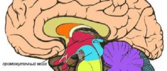
The human brain is a complex structure, consisting of different sections that are interconnected anatomically and functionally and are responsible for performing certain types of activities in the human body.
One of the most important parts of the brain, in particular its posterior part, is the cerebellum. It is responsible for the coordination of movements, regulates muscle tone, without it it is impossible to ensure normal balance of the human body in space. In addition, the involvement of the cerebellum in mental functions has been proven. That is why pathology in this part of the brain attracts the attention of not only neurologists, but also psychiatrists.
What is a pyramid system
So, the doctor detects Babinsky’s symptom on the left or right, and concludes that there are disturbances in the pyramidal system. What does it mean?
All movements that we make are controlled by parts of the nervous system - the cerebellum, pyramidal and extrapyramidal systems.
The pyramid system (which we will describe) helps us make any movements, stop or change them at will. And initially all movements are born in the cerebral cortex. As you know, it consists of 6 layers, and the 5th and 6th of them are precisely responsible for this function.
The fifth layer contains the so-called giant pyramidal cells, or Betz cells, which connect to the muscles using axons (long processes) that make up the pyramidal tract, which descends the brain stem and merges with the cells of the spinal cord. Thus, the signal generated in the cortex causes the muscles to contract. If its transmission is disrupted for some reason, then, accordingly, the reflexes are also disrupted, and this indicates the possibility of paralysis or paresis.
Ventricular septal defect
Ventricular septal defect is a congenital intracardiac anomaly characterized by the presence of communication between the right and left ventricles.
A ventricular septal defect is manifested by shortness of breath, retarded physical development, fatigue, rapid heartbeat, and the presence of a “heart hump.” Instrumental diagnosis of ventricular septal defect includes ECG, echocardiography, chest radiography, ventriculography, aortography, catheterization of cardiac chambers, MRI. For ventricular septal defects, radical (closing the defect) and palliative (narrowing of the pulmonary artery) interventions are performed.
A ventricular septal defect (VSD) is a hole in the septum separating the cavities of the left and right ventricles, the presence of which leads to pathological shunting of blood.
In cardiology, ventricular septal defect is the most common congenital heart defect (9–25% of all congenital heart defects). The incidence of critical illness with ventricular septal defect is about 21%.
The defect is found with equal frequency in newborn boys and girls.
A ventricular septal defect may be a single intracardiac anomaly (isolated VSD) or be part of the structure of complex defects (tetralogy of Fallot, truncus arteriosus, transposition of the great vessels, tricuspid valve atresia, etc.). In some cases, the interventricular septum may be completely absent - this condition is characterized as a single ventricle of the heart.
Ventricular septal defect
Most often, a ventricular septal defect is a consequence of a violation of embryonic development and is formed in the fetus due to disorders of organ formation.
Therefore, ventricular septal defect is often accompanied by other cardiac defects: patent ductus arteriosus (20%), atrial septal defect (20%), aortic coarctation (12%), aortic stenosis (5%), aortic insufficiency (2.5-4. 5%), mitral valve insufficiency (2%), less often - abnormal drainage of the pulmonary veins, pulmonary stenosis, etc.
In 25-50% of cases, the ventricular septal defect is combined with malformations of extracardiac localization - Down's disease, anomalies of the kidneys, cleft of the hard palate and cleft lip.
The direct factors causing disruption of embryogenesis are harmful effects on the fetus in the first trimester of gestation: diseases of the pregnant woman (viral infections, endocrine disorders), alcohol and drug intoxication, ionizing radiation, pathological course of pregnancy (severe toxicosis, threat of spontaneous abortion, etc.) . There is evidence of a hereditary etiology of ventricular septal defect. Acquired ventricular septal defects can be a complication of myocardial infarction.
The interventricular septum forms the inner walls of both ventricles and makes up approximately 1/3 of the area of each of them. The interventricular septum is represented by membranous and muscular components. In turn, the muscular section consists of 3 parts - inflow, trabecular and outflow (infundibular).
The interventricular septum, along with other walls of the ventricles, takes part in the contraction and relaxation of the heart. In the fetus, it is fully formed by the 4-5th week of embryonic development.
If this does not happen for some reason, a defect remains in the interventricular septum.
Hemodynamic disturbances with a ventricular septal defect are caused by communication between the left ventricle with high pressure and the right ventricle with low pressure (normally, during systole, the pressure in the left ventricle is 4–5 times higher than in the right).
After birth and the establishment of blood flow in the systemic and pulmonary circulation, due to a ventricular septal defect, a left-to-right shunt occurs, the volume of which depends on the size of the hole.
With a small volume of shunted blood, the pressure in the right ventricle and pulmonary arteries remains normal or increases slightly.
However, with a large flow of blood through the defect into the pulmonary circulation and its return to the left parts of the heart, volumetric and systolic overload of the ventricles develops.
A significant increase in pressure in the pulmonary circulation with large ventricular septal defects contributes to the occurrence of pulmonary hypertension. An increase in pulmonary vascular resistance causes the development of blood discharge from the right ventricle to the left (reverse or cross-shunt), which leads to arterial hypoxemia (Eisenmenger syndrome).
Long-term practice of cardiac surgeons shows that the best results in closing a ventricular septal defect can be achieved by shunting blood from left to right. Therefore, when planning the operation, hemodynamic parameters (pressure, resistance and volume of discharge) are carefully taken into account.
The size of the ventricular septal defect is assessed based on its absolute size and comparison with the diameter of the aortic opening: a small defect is 1-3 mm (Tolochinov-Roger disease), a medium one is equal to approximately 1/2 the diameter of the aortic opening, a large one is equal to or greater than its diameter .
Taking into account the anatomical location of the defect, the following are distinguished:
- perimembranous ventricular septal defects – 75% (inflow, trabecular, infundibular) are located in the upper part of the septum under the aortic valve and can close spontaneously;
- muscular defects of the interventricular septum - 10% (inflow, trabecular) - located in the muscular part of the septum, at a considerable distance from the valve and conduction systems;
- supracrestal defects of the interventricular septum - 5% - are located above the supraventricular crest (the muscle bundle that separates the cavity of the right ventricle from its outflow tract) and do not close spontaneously.
The clinical manifestations of large and small isolated ventricular septal defects are different. Small ventricular septal defects (Tolochinov-Roger disease) have a diameter of less than 1 cm and occur in 25-40% of all VSDs. They manifest themselves as mild fatigue and shortness of breath on exertion.
The physical development of children, as a rule, is not impaired. Sometimes they have a weakly expressed dome-shaped protrusion of the chest in the area of the heart - the “heart hump”.
A characteristic clinical sign of small defects of the interventricular septum is the presence of a rough systolic murmur over the heart area, which is detected already in the first week of life.
Large ventricular septal defects, measuring more than 1/2 the diameter of the aortic orifice or more than 1 cm, manifest themselves symptomatically already in the first 3 months of life of newborns, leading in 25-30% of cases to the development of a critical condition.
With large defects of the interventricular septum, malnutrition, shortness of breath during exercise or at rest, and increased fatigue are observed. Feeding difficulties are typical: intermittent sucking, frequent separation from the breast, shortness of breath and pallor, sweating, oral cyanosis.
Most children with ventricular septal defect have a history of frequent respiratory infections, prolonged and recurrent bronchitis and pneumonia.
At the age of 3-4 years, as heart failure increases, such children begin to complain of palpitations and pain in the heart area, a tendency to nosebleeds and fainting.
Transient cyanosis is replaced by constant oral and acrocyanosis; I am worried about constant shortness of breath at rest, orthopnea, cough (Eisenmenger syndrome).
The presence of chronic hypoxia is indicated by deformation of the phalanges of the fingers and nails (“drumsticks”, “watch glasses”).
The examination reveals a “heart hump”, developed to a lesser or greater extent; tachycardia, expansion of the boundaries of cardiac dullness, rough intense pansystolic murmur; hepatomegaly and splenomegaly. Congestive wheezing is heard in the lower parts of the lungs.
Methods for instrumental diagnosis of ventricular septal defect include ECG, PCG, chest radiography, EchoCS, cardiac catheterization, angiocardiography, ventriculography.
An electrocardiogram for a ventricular septal defect reflects ventricular overload, the presence and severity of pulmonary hypertension.
In adult patients, arrhythmias (extrasystole, atrial fibrillation), conduction disturbances (right bundle branch block, WPW syndrome) may be recorded.
Phonocardiography records a high-frequency systolic murmur with a maximum in the III-IV intercostal space to the left of the sternum.
Echocardiography allows you to detect a ventricular septal defect or suspect it based on characteristic hemodynamic disturbances.
X-ray of the chest organs with large defects of the interventricular septum reveals an increase in the pulmonary pattern, increased pulsation of the roots of the lungs, and an increase in the size of the heart.
Probing of the right cavities of the heart reveals increased pressure in the pulmonary artery and right ventricle, the possibility of inserting a catheter into the aorta, and increased oxygenation of venous blood in the right ventricle. Aortography is performed to exclude concomitant congenital heart disease.
Differential diagnosis of ventricular septal defect is carried out with an open atrioventricular canal, common truncus arteriosus, aortopulmonary septal defect, isolated pulmonary artery stenosis, aortic stenosis, congenital mitral regurgitation, thromboembolism.
The asymptomatic course of the ventricular septal defect, given its small size, allows one to refrain from surgical intervention and carry out dynamic monitoring of the child.
In some cases, spontaneous closure of the ventricular septal defect is possible by 1-4 years of life or at a later age.
In other cases, surgical closure of the ventricular septal defect is indicated, usually after the child reaches 3 years of age.
With the development of heart failure and pulmonary hypertension, conservative treatment is carried out with the help of cardiac glycosides, diuretics, angiotensin-converting enzyme inhibitors, cardiotrophs, and antioxidants.
Cardiac surgical treatment of ventricular septal defect can be radical and palliative.
Radical operations include suturing small defects of the interventricular septum with U-shaped sutures; synthetic plastic for large defects (Teflon, Dacron, etc.)
) patch or biological (preserved xenopericardium, autopericardium) tissue; X-ray endovascular occlusion of the ventricular septal defect.
In infants with severe malnutrition, large left-to-right shunting of blood and multiple defects, preference is given to palliative surgery aimed at creating artificial stenosis of the pulmonary artery using a cuff. This stage allows you to prepare the child for radical surgery to eliminate the ventricular septal defect at an older age.
Women with a small ventricular septal defect are usually able to carry and give birth to a child normally.
However, with a large defect size, arrhythmia, heart failure or pulmonary hypertension, the risk of complications during pregnancy increases significantly. The presence of Eisenmenger syndrome is an indication for artificial termination of pregnancy.
Women with a ventricular septal defect have an increased likelihood of having a child with the same or another congenital heart defect.
Before planning a pregnancy, a patient with a heart defect (operated or not) needs to consult with an obstetrician-gynecologist, cardiologist, or geneticist. Management of pregnancy in women with ventricular septal defect requires increased attention.
The natural history of ventricular septal defect as a whole does not allow one to hope for a favorable prognosis. Life expectancy with this defect depends on the size of the defect and averages about 25 years.
With large and medium defects, 50-80% of children die before the age of 6 months. or 1 year from heart failure, congestive pneumonia, bacterial endocarditis, cardiac arrhythmias, thromboembolic complications.
In rare cases, VSD does not have a significant impact on the duration and quality of life.
Spontaneous closure of the ventricular septal defect is observed in 25-40% of cases, mainly when it is small.
However, even in this case, patients should be under the supervision of a cardiologist due to possible complications from the conduction system of the heart and a high risk of infective endocarditis.
Source: https://www.KrasotaiMedicina.ru/diseases/zabolevanija_cardiology/ventricular-septal-defect
How is Babinski's sign examined?
Named after the French neurologist who described it back in the 19th century, Babinski’s symptom is a very reliable indicator of the state of the patient’s nervous system. To do this, the doctor, using the handle of a medical hammer, applies light pressure along the outer edge of the sole (touching should not cause pain), bending the trajectory in an arc under the fingers.
Normally, in both adults and children over 2 years of age, a reflex flexion of the toes appears in response to such irritation. If the big toe straightens, slowly pulling up, and the rest either remain motionless or fan out, one can suspect that the patient has damage to the cerebral cortex.
The described Babinski symptom may indicate that the patient has serious pathological conditions - stroke, brain tumor, multiple sclerosis, traumatic brain injury and other disorders of the pyramidal system.
Clinical manifestations of asynergia
This pathology was described by the French scientist Babinsky, according to his ideas there are:
- frontal asynergia (with pathology of the corresponding lobes of the brain) - disturbances when walking are manifested by the fact that the legs lag behind the body, which gradually leans back, and the person falls on his back due to a shift in the center of gravity of the body;
- cerebellar type 1 - a person experiences difficulty when trying to get up from a horizontal position without the help of hands (with arms crossed on the chest), due to the lack of symmetrical contraction of the muscles of the buttocks, the pelvis and legs are not pressed against the support, but rise upward when standing up. Cases of unilateral violation - hemiasynergy - have been described.
- cerebellar asynergia type 2 - pathology of the cerebellar vermis - the patient, when throwing his head back and trying to bend, falls, losing balance, on his back, since there is no symmetrical muscle contraction and flexion of the legs at the knees and extension at the hip joints.
It should be noted that the rapidly growing clinical picture of ataxia and asynergia is characteristic of acute situations (cerebral hemorrhage, cerebellar infarction), and a rapidly developing symptom complex with a tumor, brain abscess, and alcoholism.
Episodic manifestations of incoordination may occur when certain medications (anticonvulsants) enter the body or with hereditary pathology. Ataxia and asynergia slowly progress in idiopathic lesions and degenerative forms of diseases, multiple sclerosis.
Is the presence of Babinski's sign in children normal or pathological?
In the maternity hospital, newborns are necessarily checked for the presence of the Babinski reflex. In healthy babies, it should be positive (that is, the fingers straighten and fan out) and symmetrical on both legs.
A negative result indicates that the baby has neurological disorders - cerebral palsy, cerebrovascular disorders, brain tumors, etc.
As the child grows up, with the development of upright walking, namely after 1.5-2 years, Babinsky's symptom disappears. If it persists even after four years, then this time its presence indicates pathology in the central nervous system.
The concept of pathological reflex
When the main neuron of the brain or neural pathways are damaged, pathological reflexes occur.
They are manifested by new connections between external stimuli and the body’s response to them, which cannot be called the norm. This means that the human body reacts inadequately to physical contact, compared to a normal person without pathologies. Such reflexes indicate any mental or neurological diseases in a person. In children, many reflexes are considered normal (extensor-plantar, grasping, sucking), while in adults the same ones are considered a pathology. At the age of two years, all reflexes are caused by an immature nervous system.
Pathological reflexes are divided into the following groups:
- Reflexes of the upper limbs. This group includes pathological carpal reflexes, an unhealthy response to external stimuli of the upper extremities. May manifest as involuntary grasping and holding of an object. They occur when the skin of the palms at the base of the fingers is irritated.
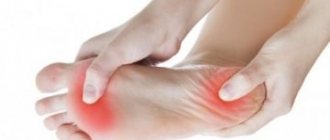
- Reflexes of the lower extremities. These include pathological foot reflexes, reactions to tapping with a hammer in the form of flexion or extension of the phalanges of the toes, and flexion of the foot.
- Reflexes of the oral muscles are pathological contractions of the facial muscles.
What does the presence of a bilateral Babinski sign indicate?
The presence of the described reflex in an adult, as mentioned above, is a sign of disruption of relationships at different levels of the spinal cord and brain. If this symptom is found on one side - left or right, then this is evidence of unilateral brain damage (myelopathy, stroke, etc.), and in situations where Babinski's symptom is present on both sides, we are usually talking about diffuse damage occurring in encephalopathy. It should be noted that this reflex remains quite persistent, with the exception of the diffuse form of the disease - in which case it appears and then disappears again.
By the way, the appearance of Babinski’s symptom in an adult can also be considered as an improvement, but, however, only if the patient has recovered from a state of shock or if the manifestations of the inflammatory process in the peripheral nervous system are subsiding.
What are asynergy tests?
They are carried out by specialists if cerebellar pathology is suspected as follows:
- from the initial position “lying on his back,” the patient is asked to cross his arms on his chest and rise, while a healthy person easily sits down, and with cerebellar disorders, a person’s legs or one leg rises upward, but he himself cannot sit down without help;
- from a “standing” position, a person is asked to lean on the palms of the doctor standing in front; when quickly removing his hands, the patient falls forward (a symptom described by Orzechowski). If a person is healthy, he will lean back slightly or stand exactly in place;
- the patient is unable to rise from a chair on his own due to the fact that the legs do not bend symmetrically at the knee joints;
- in a vertical position, a person cannot bend back and throw back his head, but falls on his back;
- When moving, the body lags behind the legs, and the patient gradually falls onto his back.
It should be noted that all of the above refers to manifestations of asynergy caused by the presence of organic brain lesions, but there is also asynergy of a functional nature, for example, dissociative motor disorders in patients with schizophrenia or other psychiatric diseases.
Therapeutic measures for asynergia boil down to finding out the cause of this condition and symptomatic therapy. It is necessary to carry out adequate treatment of the underlying disease.
Hereditary and idiopathic forms are not treated; the issues of social adaptation of patients and therapeutic exercises come to the fore. The prognosis is usually unfavorable.
What to do if Babinski reflex is detected
If an adult has a positive Babinski sign (that is, the thumb straightens), then additional studies are prescribed to determine the exact cause of this reflex and establish a diagnosis. In such cases, as a rule, the following is prescribed:
- clinical blood test;
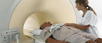
- CT and MRI of the spine and head;
- angiographic studies of the head.
If necessary, the patient undergoes a spinal puncture with the collection of cerebrospinal fluid for analysis, and other examinations. Only after studying all the results will the neurologist determine the true cause of the pathology and prescribe therapeutic measures.
It should be noted that ignoring the presence of Babinski's symptom in an adult can lead to his eventual development of paralysis, so the significance of this reflex is difficult to exaggerate.
A word about treatment
There is no treatment for asynergia syndrome as such, since it is not a disease, but a symptom of a cerebellar disease.
Specialists, as a rule, prescribe medications rich in vitamins, as well as massage procedures. This course helps maintain the overall tone of the body, strengthen the immune system, and warm up muscle tissue, but does not lead to absolute recovery of the patient.
You should not visit dubious home doctors when a symptom appears. After all, if modern medicine does not know how to eliminate a disorder, then a self-taught healer even more so. Take care of yourself!
Special role
Babinski's sign on the right or left is classified as pathological reflexes. That is, those that manifest themselves with structural or functional damage to different parts of the central nervous system. They, as you already understood, are used to diagnose nervous diseases.
When the intensity of manifestations of such reflexes is high, we talk about hyperreflexia, but if it is reduced to the point of loss, then we are talking about hyporeflexia, and if the manifestation of the reflex is uneven, then about anisoreflexia. But, by the way, if the decrease or increase in reflexes is symmetrical, then often this is not a sign of central nervous system diseases.
What are the reflexes?
In a normal situation, all reflexes appear and disappear in their own time. They are divided into congenital (unconditional) and acquired (conditional). The first ones are always with us, their loss means illness. As for the second group, some of them are lost with age, others appear. If the presence of a reflex is not typical for a given period of a person’s life or there is an increase (weakening) of it, this is a pathology that falls within the scope of activity of a neurologist.
Both unconditioned and conditioned reflexes can be pathological. Acquired (conditioned) reflexes are considered a pathology if they cause an inadequate response to a simple stimulus. The pathological nature of congenital reflexes is said if they do not fit into the neurological status of a given age or are inappropriate from a biological position.
In the practical work of neurologists, various unconditioned pathological reflexes are studied, indicating damage to the connections between the brain and spinal cord. Most often these are signs from the lower extremities. The response to the stimulus is manifested in the extension of the first toe (extensor reflexes) or the flexion of all toes (flexion group). The main pathological extensor reflex is the Babinski reflex.
Causes
Pathological reflexes can result from brain lesions and pathologies of the central nervous system, such as:
- damage to the cerebral cortex by infections, spinal cord diseases, tumors;
- hypoxia - brain functions are not performed due to lack of oxygen;
- stroke – damage to the blood vessels of the brain;
- Cerebral palsy (cerebral palsy) is a congenital pathology in which the reflexes of newborns do not fade over time, but develop;
- hypertension;
- paralysis;
- coma state;
- consequences of injuries.
Any diseases of the nervous system, damage to neural connections, or diseases of the brain can cause incorrect, unhealthy reflexes.
Notes
- Asynergy // Great Medical Encyclopedia / Ch. ed. B.V.Petrovsky. — 3rd ed. - M.: Soviet Encyclopedia, 1975. - T. II (Antibiotics - Becquerel). - P. 259. - 608 p.
- Pulatov A.M., Nikiforov A.S.
Propaedeutics of nervous diseases. - T.: "Medicine", 1979. - P. 108-120. — 368 p. — 20,000 copies. - Gusev E. I., Artaryan A. A., Lozhnikova S. M., Morgunov V. A., Speransky V. S., Fanarzhin V. V.
Cerebellum // Great Medical Encyclopedia / Ch. ed. B.V.Petrovsky. — 3rd ed. - M.:: Soviet Encyclopedia, 1981. - T. XV (Melanoma-Mudrov). - P. 359. - 576 p. — 150,000 copies.
| This is a draft article on neurology. You can help the project by adding to it. |
Tonic reflexes
Normally, tonic reflexes appear in children from birth to three months. Their continued manifestation even in the fifth month of life may indicate that the child has cerebral palsy. With cerebral palsy, congenital motor automatisms do not fade away, but continue to develop. These include pathological tonic reflexes:
- Labyrinthine tonic reflex. It is checked in two positions - on the back and on the stomach - and manifests itself depending on the location of the child’s head in space. In children with cerebral palsy, it is expressed in increased tone of the extensor muscles when lying on the back and flexor muscles when the child lies on the stomach.
- Symmetrical cervical tonic reflex. In cerebral palsy, it is manifested by the influence of head movements on the tone of the muscles of the limbs.
- Asymmetrical cervical tonic reflex. It manifests itself as increased muscle tone in the limbs when turning the head to the side. On the side where the face is turned, the extensor muscles are activated, and on the side of the back of the head, the flexor muscles.
With cerebral palsy, a combination of tonic reflexes is possible, which reflects the severity of the disease.