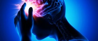
Unilateral focal lesions of half of the brainstem are accompanied by alternating syndromes (AS): dysfunction of the cranial nerves on the affected side and conduction disorders (motor, sensory) on the opposite side. Weber's syndrome (lesion in the area of the nuclei or fibers of the III nerve): symptoms of damage to the oculomotor nerve on the side of the lesion, contralateral central hemiplegia and central paralysis of the muscles of the face and tongue (involvement of the corticonuclear pathways to the nuclei of the VII and XII nerves). Benedict's syndrome (the lesion is located at the same level, but more dorsally, with the involvement of the nigra and red nucleus in the process with relative preservation of the pyramidal tract): on the side of the lesion - peripheral oculomotor palsy, on the opposite side - intentional temiteremor. With a more extensive lesion, damage to the conductors of the lemniscus medialis, passing outward from the nuclei of the oculomotor nerve, is possible, with the addition to the Benedict symptom complex of violations of superficial and deep sensitivity according to the hemitype on the side opposite to the lesion. Claude's syndrome is a combination of peripheral paralysis of the oculomotor muscles (nucleus of the third nerve) with impaired coordination of movements, hemihyperkinesis and muscle hypotonia on the opposite side (superior cerebellar peduncle). Nothnagel syndrome is observed with extensive lesions of the midbrain involving the nuclei of the oculomotor nerve, superior cerebellar peduncles, lateral lemniscus, pyramidal and corticonuclear tract and is characterized on the affected side by ataxia, peripheral paresis of m. oculomotorius, mydriasis and hearing impairment (usually on both sides), hemiparesis with central paresis of the muscles innervated by the VII and XII nerves. AS when the bridge is damaged. Millard-Hübler syndrome (damage to the nucleus or fibers of the VII nerve and pyramidal tract): peripheral paralysis of the facial muscles on the affected side and central hemiplegia on the opposite side. Fauville syndrome (a more extensive lesion involving a pathological process of the nucleus or fibers of the VI nerve): the Millard-Hubler symptom complex and paralysis of the abductor muscle of the eye (convergent strabismus, diplopia, failure to bring the eyeball outward). Brissot-Sicart syndrome is characterized by spasm of the facial muscles on the affected side (irritation of the nuc. fascialis), on the contralateral side - spastic hemiparesis (damage to the pyramidal tract). Raymond-Sestan syndrome is caused by a combined lesion of the posterior longitudinal fasciculus and the pontine center of gaze, the middle cerebellar peduncle, the medial lemniscus and the pyramidal tract, gaze paresis towards the lesion, ataxia, choresothetoid hyperkinesis, contralateral spasticity are observed
hemiparesis and hemianesthesia. Grene's syndrome (damage to the nucleus of the superficial sense of the V nerve and spinothalamic tract): prolapse on top of the senses
(pain and temperature) on the face according to a segmental type on the side of the lesion, contralaterally - prolapse on top. conductive type feelings on the torso and limbs. AS with damage to the medulla oblongata. Jackson syndrome is a lesion at the level of the nucleus of the hypoglossal nerve: on the side of the lesion there is peripheral paralysis of the tongue muscles, contralaterally there is central hemiplegia. Avellis syndrome is caused by a combined lesion of nuc. ambiguus or associated fibers of the IX, X nerves and pyramidal tract: on the side of the lesion there is paresis of the vocal cord, soft palate, trapezius and sternocleidomastoid muscles, on the contralateral side - spastic hemiparesis. Wallenberg-Zakharchenko syndrome: on the affected side - symptoms of nuc involvement in the process. ambiguus (paralysis of the soft palate and vocal cord), descending sympathetic fibers to the smooth muscles of the eye (p. Bernard-Horner), rope body (vestibular-cerebellar distribution), nuc. spinalis (loss of feelings on the face), on the opposite side loss of pain and temperature senses (damage to the fibers of the spinothalamic tract). The syndrome is observed when there is a violation of blood circulation in the posterior inferior cerebellar artery. Tapia syndrome is caused by a combined lesion of the nuclei or fibers of the XI, XII nerves and the pyramidal tract: on the side of the lesion, paralysis of the trapezius and sternocleidomastoid muscles and half of the tongue, contralaterally spastic hemiparesis. Volestein's syndrome is caused by a combined lesion of the oral part of the nuc. ambiguus and spinothalamic tract: on the side of the lesion there is paresis of the vocal cord, contralaterally there is hemianesthesia of the superficial sense. AS associated with damage to several parts of the brain stem include Gluck syndrome, which is characterized by combined damage to the II, V, VII, X nerves and the pyramidal tract; on the side of the lesion, paresis of the facial muscles with spasm, pain in the supraorbital region, decreased vision or amaurosis, difficulty swallowing, contralaterally - spastic hemiparesis.
Alternating syndromes
(lat. alternans - alternating; alternating paralysis, cross paralysis) - symptom complexes characterized by a combination of damage to the cranial nerves on the side of the lesion with conduction disturbances of movement and sensitivity on the opposite side. They occur with damage to one half of the brain stem, spinal cord, as well as with unilateral combined damage to the structures of the brain and sensory organs. Various AS can be caused by cerebrovascular accident, tumor, traumatic brain injury, etc. A gradual increase in symptoms is possible even without impairment of consciousness, with the spread of edema or progression of the process itself.
Bulbar alternating syndromes
- Avellis syndrome
(palatopharyngeal paralysis) develops when the nuclei of the glossopharyngeal and vagus nerves and the pyramidal tract are damaged. It is characterized on the side of the lesion by paralysis of the soft palate and pharynx, on the opposite side by hemiparesis and hemihypesthesia. (in the diagram - A) - Jackson syndrome
(medial medullary syndrome, Dejerine syndrome) occurs when the nucleus of the hypoglossal nerve and the fibers of the pyramidal tract are damaged. It is characterized by a paralytic lesion of half the tongue on the side of the lesion (the tongue “looks” at the lesion) and central hemiplegia or hemiparesis of the limbs on the healthy side. (in the diagram - B)
- Babinski-Nageotte syndrome
occurs with a combination of lesions of the inferior cerebellar peduncle, olivocerebellar tract, sympathetic fibers, pyramidal, spinothalamic tracts and medial lemniscus. It is characterized on the side of the lesion by the development of cerebellar disorders, Horner's syndrome, on the opposite side - by hemiparesis, loss of sensitivity (In the diagram - A). - Schmidt's syndrome
is characterized by combined damage to the motor nuclei or fibers of the glossopharyngeal, vagus, accessory nerves and pyramidal tract. It manifests itself on the side of the lesion as paralysis of the soft palate, pharynx, vocal cord, half of the tongue, sternocleidomastoid and upper part of the trapezius muscle, on the opposite side - hemiparesis and hemihypesthesia. (In the diagram - B).
Wallenberg-Zakharchenko syndrome
(dorsolateral medullary syndrome) occurs when the motor nuclei of the vagus, trigeminal and glossopharyngeal nerves, sympathetic fibers, inferior cerebellar peduncle, spinothalamic tract, and sometimes the pyramidal tract are damaged. On the side of the lesion, paralysis of the soft palate, pharynx, vocal cord, Horner's syndrome, cerebellar ataxia, nystagmus, loss of pain and temperature sensitivity of half the face are noted; on the opposite side - loss of pain and temperature sensitivity on the torso and limbs. Occurs when the posterior inferior cerebellar artery is damaged. Several options are described in the literature.
Prevention
In order for a child to be born without this pathology, the mother needs to be responsible about her lifestyle. It is known that Weber's disease develops if, during pregnancy, the fetus was exposed to negative influences, such as:
- smoking;
- alcohol and drug use;
- sexually transmitted infections (especially acquired during pregnancy);
- intoxication of various types;
- metabolic problems in the expectant mother.
So, the prevention of disease in a pregnant woman includes:
- Healthy eating, proper lifestyle.
- Taking into account medical recommendations.
- Examination before and during pregnancy.
- Walks, sleep, gymnastics.
Pontine alternating syndromes
- Raymond-Sestan syndrome
is observed with damage to the posterior longitudinal fasciculus, middle cerebellar peduncle, medial lemniscus, and pyramidal tract. It is characterized by paralysis of gaze towards the lesion, on the opposite side - hemihypesthesia, sometimes hemiparesis. (On the diagram - A) - Millard-Gubler syndrome
(medial pontine syndrome) occurs when the nucleus or root of the facial nerve and pyramidal tract are affected. It manifests itself on the side of the lesion as paralysis of the facial nerve, on the opposite side as hemiparesis. (In the diagram - B)
Brissot-Sicard syndrome
occurs when the nucleus of the facial nerve is irritated and the pyramidal tract is damaged.
It is characterized by facial hemispasm on the side of the lesion and hemiparesis on the opposite side (In the diagram - A). Foville syndrome
(lateral pontine syndrome) is observed with combined damage to the nuclei (roots) of the abducens and facial nerves, the medial lemniscus, and the pyramidal tract. Characterized from the side of the lesion by abducens nerve palsy and gaze paralysis towards the lesion, sometimes by facial nerve paralysis; on the opposite side - hemiparesis and hemihypesthesia (In the diagram - B).
Peduncular alternating syndromes
- Benedict's syndrome
(superior red nucleus syndrome) occurs when the nuclei of the oculomotor nerve, red nucleus, red nucleus-dentate fibers, and sometimes the medial lemniscus are affected. On the side of the lesion, ptosis, divergent strabismus, and mydriasis occur, on the opposite side - hemiataxia, eyelid trembling, hemiparesis without Babinsky's sign (In the diagram - B). - Foix syndrome
occurs when the anterior parts of the red nucleus and fibers of the medial lemniscus are affected without involving the oculomotor nerve. The syndrome includes choreoathetosis, intention tremor, and hemitype sensitivity disorder on the side opposite to the lesion. (in the diagram - A)
- Weber's syndrome
(ventral mesencephalic syndrome) is observed when the nucleus (root) of the oculomotor nerve and the fibers of the pyramidal tract are damaged. On the affected side, ptosis, mydriasis, and divergent strabismus are noted, on the opposite side - hemiparesis. (In the diagram - B) - Claude's syndrome
(dorsal mesencephalic syndrome, inferior red nucleus syndrome) occurs when the nucleus of the oculomotor nerve, superior cerebellar peduncle, or red nucleus is damaged. It is characterized on the affected side by ptosis, strabismus, mydriasis, and on the opposite side by hemiparesis, hemiataxia or hemiasynergia. (On the diagram - A)
Nothnagel syndrome
occurs with combined damage to the nuclei of the oculomotor nerves, the superior cerebellar peduncle, the lateral lemniscus, the red nucleus, and the pyramidal tract. On the side of the lesion, ptosis, divergent strabismus, and mydriasis are noted, on the opposite side - choreoathetoid hyperkinesis, hemiplegia, paralysis of the facial and tongue muscles.
Alternating syndromes associated with damage to several parts of the brain stem.
Glick syndrome
caused by damage to the optic, trigeminal, facial, vagus nerves and pyramidal tract. On the affected side - peripheral paralysis (paresis) of the facial muscles with their spasm, pain in the supraorbital region, decreased vision or amaurosis, difficulty swallowing, on the opposite side - central hemiplegia or hemiparesis.
Cross hemianesthesia
observed when the nucleus of the spinal tract of the trigeminal nerve is damaged at the level of the pons or medulla oblongata and the fibers of the spinothalamic tract. On the affected side there is a disorder of superficial sensitivity on the face of a segmental type, on the opposite side there is a disorder of surface sensitivity on the trunk and limbs.
Diagnostic measures
At the first slightest manifestation of Sturge-Weber syndrome, you should consult a doctor.
It is the specialist who will prescribe the correct diagnostic tests and make the most accurate diagnosis, and then prescribe effective treatment.
The diagnostic complex for encephalotrigeminal angiomatosis consists of several stages and procedures:
- First, the medical history and complaints of the disease are analyzed. It is important to know at what age symptoms began (seizures, weakness, headache, etc.). It is necessary to clarify whether any other family members had this disease.
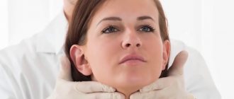
- A neurological examination of the patient will be required. At the same time, skin sensitivity and muscle strength of all extremities of the body are carefully assessed.
- You'll have to go see an ophthalmologist. Only he will measure intraocular pressure, examine the eyes, and study their internal structure. This is necessary to detect angiomas that may appear on the lining of the eye. And if the optic nerve is swollen, then the doctor will be able to determine the presence of increased intracranial pressure.
- Often, the attending physician refers the patient to a consultation with a medical geneticist.
- Computed tomography and MRI will help to see all the changes in the brain (those areas where the cortex is most thinned, where tumors and angiomas are present).
- Electroencephalography is a technique that allows you to evaluate the electrical conductivity and activity of the brain in various areas. Depending on the disease, this parameter may change. Especially if there is an epileptic focus (this is the main cause of convulsive seizures).
If surgical intervention is necessary to treat the disease, you will definitely need to see a neurosurgeon. He will not only give advice on preparing for surgery, but will also tell you all the stages and consequences of such treatment.
Slowly progressive Charcot-Marie amyotrophy is nevertheless very dangerous for human health and life. Tabes dorsalis is the most severe complication of neurosyphilis. What happens if you don’t start timely treatment for the disease.
Extracerebral alternating syndromes.
Alternating syndrome at the level of the spinal cord - Brown-Séquard syndrome
- a combination of clinical symptoms that develop when half the diameter of the spinal cord is affected. On the affected side, spastic paralysis, conduction disorders of deep (muscular-articular sense, vibration sensitivity, sense of pressure, weight, kinesthesia) and complex (two-dimensional-spatial, discriminatory, sense of localization) sensitivity, and sometimes ataxia are observed. At the level of the affected segment, radicular pain and hyperesthesia, the appearance of a narrow zone of analgesia and termanesthesia, are possible. On the opposite side of the body, there is a decrease or loss of pain and temperature sensitivity, and the upper level of these disorders is determined several segments below the level of the spinal cord lesion. When lesions occur at the level of the cervical or lumbar enlargements of the spinal cord, peripheral paresis or paralysis of the muscles innervated by the affected anterior horns of the spinal cord develops (peripheral motor neuron damage). Brown-Séquard syndrome occurs with syringomyelia, spinal cord tumor, hematomyelia, ischemic disorders of the spinal circulation, injury, spinal cord contusion, epidural hematoma, epiduritis, multiple sclerosis, etc. True half-damage to the spinal cord is rarely observed. Most often, only part of half of the spinal cord is affected - a partial variant in which some of its constituent signs are absent. In the development of different clinical variants, the localization of the pathological process in the spinal cord (intra- or extramedullary), its nature and characteristics of the course, the different sensitivity of the afferent and efferent conductors of the spinal cord to compression and hypoxia, individual characteristics of vascularization of the spinal cord, etc. play a role. - diagnostic value. The localization of the lesion in the spinal cord is determined by the level of impairment of surface sensitivity.
Asphymohemiplegic syndrome
(brachiocephalic arterial trunk syndrome) is observed with unilateral irritation of the nucleus of the facial nerve, vasomotor centers of the brain stem, and damage to the motor zone of the cerebral cortex. On the affected side there is a spasm of the facial muscles, on the opposite side there is central hemiplegia or hemiparesis. There is no pulsation of the common carotid artery on the affected side.
Vertigohemiplegic syndrome
is caused by unilateral damage to the vestibular apparatus and the motor zone of the cerebral cortex due to circulatory disorders in the system of the subclavian and carotid arteries with impaired circulation in the labyrinthine (vertebrobasilar basin) and middle cerebral arteries. On the affected side - noise in the ear, horizontal nystagmus in the same direction; on the opposite side - central hemiplegia or hemiparesis.
Optical-hemiplegic syndrome
occurs with unilateral damage to the retina, optic nerve, motor area of the cerebral cortex due to circulatory disorders in the internal carotid artery system (in the basin of the ophthalmic and middle cerebral arteries). On the affected side there is amaurosis, on the opposite side there is central hemiplegia or hemiparesis.
It is an alternating syndrome (a combination of cranial nerve damage on the side of the pathological process with a sensory disorder on the other side) that develops with focal damage to the lower part of the pons. Into the clinical picture. Thus, the nuclei and roots of the facial are involved. Abducens nerves, pyramidal tract, and also (not always) the medial lemniscus.
Types of angiomatosis
Vascular changes can occur in the liver, face, respiratory, and digestive organs. In addition, the disease can affect the skin, bones, nervous system
Please note that the local form of the disease is not dangerous to human life, it is benign
Scientists are still trying to unravel the reasons for the development of the syndrome. Most of all, scientists are inclined to believe that the disease is triggered by toxic influences when tissues and organs are formed, as well as gene mutations.
Often the pathology is caused by the fact that the expectant mother:
- He is fond of alcohol and drugs.
- Smokes a lot.
- Uses illegal drugs.
- Has extragenital pathology of the thyroid gland and diabetes mellitus.
Signs of the disease depend on where the disease is located. If the central nervous system is affected, it ends in blindness and dysfunction of internal organs.
The prognosis in most cases depends on the form of the disease. Most often it is favorable, but it happens that the disease progresses significantly and ends in severe disability.
The following types of angiomatosis can be distinguished:
- Diffuse – accompanied by systemic damage.
- Isolated – the retina is affected.
- Bacillary occurs in case of bacterial infection.
Sturge-Weber syndrome affects the eye vessels, facial skin, and lining of the brain. The pathology is considered congenital and quite rare. It is dangerous that the syndrome affects the eyes and brain - angiomas appear here, leading to disability.
The classic version is characterized by damage to the nervous system, eyes, and the appearance of bright red spots on the face. The disease can occur in the following ways:
- Skin angioma, leading to glaucoma, damage to the membranes of the brain.
- Damage to the nervous system.
Typically, one part of the face is affected. The spot is located under the eye on the forehead and can go down to the lip. When you press on the skin, it turns pale. In newborns, the spot on the face is pink, after a while it becomes cherry. Gradually, the formation begins to increase and completely affects one half of the face.
Symptoms
On the side of the pathological process, peripheral paresis/paralysis of the facial muscles, as well as the external rectus muscle of the eye, develops; on the opposite side, hemiparesis/hemiplegia of a central nature develops, sometimes in combination with impaired sensitivity (pain and temperature) according to the hemitype. The syndrome was described in 1858 by the neurologist A. Foville (1799-1879 years of life, country of residence - France).
Symptoms of this lesion are peripheral paresis (up to paralysis) of the facial muscles (on the side of the pathological focus) in combination with central hemiparesis (up to plegia). This pathological condition was described by the Frenchman A. Villard (years of life 1830-1915) in 1856, and also, somewhat later, in 1896 by the German doctor (regardless of the research data of his colleague) A. Gubler (years of life 1821-1897).
Alternating syndromes - dysfunction of the cranial nerves (III, YII, IX, X, XI, XII) and the cerebellum on the affected side in combination with hemiparesis or hemihypesthesia on the opposite side - alternating syndromes
or tetraparesis and internuclear ophthalmoplegia (brain stem damage).
Classification depends on the level at which the nuclei are affected:
- Bulbar (damage at the level of the medulla oblongata):
— Jackson syndrome is clinically manifested by a combination of peripheral paralysis of the hypoglossal nerve (XII) on the side of the brain lesion with spastic hemiplegia on the opposite side, in some cases, loss of muscle-articular and vibration sensitivity. Dysarthria, paresis of the laryngeal muscles, swallowing disorders, and cerebellar ataxia on the side of the lesion may be observed. Deviation of the tongue in the direction of the lesion, atrophy and tuberosity of half of the tongue, and fascicular twitching in it are observed. A common cause of the syndrome is thrombosis of the branches of a. spinalis anterior, so-called ah. Sulci bulbaris.
Wallenberg-Zakharchenko syndrome (facial hypoesthesia, Horner's sign (ptosis, miosis and enophthalmos), paresis of the pharyngeal muscles, cerebellar ataxia, weakness of the sternocleidomastoid and trapezius muscles on the affected side and hemihypesthesia (without facial involvement) on the opposite side (lateral medulla oblongata infarction)
2. Peduncular (lesion at the level of the cerebellar peduncles):
- Weber's syndrome is characterized by paralysis of the oculomotor nerve (III) on the affected side, contralaterally - central hemiplegia or hemiparesis of the limbs and trunk, as well as central paresis of the facial (VII) and hypoglossal (XII) nerves. It is possible that hemianopsia may occur if the lateral geniculate body is involved in the pathological process. Weber's syndrome is observed when the base of the cerebral peduncles is damaged due to stenosis of the posterior cerebral artery and its branches, with syphilitic endarteritis, aneurysms of the posterior cerebral artery, with tumors, basal leptopachymeningitis. It is possible that the pathological process, for example, syphilitic gumma, may be localized in the meninges with subsequent spread to the cerebral peduncle.
3. Pontine (damage at the level of the bridge):
- Foville syndrome. In this syndrome, damage to the nuclei of the facial and abducens nerves (VII, VI) is observed with phenomena of peripheral paresis of the facial muscles and external rectus muscle of the eye on the affected side, as well as hemiparesis or hemiplegia of the central type on the opposite side. Characterized by convergent strabismus due to the eye on the affected side. It occurs due to obstruction of the circumferential branches of the basilar artery.
- Millard-Gubler syndrome: damage to the 7th pair of the cranial nerve and the pyramidal tract.
Jackson's syndrome (medial medullary syndrome, Dejerine's syndrome) occurs when the nucleus of the hypoglossal nerve and the fibers of the pyramidal tract are damaged. It is characterized by a paralytic lesion of half the tongue on the side of the lesion (the tongue “looks” at the lesion) and central hemiplegia or hemiparesis of the limbs on the opposite side.
Avellis syndrome (palatopharyngeal paralysis) develops when the nuclei of the glossopharyngeal and vagus nerves and the pyramidal tract are damaged. It is characterized on the side of the lesion by paralysis of the soft palate and pharynx, on the opposite side by hemiparesis and hemihypesthesia.
Schmidt syndrome is characterized by combined damage to the motor nuclei or fibers of the glossopharyngeal, vagus, accessory nerves and pyramidal tract. It manifests itself on the side of the lesion as paralysis of the soft palate, pharynx, vocal cord, half of the tongue, sternocleidomastoid and upper part of the trapezius muscle, on the opposite side - hemiparesis and hemihypesthesia.
Wallenberg-Zach-Rchenko syndrome (dorsolateral medullary syndrome) occurs when the motor nuclei of the vagus, trigeminal and glossopharyngeal nerves, sympathetic fibers, inferior cerebellar peduncle, spinothalamic tract, and sometimes the pyramidal tract are damaged. On the side of the lesion, paralysis of the soft palate, pharynx, vocal cord, Horner's syndrome, cerebellar ataxia, nystagmus, loss of pain and temperature sensitivity of half the face are noted; on the opposite side - loss of pain and temperature sensitivity on the torso and limbs. Occurs when the posterior inferior cerebellar artery is damaged.
Babinski-Najaut syndrome occurs with a combination of damage to the inferior cerebellar peduncle, olivocerebellar tract, sympathetic fibers, pyramidal, spinothalamic tracts and medial lemniscus. It is characterized on the side of the lesion by the development of cerebellar disorders, Horner's syndrome, on the opposite side - by hemiparesis, loss of sensitivity.
Diagnostics
The presence and type of cross syndrome can be determined by examination by a neurologist. The data obtained make it possible to determine the topical diagnosis, that is, the localization of the pathological process. The etiology can be roughly judged by the course of the disease. Tumor processes are characterized by a progressive increase in symptoms over several months, sometimes days. Inflammatory lesions are often accompanied by general infectious symptoms (fever, intoxication). During a stroke, alternating symptoms occur suddenly, increase rapidly, and occur against the background of changes in blood pressure. Hemorrhagic stroke differs from ischemic stroke by a blurred atypical picture of the syndrome, which is due to the absence of a clear boundary of the pathological focus due to pronounced perifocal processes (edema, reactive phenomena).
To determine the cause of neurological symptoms, additional studies are carried out:
- MRI of the brain.
Allows you to visualize the inflammatory focus, hematoma, tumor of the trunk, area of the stroke, differentiate hemorrhagic and ischemic stroke, determine the degree of compression of the trunk structures. - TCD of cerebral vessels
. The most accessible, fairly informative method for diagnosing cerebral blood flow disorders. Detects signs of thromboembolism, local spasm of intracerebral vessels. - Doppler ultrasound of extracranial vessels
. Necessary in the diagnosis of occlusion of the carotid and vertebral arteries. - MRI of cerebral vessels
. The most informative way to diagnose acute cerebrovascular accidents. Visualization of blood vessels helps to accurately diagnose the nature, location, and extent of their damage. - Cerebrospinal fluid examination.
Lumbar puncture is performed if an infectious-inflammatory nature of the pathology is suspected, as evidenced by inflammatory changes in the cerebrospinal fluid (turbidity, cytosis due to neutrophils, the presence of bacteria). Bacteriological and virological studies can identify the pathogen.
Pontine alternating syndromes
Miya-Hubler syndrome (medial pontine syndrome) occurs when the nucleus or root of the facial nerve and the pyramidal tract are affected. It manifests itself on the side of the lesion as paralysis of the facial nerve, on the opposite side - hemiparesis.
Foville syndrome (lateral pontine syndrome) is observed with combined damage to the nuclei (roots) of the abducens and facial nerves, the medial lemniscus, and the pyramidal tract. Characterized from the side of the lesion by abducens nerve palsy and gaze paralysis towards the lesion, sometimes by facial nerve palsy; on the opposite side - hemiparesis and hemihypesthesia.
Raymond-Se Stan syndrome is observed with damage to the posterior longitudinal fasciculus, middle cerebellar peduncle, medial lemniscus, and pyramidal tract. It is characterized by paralysis of gaze towards the lesion, on the opposite side - hemihypesthesia, sometimes hemiparesis.
Brissot's syndrome occurs when the nucleus of the facial nerve is irritated and the pyramidal tract is damaged. It is characterized by facial hemispasm on the side of the lesion and hemiparesis on the opposite side.
Causes of the disease
There can be many reasons for this type of disease. Among the most common are:
- Blood supply disorders. This includes hemorrhagic and ischemic strokes, which are the most common causes of alternating syndromes.
- Neoplasms in the brain. If the brain stem is affected by a tumor, compression of the nerve endings occurs, which provokes the development of the disease.
- Inflammatory diseases that are localized in the trunk and act as an impetus for the development of pathology.
- Traumatic brain injury. Very often the disease occurs after damage to the skull.
Treatment
Treatment is aimed at eliminating the underlying pathology and includes many techniques:
- Conservative therapy. Patients are prescribed neuroprotective, decongestant and neuroprotective agents. For ischemic strokes, vascular therapy is carried out, for hemorrhagic strokes, calcium and aminocaproic acid preparations are prescribed.
- Neurosurgical treatment. In some particularly severe cases, it is performed for hemorrhagic stroke. The advisability of such an operation should be assessed by a neurosurgeon.
- Rehabilitation. After conservative therapy, patients must undergo a rehabilitation stage, which includes physical therapy, massage and other procedures.