At-risk groups
Typically, focal changes in the white matter of the brain of a dystrophic nature most often occur in old age. Most lesions appear during life and as a result of hypoxia and ischemia. People who lead a sedentary and unhealthy lifestyle are also susceptible to the disease. Genetic predisposition also plays a role. The risk group includes people suffering from high or low blood pressure, diabetes, rheumatism, obesity and atherosclerosis. In addition, emotional individuals prone to stress are at risk of developing pathology.
You may be interested: Inhalations with Miramistin in a nebulizer
The white matter of the brain coordinates all human activity. It is what connects different parts of the nervous system. White matter is necessary for the two hemispheres to work together.
Causes
Focal changes in the brain substance of a dystrophic nature occur in a number of diseases of different origins:
- Changes in vascular origin: atherosclerosis, migraine, hypertension, etc.
- Inflammatory diseases. Multiple sclerosis, Behcet's disease, Sjögren's disease, inflammatory bowel disease (Crohn's disease).
- Infectious diseases. HIV, syphilis, borreliosis.
- Intoxication and metabolic disorders, carbon monoxide poisoning, B12 deficiency.
- Traumatic processes associated with radiation therapy.
- Congenital diseases caused by metabolic disorders.
Channel PROGRAMMER'S DIARY
The life of a programmer and interesting reviews of everything. Subscribe so you don't miss new videos.
The occurrence of pathology is caused by impaired blood supply in any part of the brain. It is fraught with loss of function of brain tissue. The more blood flow has decreased, the more noticeable the consequences. An example is damage to the spinal or cerebral blood flow. Such violations progress slowly, but entail serious consequences.
Signs
The signs of focal changes in the brain substance of a dystrophic nature are also different. With focal changes, not the entire brain suffers, but only its individual parts. Tissue degeneration occurs when there is insufficient supply of nutrients necessary for the normal functioning of the body’s nervous systems. We are talking about proteins - the building material of the human body. Proteins break down into amino acids, which, in turn, stimulate the nervous system. It also requires fats and carbohydrates - the main sources of energy needed by every living creature.
You may be interested in: The act of defecation: mechanism, regularity, causes of violations
Of the vitamins, the brain needs B1 (activates its work), B3 (provides energy at the intracellular level), B6 (it is difficult to imagine metabolic processes without it, in addition, it is also a kind of antidepressant), B12 (promotes memory preservation and helps to stay alert) . All these vitamins can be obtained in sufficient quantities by creating the right diet.
Doctors' recommendations
Quit tobacco products or get rid of addiction. Do not drink alcohol or drugs. Move more, do exercises. The permissible intensity of physical activity is determined only by a doctor. Sleep 7-8 hours a day. Doctors advise increasing sleep duration when diagnosing such disorders.
You need to eat a balanced diet; it is preferable to develop a diet together with your doctor in order to take into account all the nutritional components that help with destructive processes in the brain. Neurons need to be nourished with useful substances.
Review other bad habits. It is better to get rid of regular stressful situations. It is better to change your job if it is a little stressful. Relax more often, choose the most suitable ways for this. Visit the doctor for examinations at the specified frequency in order to timely record changes in pathological processes and timely apply therapeutic techniques.
Nervous tissues are characterized by increased vulnerability. Neurons die when there is a lack of oxygen, so you need to treat your body with special attention.
Pathologies
Focal changes in the substance of the brain of a dystrophic nature most often take the form of pathologies such as:
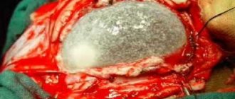
- A cyst is a small cavity that is filled with fluid. It often interferes with the normal functioning of neighboring areas of the brain, as it compresses blood vessels. Cysts are divided into intracerebral (cerebral) and arachnoid. The latter appears in the meninges. Its occurrence is promoted by the accumulation of cerebrospinal fluid and inflammatory processes. Cerebral occurs in place of dead brain tissue.
- Necrotic state of tissue - appears when the supply of important nutrients to areas of the brain for any reason deteriorates. Dead cells form so-called dead zones and are not regenerated.
- Hematomas and brain scars occur after severe trauma or concussion. Foci of this type lead to structural damage.
Vascular changes in the brain on MRI
MRI: ischemic zone during stroke (highlighted with a red oval)
If cerebrovascular pathology is suspected, special attention is paid to the condition of the arteries during MRI of the brain. The study always involves the introduction of contrast, and it is called magnetic resonance angiography. In urgent situations in case of vascular accidents, CT is performed, since X-ray diagnostics takes less time and clearly demonstrates the area of damage during hemorrhages, but after stabilization of health, magnetic resonance angiography is quite justified. The study shows atherosclerotic plaques, blood clots and aneurysms (protrusions), their localization, and wall deformation.
With an ischemic stroke, darkened and blurred areas of irregular shape are visible almost immediately on magnetic tomograms, appearing on a computer scan only at the end of the first day. The lesion is often unilateral. The outpouring of blood from a ruptured vessel gives an intense light color, but only in the first hour and a half from the moment of the disaster, and then becomes invisible on MRI, although it is clearly visualized using CT. As a consequence of a stroke, a fluid-filled pseudocyst is formed, and deformation of the nerve tissue appears. MRA is indispensable in the diagnosis of tumor angiogenesis. Increased vascularization of a pathological focus is always suspicious of a malignant neoplasm, which grows and feeds due to increased blood flow. If the vessels do not keep up with the growth of the tumor, areas of ischemia and necrosis appear.
Diagnostics
You may be interested in:Hot spring "Avan" in Tyumen: detailed information
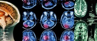
The complete picture of focal changes in the brain substance of a dystrophic nature is determined using an MRI study. This procedure allows you to see even small areas of transformation in the white matter. And they, in turn, lead to cancer and stroke.
Focal dystrophic lesions come in different sizes and differ in location. Based on this, the examination may show some types of disorders.
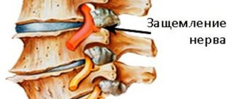
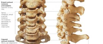
In the cerebral hemisphere, blockage of vital arteries is usually diagnosed due to abnormal development of the embryo or acquired atherosclerotic plaques. A herniated cervical spine is also detected.
Changes in the white matter of the brain indicate hypertension and congenital developmental anomalies. In other cases, numerous areas of brain pathology may indicate a pre-stroke condition, senile dementia, or epilepsy.
Sometimes doctors perform tests on a patient to detect the presence of cognitive impairment. That is, cognitive dysfunction. Such as orientation in space and time, understanding of external processes, the ability to remember information, drawing, writing, reading, etc.
Focal changes in the brain substance of a dystrophic nature can develop in three ways:
- In the first case, the disease is remitting in nature. Symptoms increase gradually, the condition worsens, and brain productivity decreases. But from time to time, remissions occur—temporary improvements in health, after which the patient becomes worse again.
- Progressive focal changes in the brain substance of a dyscirculatory dystrophic nature develop very quickly. Each stage of the disease takes no more than two years, which is considered a short period for organic brain lesions.
- Typically, the deterioration of a person suffering from focal changes lasts for many years, eventually leading to dementia.
It should be remembered that single focal changes in the brain substance of a dystrophic nature often appear in young people, and single damage to the white matter in an elderly person is considered normal. Structural disorders of the cerebral arteries of the atherosclerotic type appear in 50% of patients over 50 years of age. For the most part, hypertensive patients suffer from this. Therefore, you need to show the MRI result to a neurologist so that he can determine the severity of the disorders in the brain by comparing the MRI result and the clinical picture of the disease.
Prevention
Preventive measures should be taken by completely healthy people and when the first signs of circulatory abnormalities appear:
- Maintaining a daily routine;
- Adequate physical activity;
- Complete rest;
- Healthy lifestyle;
- Gymnastics, sports;
- Proper, balanced nutrition;
- Professional examination by a neurologist once a year.
Author of the article: neurologist Lidiya Rashidovna Magdoteva
Prevention of multiple focal changes in the brain substance includes:
- Maintaining an active lifestyle. After all, movement stimulates improved blood circulation throughout the human body and in the brain, in particular, and thereby reduces the risk of lesions in the brain.
- Proper and rational nutrition.
- Avoiding stress and other nervous situations. After all, constant nervous tension can be the cause of more than one disease. There is no need to overwork often, you should rest and relax more.
- Healthy and sound sleep is always the key to health. You need to spend at least 7-8 hours sleeping per day. If you experience insomnia or any other sleep pathologies, then your sleep time should be increased to 10 hours a day.
- It is necessary to conduct an examination in the hospital every year to identify hidden pathologies and diseases. If symptoms are detected that may indicate changes in the brain matter, then an MRI is required 2 times a year, as well as all necessary tests.
Everyone knows that it is always easier to prevent a problem in advance than to look for a right and proper solution later. The same goes for health. It is easier to carry out the necessary prevention than to treat the disease later.
Diet
In the early stages of this disease, it is enough to reconsider your lifestyle and diet, choosing a more gentle regimen and diet. In the diet, it is advised to reduce the consumption of animal fats, and it is better to completely replace them with vegetable ones. You should eat fish and seafood instead of fatty meat, and reduce the amount of salt in your diet. Fresh vegetables and fruits will be of great benefit.
Treatment
There are a huge number of focal anomalies, so each has its own cause. Treatment of brain pathologies is based on the destruction of those factors that led to the appearance of lesions in brain tissue. In addition to eliminating the underlying disease, the doctor may also prescribe vitamins and medications to help combat the deterioration of cerebral blood flow.
The treatment process depends directly on what somatic problems in the body led to the occurrence of lesions in the brain. For infections, for example, antibiotics are taken; for injuries, diuretics, decongestants, and anticonvulsants are taken. If the damage to the brain tissue was caused by a circulatory disorder, then nootropics and coagulants are prescribed.
Source
Stages of development
In comparison with other types of pathologies, focal transformations of the discircular type spread in several stages. Each has its own distinctive features. Therefore, specialists must first understand at what stage this disorder is in order to determine the optimal therapeutic technique.
In the initial stages, it is difficult to determine the presence of the disease, since the process of circulatory disorders in the head is poorly developed. In such a situation, the specific signs of the disorder are still not expressed, so diagnosis will be difficult. Patients also do not describe specific complaints.
The second stage is characterized by deterioration of the tissue in the brain, which gradually dies. Such processes are caused by problems with cerebral circulation. At the last stage, half of the brain matter dies, the functioning of the organ is disrupted, and recovery cannot be expected. In each patient, symptoms manifest themselves depending on the individual characteristics of the body.

