What is ependymoma
The human brain contains tissue called ependyma. This is a thin membrane that lines the walls of the ventricles of the brain and the spinal canal. Under the influence of various unfavorable factors, ependymal cells can undergo malignant changes. In this case, tumors form in the tissue, called ependymomas. They are divided into several types:
- Subependymoma. This is a grade 1 tumor. It is growing, but very slowly.
- Myxopapillary ependymoma. This tumor is located in the spinal cord canal. It also tends to grow slowly.
- Ependymoma grade 2. This tumor is characterized by faster growth than the previous two.
- Anaplastic ependymoma grade 3. This is a malignant tumor that grows rapidly. It can metastasize from the brain to the spinal canal. Usually its appearance is preceded by a tumor of the 2nd degree of malignancy.
We will consider the last type of ependymoma in more detail.
Ependymoma of the spinal cord and brain: treatment, rehabilitation, prognosis
The human brain is made up of a special tissue called ependyma. It is she who lines the cavities in the brain. This tissue, under the influence of various factors, tends to grow in the form of tumors. These neoplasms are called ependymomas.
This pathology can occur in any person, and this disease has no age restrictions. Most of the formations are benign in nature, that is, those that do not metastasize.
The few cases of malignant ependymomas are very insidious and life-threatening for the patient.
These tumors are characterized by slow growth. Gradually growing, they compress the brain, sometimes displacing spinal or cerebral tissues, but without penetration into their structure.
Some types of tumors can grow to other areas of the brain, entering them with the cerebrospinal fluid. Malignant varieties are characterized by very caustic metastasis, often even beyond the nervous system structures. If a relapse occurs, a new tumor appears in or near the old site.
Localization and classification of malignancy
The disease occurs due to mutation of the ependymal cells that line the spinal canal and ventricles of the brain. Consequently, the neoplasm occurs precisely in these zones.
According to statistics:
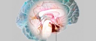
- In 60% of all cases, ependymoma is diagnosed in the region of the 4th ventricle of the brain in the posterior cranial fossa. From this location, the mass can often grow into the spinal cord, upper cervical spine, or cerebellum. infratentorial in medicine .
- Approximately 30% of such neoplasms can form in the lateral ventricles of the cerebrum or in the third ventricle of the diencephalon. This type is called supratentorial .
- In 10% of cases, the tumor occurs in the spinal canal. And this type of disease is called intraspinal .
The properties and structure of the tissue of a neoplasm (epindymoma) are very diverse. The tumor may have:
- Low degree of malignancy . Which indicates a low rate of cell division. Consequently, education itself is growing very slowly.
- High degree of malignancy . In this case, the rate of division of pathological cells is higher. The structure of the tumor is completely different. The tumor grows aggressively and rapidly.
Classification of ependymomas according to WHO (World Health Organization):
- Subepindymoma (grade I malignancy). They grow and divide very slowly.
- Myxopapillary epindymoma (grade I malignancy). The tumor grows slowly with a low level of malignancy, most often forming in the spinal canal.
- Tumor of the IInd degree of malignancy . Tumors grow slowly but tend to grow aggressively. Cancer cells come in several varieties.
- Anaplastic ependymoma of the III degree of malignancy. Pathology characterized by a high level of aggressiveness.
Unfortunately, it is very difficult to draw the exact boundary between grades II and III of malignancy. This makes it difficult to make an accurate diagnosis without appropriate testing. Doctors in such cases rely solely on tissue histology.
The reliable cause of the development of the pathology in question has not yet been fully established. Histology shows only the presence of a very active virus of the SV40 form in the tumors. However, this fact requires further scientific research in order to determine the etiology of this disease.
The risk group includes:
- patients with a genetic predisposition to the disease;
- workers whose field of activity involves hazardous substances;
- patients who received a high dose of radiological radiation;
- infected with a highly oncogenic virus.
Symptoms and manifestations
The clinical picture of the disease manifests itself in different ways, this is explained by the zone of tumor localization.
If the spinal cord area is affected
If the ependymoma is localized in the spinal cord, the patient feels:
- impaired sensitivity in relation to tactile touch, pain, heat and cold; unpleasant sensations can be felt on one side of the spine or on both;
- as the tumor enlarges, paresis or paralysis develops;
- there is no radicular syndrome;
- due to impaired functionality and sensitivity, the patient may exhibit clumsiness, changes in motor skills, gait, and so on.
The tumor has affected the brain
If the localization of ependymoma is the brain, then the patient begins to be bothered by the following symptoms:
- headaches of a paroxysmal nature, especially often associated with physical activity, changes in temperature or conditions, changes in posture, sometimes occurring for no reason;
- vomiting, most often unfounded, accompanied by or without headache;
- the patient begins to be forced to change his position, in which his headaches become less intense;
- Bruns syndrome – dizziness occurs with sudden turns of the neck or changes in body position;
- sometimes a convulsive muscle contraction may be visible, a bit like a banal twitch;
- in advanced phases of pathology development, symptoms are supplemented by mental disorders in the form of stupor, psycho-emotional inhibition, lethargy, and so on.
Tumor process in the cerebral ventricles
When the ependymoma is localized in the lateral ventricles (brain), the course of the disease can be latent for a long time. The first signs may become apparent only in the later stages.
In this case, the patient also develops concomitant diseases, such as intracranial hypertension and the like.
Symptoms manifest as:
- disorders of consciousness, psyche or memory;
- the appearance of hallucinations;
- spatial disorientation;
- apathy or lethargy.
Diagnosis and treatment
Diagnosis of the tumor in question is based on a thorough examination of the patient, which includes:
- assessment of reflex function;
- skin sensitivity;
- some types of neurological tests;
- A vision assessment may also be required;
- electroneuromyography;
- Ultrasound;
- MRI and other related brain studies.
Treatment of ependymoma can be complex - surgical and (or) radiation. The use of a specific method of therapy depends on the severity and stage of development of the tumor process at the time of diagnosis.
Chemotherapy in this case is considered ineffective and is used exclusively in extreme cases, when it is not about treatment, but about prolonging the life of a patient with a relapse.
Surgery is considered the standard of treatment. It involves craniotomy, where the skull is opened to gain access to the area affected by the tumor. After this, the tumor is excised. In fact, this method is also not considered ideal.
It does not give an absolute guarantee that new formations will not appear in the same place. For this reason, surgery is combined with radiation therapy. Thanks to this, it is possible to reduce the size of the formation.
Radiation therapy can be used before and after surgery. In cases where surgery cannot be used, radiation therapy is considered the only treatment option.
Be careful, video of the operation! Click to open
Problems of the recovery phase
Brain surgery in itself is considered a difficult ordeal for the patient; not all patients manage to survive after it. The rehabilitation period is even more difficult, especially if the patient is a child.
Problems that patients may encounter after surgery:
- for children – growth retardation is possible;
- disability;
- depression, fatigue;
- development of hydrocephalus;
- hearing and speech disorders;
- convulsions are possible;
- change in perception of the environment.
If similar signs appear, the patient is recommended to undergo special treatment and regularly visit doctors to detect metastases or other pathologies.
Prognosis and survival
Statistics show that the mortality rate in the first days after surgery in patients who have had a cerebral ependymoma removed is approximately 8%, but the overall prognosis for survival is very negative.
If we take into account the five-year survival rate, then its percentage increases to 60, and only if surgical treatment is performed.
The prognosis is influenced primarily by the degree of resection. Successfully performed resection improves the prognosis, its percentage is approximately 80%.
If the removal of cancer cells is unsuccessful or incomplete, the tumor will form again.
Causes
Experts cannot determine the exact cause of the development of anaplastic ependymoma of the brain and spinal cord. It is possible to identify only risk factors that increase the likelihood of malignant tumors. These include:
- contact with carcinogenic substances;
- work in hazardous production;
- exposure to radiation;
- infection with oncogenic microorganisms (some strains of HPV, herpes virus, cytomegalovirus);
- excessive exposure to sunlight;
- hereditary predisposition to cancer.
Medical scientists have discovered a special type of virus, SV40, in anaplastic ependymoma cells. This microorganism was in an active state. However, at present, science does not know how pathogenic such a virus is and whether it plays any role in the occurrence of tumors.
Symptoms
The symptoms of the disease directly depend on the size and location of the tumor. The severity of symptoms is also influenced by the degree of damage to healthy tissue. Among the characteristic symptoms are:
- gagging, regardless of food intake,
- pressing headaches of varying degrees of intensity,
- uneven gait
- dysfunction of the lower and upper extremities,
- fog,
- increased eye pressure, manifested by pain in the eyeballs,
- attacks of irritability, outbursts of anger,
- mental disorders,
- lethargy, excessive activity.
Important! Symptoms of the third and fourth degrees of ependymoma are considered the most dangerous.
Symptoms
Manifestations of the disease depend on the location of anaplastic ependymoma. If the tumor is located in the spinal canal, the following symptoms are noted:
- Various areas of the body lose sensitivity to heat and cold, as well as pain.
- There is pain in the spine.
- The patient's gait changes. Movements become awkward and clumsy.
- If the tumor is large, paralysis of the limbs is possible.
If the tumor is located in the brain, two types of symptoms may occur:
- General cerebral. These manifestations are associated with intracranial hypertension due to compression of brain tissue by ependymoma and accumulation of cerebrospinal fluid.
- Focal. Depending on the location of the tumor, signs of dysfunction of one or another part of the brain appear.
With any location of anaplastic ependymoma of the brain, the patient experiences the following cerebral symptoms:
- attacks of severe headache accompanied by vomiting;
- dizziness with sudden movements;
- increased pain with changes in body position and physical activity;
- seizures.
This clinical picture indicates intracranial hypertension.
Focal symptoms are varied and depend on the location of the tumor. If anaplastic ependymoma compresses the cranial nerves, then the patient experiences deterioration of hearing and smell, slurred speech, numbness of part of the face, impaired balance and coordination of movements.
If ependymoma is located in the lateral ventricles of the brain, then in the early stages the disease can be asymptomatic for a long time. Signs of increased intracranial pressure appear already at a late stage of the pathology. Patients also experience mental disorders:
- hallucinations;
- memory impairment;
- apathy;
- depression;
- poor spatial orientation.
Very often, the location of the tumor is the posterior fossa of the skull. The patient complains of double vision. Signs of vestibular ataxia appear. It is difficult for a person to maintain balance not only when walking, but also when sitting. The patient experiences dizziness even when at rest.
Ependymoma of the brain - prognosis, treatment, causes
Ependymoma of the brain occurs regardless of age. More often it manifests itself as benign, but cases of malignancy are known.
Ependyma is brain tissue that grows under the influence of certain factors and can develop into a neoplasm.
Causes
There is a theory that the development of the disease is caused by the SV40 virus. Polyomavirus has been found in a subset of patients with ependymoma. However, the US National Institute conducted a number of studies and proved that the SV40 virus is not capable of causing the development of ependymoma in humans.
The following factors are identified that influence the occurrence of ependymoma:
- Complicated genetic background.
- Chronic toxemia.
- Hormonal imbalance.
- Radioactive radiation.
- Various types of infections.
- Injuries.
Localization
The tumor is localized in that part of the brain that can easily provoke damage to nearby and distant parts of the brain, which is why the disease is considered dangerous.
Since, under the influence of an increase in the size of the tumor, destruction and compression of tissue occurs, the first symptoms begin to appear. If the disease begins to progress, general cerebral symptoms will occur: cerebral edema, generalization of hemodynamic disorders. There are cases when the tumor is located in a “silent” area. Then the pathology occurs latently, but the disease progresses.
Ependymoma of the brain
Ependymoma of the brain is accompanied by characteristic symptoms:
- The patient feels a severe headache, which occurs with increased physical activity, changes in climatic conditions and sleeping positions. Please note: an attack may occur for no reason.
- Sudden onset of vomiting, severe headache.
- When you try to change your body position, you experience severe dizziness.
- Development of seizures.
- If the disease is in its final stage, mental abnormalities may occur. The patient's actions are slowed down, the patient feels lethargy, drowsiness, and apathy.
We recommend reading Pituitary tumor - causes, types, treatment
Ependymomas of the spinal cord
If a spinal cord ependymoma is diagnosed, the patient experiences:
- backache;
- decreased sensitivity of the spinal conduction type;
- leg paralysis;
- dysfunction of the pelvic spine.
Ependymomas in the cauda equina region
With the development of ependymoma in this part, the pain is similar to the symptoms of spinal diseases:
- an unpleasant tingling sensation in the legs, accompanied by a decrease in skin sensitivity;
- lumbar pain;
- pathological change in gait due to impaired functionality of the lower extremities;
- radicular syndrome;
- pain in the leg, buttock;
- increased pain when trying to take a horizontal position;
- decreased functionality of the intestines and bladder.
Ependymomas of the lateral ventricles
The disease has a latent course for a long time. Symptoms appear only in the fourth stage, which is very dangerous. Let's look at what factors can arise as a consequence of ependymoma in the area of the lateral ventricles:
- the patient does not remember well;
- there are mental disorders;
- hallucinations occur;
- spatial orientation is impaired;
- apathy, lethargy.
Classification
The World Health Organization has put forward the following classification of the development of the disease:
- Subepindymoma (first stage of malignant tumor). The neoplasm grows slowly and looks like a node. Localized under the ependymal tissue.
- Myxopapillary ependymoma (the degree of malignancy corresponds to the previous paragraph). Practically does not appear, develops slowly. The level of malignancy is low.
- Tumor of the second degree of malignancy. The disease tends to progress.
- Anaplastic ependymoma stage 3 malignancy. A dangerous period characterized by rapid growth of the tumor. Has a tendency to metastasize.
- True ependymoma is the most common type. Characterized by the formation of pathogenic cells around blood vessels.
Symptoms
The symptoms of the disease directly depend on the size and location of the tumor. The severity of symptoms is also influenced by the degree of damage to healthy tissue.
Among the characteristic symptoms are:
- gagging, regardless of food intake;
- pressing headaches of varying degrees of intensity;
- uneven gait;
- dysfunction of the lower and upper extremities;
- fog;
- increased eye pressure, manifested by pain in the eyeballs;
- attacks of irritability, outbursts of anger;
- mental disorders;
- lethargy, excessive activity.
Important! Symptoms of the third and fourth degrees of ependymoma are considered the most dangerous.
Diagnostics
If the patient shows the first signs of the disease, the doctor issues a referral for an MRI or CT scan of the brain. If there are symptoms in infants, babies are prescribed neurosonography.
Adult patients are referred for electroencephalography and ophthalmoscopy. The doctor may additionally prescribe a number of examinations:
- Ultrasound;
- vision test;
- collection of data on skin sensitivity;
- diagnostics of reflex function;
- examination by a neurologist.
The final stage of diagnosis is histological examination of pathological tissues. The biomaterial is collected intraoperatively.
Treatment
Doctors recommend several methods of therapy, but note in advance that each person needs a different approach.
Surgical intervention
In more than half of the cases, experts advise using the surgical method. The patient has the right to refuse surgery only if the tumor is in a hard-to-reach place, because then the risk to life would be too high.
The goal of surgery is to remove most of the tumor while preserving healthy tissue, that is, performing the operation in such a way that the patient’s condition does not worsen afterward. But here, too, some problems arise, since individual parts of the tumor can affect the further progression of the ependymoma.
Chemotherapy
This method is a combination of medications that will help remove the tumor. This treatment takes place in a hospital after surgery.
This method is recommended for children under three years of age, since surgery cannot yet be performed at this time.
Radiation therapy technique
Modern technologies will help get rid of malignant cells. Such therapy is needed if it was not possible to get rid of ependymoma surgically. The course of treatment is drawn up by a professional for each patient individually, since age, stage of tumor development and other factors are important here.
After using radiation therapy, the following consequences may occur:
- nausea, lack of appetite, vomiting;
- hair loss;
- change in speech.
Radiosurgery
Among the positive properties of radiosurgery are:
- During the procedure, low blood loss is observed, which cannot be said about surgical methods, after which a long rehabilitation period is required. The procedure takes 1 day and is performed on an outpatient basis.
- The operation is painless.
- Healthy tissues will not be affected or damaged.
- The therapy is effective. Pathological cells die, and the body does not suffer from the effects of radiation.
We recommend reading: What is a teratoma, its symptoms, types and location
Prognosis and survival
The prognosis for adults with successful therapy is favorable: five-year survival rate is observed in 67-80%.
With anaplastic ependymoma, the situation is different, since the tumor is characterized by rapid development. Child survival rate is low.
Brain ependymoma is a rather complex disease, since the exact causes of the development of the pathology are unknown. This is considered to be a polyetiological disease. If signs of pathology appear, you should immediately visit a doctor.
Ependymoma of the brain - prognosis, treatment, causes Link to main publication
Source: https://rakuhuk.ru/opuholi/ehpendimoma-golovnogo-mozga
Features of the disease in children
Anaplastic ependymoma of the brain is more common in children than in adults. More than half of the cases occur under the age of 5 years. In children, the pathology is usually more severe than in adults.
Anaplastic ependymoma in a child is accompanied by the following symptoms:
- movement coordination disorder;
- headache with nausea and vomiting;
- unsteady gait;
- tearfulness, capriciousness;
- hearing loss;
- delayed growth and development.
Such manifestations should alarm parents. In childhood, it is very important to diagnose the disease as early as possible, since the tumor grows quickly.
Problems of the recovery phase
Brain surgery in itself is considered a difficult ordeal for the patient; not all patients manage to survive after it. The rehabilitation period is even more difficult, especially if the patient is a child.
Problems that patients may encounter after surgery:
- for children – growth retardation is possible;
- disability;
- depression, fatigue;
- development of hydrocephalus;
- hearing and speech disorders;
- convulsions are possible;
- change in perception of the environment.
If similar signs appear, the patient is recommended to undergo special treatment and regularly visit doctors to detect metastases or other pathologies.
Diagnostics
Treatment of grade 3 anaplastic ependymoma is carried out by an oncologist and a neurologist. The reason for the diagnosis is the patient’s complaints of headaches with vomiting and seizures. The following examinations are prescribed:
- MRI and CT scan of the brain or spinal cord;
- electroencephalogram;
- angiography of the vessels of the head and spine;
- myelography (study of the movement of cerebrospinal fluid using a contrast agent).
Ventriculoscopy is also performed. This is a complex endoscopic procedure that allows you to evaluate the condition of the 3rd and 4th ventricles. It is in these areas that anaplastic ependymoma is most often localized. This study is done under general anesthesia. Thin tubes are inserted into the cranial cavity, with cameras attached to the ends. The image is shown on a large screen. In this way, the doctor can examine in detail the condition of the ventricles of the brain.
In childhood, MRI and CT scans of the brain are most often performed. These methods do not involve radiation. Infants undergo ultrasonography and neurosonography through an open fontanel. Additionally, a consultation with an ophthalmologist with an examination of the fundus is prescribed. If necessary, a lumbar puncture is performed to collect cerebrospinal fluid for analysis. This allows you to determine the area of tumor spread.
Diagnosis
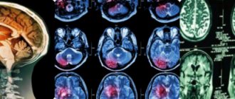
Ependymomas are diagnosed using imaging tests such as computed tomography (CT) or magnetic resonance imaging (MRI). If a child is admitted to the emergency department, they will first have a CT scan. A CT scan uses X-rays to take a series of pictures of the brain from different angles. Before the CT scan is performed, the child may be given intravenous contrast or anesthesia. CT scans are very quick, but young children may need to be sedated to lie still during the test. If a CT scan shows a possible tumor, the child is given an MRI to confirm the diagnosis. It provides clearer images for diagnosing ependymoma. MRI scans use magnets to take detailed pictures of the brain from different angles. A chemical called gadolinium is injected into a vein before an MRI to help certain parts of the brain show contrast.
On MRI, ependymoma is visible due to contrast. The tumor is usually located along the ventricles and may extend through the openings between the ventricles. There may also be evidence of obstructive hydrocephalus on MRI due to blockage of CSF flow by the tumor.
While most ependymomas are localized, sometimes tumors can spread to other parts of the brain or spine. Children with ependymomas usually have an MRI of the spine to make sure there is no tumor along the spine. Children usually also require a lumbar puncture to look for tumor cells in the cerebrospinal fluid. A lumbar puncture is a small needle inserted into the lower back to obtain a sample of cerebrospinal fluid. A specially trained doctor, a pathologist, will look at cells from the cerebrospinal fluid under a microscope to see if the tumor has spread.
Although ependymomas are usually detected on MRI, the diagnosis cannot be officially made until a biopsy or surgical removal of the tumor is performed. A biopsy is when the surgeon takes a small piece of tissue from the tumor. The pathologist will look at the tumor cells under a microscope to determine the type of tumor.
Treatment
Ependymoma is not subject to conservative therapy. The tumor must be completely removed. Therefore, the patient is indicated for neurosurgical surgery with craniotomy. This is a rather difficult intervention.
The tumor is often located in such a way that it is difficult for a neurosurgeon to get to it. If complete removal of the tumor cannot be done, bypass surgery is performed. Drainage tubes are installed for the outflow of cerebrospinal fluid. This helps reduce the manifestations of intracranial hypertension.
In some cases, the CyberKnife device is used to remove the tumor. This is a non-invasive radiosurgery method. The tumor is destroyed using radioactive radiation. There is no need to make an incision or open the skull.
Cerebral ependymoma is prone to recurrence. Therefore, in order to prevent re-growth of the tumor, it is necessary to undergo radiation therapy sessions.
Irradiation is contraindicated for children. Therefore, after removal of the tumor, they are prescribed a course of chemotherapy with cytostatics. The drugs Carboplatin and Cisplatin are used.
Consequences of surgery and radiation therapy
Rehabilitation after tumor removal and radiation therapy is usually long and difficult. In the postoperative period, patients may experience depression, seizures, memory, vision and hearing impairments, and changes in gait. Children experience delayed growth and development. Most patients experience nausea and hair loss. The human body is usually severely weakened by surgery and radiation.
The recovery period should be supervised by a specialist. During rehabilitation, you must regularly visit your oncologist and inform him of any changes in your health.
How to reduce risks?
Unfortunately, there are currently no preventive measures to prevent the development of this disease. But you can reduce the likelihood of the formation of any tumor by generally accepted principles of a healthy, fulfilling lifestyle:
- give up harmful habits;
- review your diet, start eating right;
- reduce exposure to harmful environmental influences to a minimum;
- undergo medical examinations in a timely manner;
- Work regularly to improve your immune system.
All patients, without exception, who have been treated for this pathology are required to regularly visit oncology centers in order to timely detect new tumors or relapse of existing ones.
Forecast
The prognosis of anaplastic ependymoma is always very serious. The outcome of the disease largely depends on the chosen method of therapy. If treatment is limited to surgery only, then about 8% of patients die immediately after surgery. Then, during the first 5 years after tumor removal, about 40% of patients die.
However, the prognosis for life becomes more favorable with complex treatment. If surgery is supplemented with chemotherapy and radiation therapy, the survival rate is about 80%.
From this we can conclude that after removal of the tumor, the patient must undergo an additional course of treatment with chemotherapy drugs and attend radiation therapy sessions. In this case, patients may experience side effects from aggressive treatment methods, but only an integrated approach can save the patient’s life.
What are the chances of being cured from ependymoma?
The chances of recovery depend to a very large extent on how much of the tumor could be removed during surgery.
In children and adolescents who have the entire tumor removed during surgery and then irradiated to the tumor region, survival rates reach 60 to 75% five years after the end of treatment. 10 years after the end of treatment, medical statistics give figures from 50 to 60%, if the disease did not continue to grow. If the tumor could not be completely removed and the child also received radiation to the tumor region, then medical statistics on 10-year survival rate indicate only 30 to 40% of cured children.
If the disease returns, that is, the child experiences a relapse of ependymoma, then specialists, as a rule, once again check whether it is possible to undergo a new operation and radiation. Practice shows that today a new special irradiation technique is available (more precisely, radiosurgical irradiation, or stereotactic radiosurgery [stereotactic method]), which can prolong life time. In addition, recurrent ependymomas are sensitive to chemotherapy drugs. Therefore, this form of treatment can also improve the prognosis of children and adolescents with recurrent ependymoma.
A necessary note: when in the text we give figures on the survival rate of children with ependymoma, we present only statistics. Data from statistics accurately and reliably describe in numbers all children and adolescents with this type of brain tumor. But no statistics can predict whether a particular child will recover or not.
When we talk about recovery, here this term must first of all be understood as “absence of a tumor.” Because modern treatment methods, although they can save a child from a malignant tumor, are usually associated with unwanted complications and late consequences. Therefore, doctors usually monitor all children for a long time after treatment. If necessary, children undergo intensive rehabilitation [rehabilitation].
Prevention
Specific prevention of ependymoma has not been developed. Medicine does not know the exact reasons for the formation of such a tumor. You can only reduce the risk of malignant neoplasms by using the following measures:
- When working in hazardous industries, undergo regular preventive inspections.
- Avoid excessive sun exposure.
- Timely detect and cure papillomatosis, herpetic diseases, cytomegaly and other pathologies caused by oncogenic viruses.
- If the patient’s closest relatives have had cerebral tumors, then the person needs to be regularly examined by a neurologist, as well as have an MRI of the brain.
It should be remembered that attacks of headaches with nausea can be a sign of a dangerous cancer disease. Therefore, if such symptoms occur, you should immediately consult a doctor.
Factors of occurrence
The development of ependymoma in the brain occurs under the influence of general factors that provoke tumor formation. A type of virus called SV40 has been found in malignant cells, but its role is not exactly clear.
Probable causes:
- genetic predisposition;
- long and regular contact with chemicals and metals;
- the presence of oncogenic viruses in the body;
- irradiation.
The genetic factor has been proven by the frequent combination of ependymomas with familial intestinal polyposis, endocrine neoplasia syndrome and Recklinghausen's neurofibromatosis.