Symptoms
Common to all types of edema, they are divided into two groups: focal and stem.
Focal symptoms appear when swelling is localized in a specific area of the spinal cord. In this case, the functions of this area are disrupted.
Stem symptoms are manifested by impaired blood circulation and breathing, decreased pupillary response.
A characteristic sign of shock is flaccid plegia of the limbs. Hypothermia and bradycardia develop.
The consequences of shock are acts of urination and defecation, a drop in blood pressure, and the absence of vascular reactions. The duration of the shock can range from several hours to several weeks.
With such a serious condition as spinal cord edema, symptoms can be varied, often protracted.
Treatment
First of all, eliminate the cause of the swelling.
It should be taken into account that morphological and physiological changes do not stop after the end of exposure to the traumatic factor. Specific treatment is aimed mainly at reducing the amount of edema. This is achieved by using drugs that normalize electrolyte balance (Brinaldix, Veroshpiron), drugs for forced diuresis (Lasix, Furosemide), drugs that reduce the production of CSF (Diacarb, Acetazolamide). You should not use osmotic diuretics (Mannitol), because there is a high probability of “recoil” symptoms.
The use of glucocorticosteroids (Dexamethasone, Hydrocortisone, Dexaven, Cortef). The dosage depends on the severity of symptoms - 8 - 20 mg per day. These drugs stabilize cell membranes, the endothelium of small vessels, preventing the accumulation of catecholamines and lysosomal disorders in the cells of the damaged brain.
The use of nootropic agents as membrane protectors. These include Vinpotropil, Gammalon, Lucetam, Nootropil, Piracetam.
The use of hyperbaric oxygen therapy increases oxygen pressure into the cells and tissues of the spinal cord and improves blood flow to the affected areas.
Some clinics use lidocaine, dopamine, barbiturates, and hyaluronidases to suppress auto-destructive processes.
Vitamins - Cyanocobalamin, Thiamine, Pyridoxine, Ascorbic acid - normalize metabolism, improve blood circulation in capillaries , and as a result, increase the transport of oxygen and nutrients to brain tissue.
For paralysis, muscle relaxants (Pancuronium, Tubocurarine, Metokurine) will be used.
You cannot use vasodilators (Dibazol, Nitroglycerin), calcium ion antagonists, drugs such as Aminazine, Reserpine, Droperidol.
Prevention
When spinal cord swelling occurs, the consequences are varied.
Changes and disturbances may occur in any part or organ of the body. The ability to restore the functionality of these organs depends on many factors. Rehabilitation after treatment is of great importance. Prevention is to prevent the development of complications. During bed rest, you need to determine the state of urination and find an adequate method for removing urine. In this case, the rules of antisepsis and asepsis must be observed to avoid infection.
Prevention of contractures is carried out by therapeutic exercises and massage, and the use of orthopedic techniques.
Prevention of pulmonary inflammatory complications involves normalizing respiratory functions, inhalations, therapeutic exercises, and vibration massage.
Experts advise not to neglect physiotherapeutic procedures to stimulate recovery processes and therapeutic exercises even after discharge from the hospital.
source
Purpose of joint water
Fluid begins to accumulate in the synovium due to an inflammatory process; doctors call this accumulation synovitis. This occurs due to a blow, bruise, accompanying joint disease, problems with metabolism, or overwork.
The following conditions can lead to the appearance of water:• Injury to the knee: blows, bruises, dislocations, sprains and ruptures of ligaments, fractures.• Diseases of the joints: arthritis and arthrosis.• Other diseases: neoplasms, blood flow disorders.
Among the causes of risk are the following: • Natural aging. Elderly people are more often affected by synovitis, since their joints are already extremely susceptible to various diseases.• Professional sports. Football and basketball players more often turn to the doctor with similar complaints. After all, the overload on the knees to which their legs are exposed is even higher than normal.• Excess weight. Excessive kilograms give additional overload and serve as a prerequisite for a host of different diseases.
Fluid in the knee joint, the healing of which depends on the symptoms, has manifestations corresponding to most joint diseases. These may be the following symptoms of water in the knee joint: a swollen knee, the inability to make most of the movements of the leg, severe pain, redness and an increase in the temperature of the skin.
To make a diagnosis, the doctor only needs to examine the patient and collect an anamnesis, so a whole list of tests will be prescribed: • X-ray. The specific image makes it possible to detect joint deformation.• Ultrasound and MRI. Both methods have similar effectiveness; they differ only in cost.• Urine and blood tests.
Regardless of the diagnosis of the circumstances surrounding the occurrence of water in the knee, the first step of healing will consist of draining the knee and taking non-steroidal anti-inflammatory products. The procedure for pumping out water is carried out without anesthesia or under local anesthesia. Using a syringe and a special needle, excess fluid is removed, and medications are injected into the joint cavity to prevent infection.
Afterwards, the knee joint is immobilized using a knee brace or an orthopedic bandage. In the future, the complex for healing water in the knee joint may vary. But the approximate scheme is as follows: • Painkillers (Ibuprofen, Diclofenac, Nimesil); • Taking medications of a wide range of actions.
In addition to the list above, we can recommend several common measures to alleviate the general condition: • Relax the unhealthy joint, limit overload, avoid overwork. • A cool compress will help to cope with pain and reduce swelling. • You need to know such a position so that the unhealthy leg is above the level bodies.
You can use a pad or roller.• You can buy the most common painkillers (Ibuprofen, Analgin) at the pharmacy without a prescription. But their intake also needs to be controlled, because addiction occurs quite quickly. In order not to wait until fluid begins to accumulate in the knee, the healing of which is a labor-intensive and costly process, you need to stop being negligent about your own health, because acquired diseases and damage to the joints directly contribute to the accumulation of effusion.
There are many reasons for the occurrence of excess water in the knee joint:
- inflammatory conditions (aseptic, purulent, immune);
- resulting in joint injury;
- for other joint diseases.
Spinal cord edema: how it manifests itself, treatment, rehabilitation
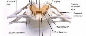
The spinal cord has cells and intercellular space, which in no case should contain excess fluid. Therefore, when it accumulates, it is referred to in medical terminology as spinal cord edema. This concept refers to the expansion of cells by fluid, as a result of which the spinal cord begins to rapidly increase in volume. Due to the accumulation of fluid, intercellular exchange is disrupted, oxygen starvation occurs, and therefore the cells simply begin to die.
Edema of the spinal cord of the spine has several forms, which have certain characteristics and causes. The main classification depends on the reasons. For example, a tumor, a complication after surgery, post-traumatic stress, toxic effects, ischemia, inflammation or hypertension.
Prerequisites
If we talk about certain diagnoses that a doctor can make after diagnosis, these include:
- Lipomas;
- Hematomas;
- Atheromas;
- Osteochondrosis;
- Hemangiomas.
Lipoma is a high-quality tumor formed from human adipose tissue. The formation is not solid to the touch, is perfectly mobile, and no pain is observed on palpation. The development of lipoma can be rapid and relatively slow. The cause is a violation of fat metabolism or improper functioning of the gallbladder. Sizes vary individually, as they can reach extremely huge sizes, but are traditionally up to 4 cm.
Hematomas are also a common diagnosis when a lump appears in the lumbar region. They are benign formations that are formed from proliferated blood vessels. It is unsafe because it destroys healthy tissue and can grow to large sizes.
Atheromas (cysts) are tumors formed due to blockage of the sebaceous gland ducts. The formation is painless, round in shape, contains epithelial cells and fatty substances. Sizes from centimeter to 7. The danger is infection of the contents, which leads to swelling and a rise in temperature.
Osteochondrosis (salt deposition) is a common factor for the development of cones. The main reasons for the development can be sedentary work, little physical activity, metabolic disorders in the body, poor nutrition and various traumatic events.
Hemangiomas are characterized, like hematomas, by pathological proliferation of blood vessels. To determine the nature of the formation, it is important to contact a doctor, who should prescribe diagnostic procedures.
The most common patients who complain of the development of lumps in the lumbar region are men. The work of the sebaceous glands in guys differs quite significantly from that of women. Specificity describes the development of lipomas to an even greater extent.
Ladies during menopause are also most susceptible to the appearance of similar symptoms. The stress and hormonal imbalance that the body experiences during menopause affects the formation of benign tumors. A person who leads a sedentary lifestyle (especially after the age of 35) is more at risk of developing a tumor. Spinal injuries, incorrect back alignment when walking and scoliosis, as well as very little physical activity, put a person at risk of illness.
- We recommend reading: hemangioma of the cervical spine.
Types of cerebral edema depending on pathogenesis
- A cytotoxic type of spinal cord edema occurs after spinal injury. As a result of hypoxia, metabolic processes are disrupted directly in the cells, causing sodium to accumulate, which contributes to the accumulation of water in the tissues. After this, astrocytes located near the circulatory system begin to swell and die, so neurons are affected.
- The interstitial type of edema develops with hydrocephalus. In this case, there is a violation of the outflow of cerebrospinal fluid, as a result of which intracranial pressure increases significantly, and water accumulates in the tissues.
Vasogenic edema occurs when the blood-brain barrier is disrupted. As a rule, osmotic pressure changes for the worse, and positive ions are responsible for it. Consequently, the cellular barrier can no longer fully perform its functions, which leads to the passage of ions. Further, the intercellular space accumulates unnecessary fluid. It turns out that if the permeability of the blood-brain barrier increases, then this intercellular fluid will accumulate depending on blood pressure. That is, the more it rises, the faster the liquid fills the free space. The consequence of the occurrence of vasogenic edema is tumor neoplasms in the spinal cord, micro-embolism of blood vessels and occlusion.
Symptoms of spinal cord edema
Symptoms of cerebral edema of the back can be of 2 types: focal and stem.
So, spinal cord edema symptoms are stem:
Over time, pain and crunching in the back and joints can lead to dire consequences - local or complete restriction of movements in the joint and spine, even to the point of disability. People, taught by bitter experience, use a natural remedy recommended by orthopedist Bubnovsky to heal joints. Read more"
- Circulatory disorders.
- Change in breathing.
- Decreased reactions of the visual apparatus.
- Decrease in excitability.
- Decreased reflex functions.
- Loss of pain, temperature, proprioceptive and tactile sensitivity.
- Flaccid plegia of the limbs.
- Bradycardia.
- Hypothermia.
With focal symptoms, there is a violation of the functionality of the organ at the site of localization.
Causes of spinal cord edema
- Tumor formations.
- High blood pressure.
- Spinal cord injury due to fractures, compression, ruptured discs, bruises.
- Inflammatory process.
- Infection of the body.
- Hemorrhage.
- Intoxication.
- Ischemic condition.
Treatment of spinal cord edema
Have you ever experienced constant back and joint pain? Judging by the fact that you are reading this article, you are already personally familiar with osteochondrosis, arthrosis and arthritis. You've probably tried a bunch of medications, creams, ointments, injections, doctors and, apparently, none of the above has helped you. And there is an explanation for this: it is simply not profitable for pharmacists to sell a working product, as they will lose customers! Nevertheless, Chinese medicine has known the recipe for getting rid of these diseases for thousands of years, and it is simple and clear. Read more"
- For successful treatment, the cause of the pathology is always initially eliminated.
- Next, the amount of swelling is reduced with the help of drugs that normalize electrolyte balance. For example, “Veroshpiron”, “Brinaldix”.
- Forced diuresis medications are prescribed: Furosemide, Lasix.
- Drugs that reduce the production of CSF: “Acetazolamine”, “Diacarb”.
- Glucocorticosteroids: Hydrocortisone, Cortef, Dexamethasole, Dexaven. Thanks to these drugs, it is possible to stabilize the endothelium of small vessels and cell membranes.
- Nootropic agents are used as membrane protectors: “Lucetame”, “Vinpotropil”, “Gammalon”, “Nootropil”, “Piracetam”.
- To reduce swelling and improve blood microcirculation, active rheological drugs are used: “Reopoliglyukin”, “Trental”, “Curantil”.
- In some cases, barbiturates, lidocaine, and hyaluronidases can be used.
- Vitamin therapy: Thiamine, Ascorbic acid, Cyanocobalamin, Pyridoxine. These drugs accelerate metabolic processes at the cellular level and blood circulation. As a result, the cells are saturated with the necessary oxygen and nutrients.
- If there is a problem of paralysis, then muscle relaxants are prescribed: “Tubocurarine”, “Pancuronium”.
- If the pathology: swelling of the spinal cord cannot be treated, or is severe, surgical intervention may be used.
It is strictly forbidden to take drugs that dilate blood vessels, potassium ion antagonists and osmotic diuretics. The fact is that when using them, a symptom of recoil may be observed.
Prevention, rehabilitation, consequences
Edema of the spinal cord: the consequences can be of a different nature and pathological changes can even appear in absolutely any organ. Preventive measures after treatment are mandatory because this will avoid complications. Since the patient will be in a supine position during this period, it is important to provide artificial urination and treatment of bedsores. It is imperative to do both breathing and physical therapeutic exercises. Vibromassage and physiotherapeutic measures can be used. It is advisable to refer the patient to a specialized rehabilitation center, where they will be helped to recover.
We all know what pain and discomfort are. Arthrosis, arthritis, osteochondrosis and back pain seriously spoil life, limiting normal activities - it is impossible to raise an arm, step on a leg, or get out of bed.
These problems begin to manifest themselves especially strongly after 45 years. When you are face to face with physical weakness, panic sets in and it is hellishly unpleasant. But there is no need to be afraid of this - you need to act! Which product should be used and why, says leading orthopedic surgeon Sergei Bubnovsky. Read more >>>
source
Replenishing hyaluronan reserves with food
First, let's look at how to restore synovial fluid using the simplest and most accessible methods, which primarily include a balanced diet. In a healthy adult, the synovium should contain from 2.45 to 3.97 g/l of hyaluronan. The body begins to reduce its production due to natural reasons (without diseases) from about 30 years of age. You can find out how much hyaluronic acid is contained in the synovium in a medical institution using a puncture. But you don’t have to resort to such drastic traumatic methods. Hyaluronan not only knows how to retain water, it is able to change its liquid state to a gel-like state, thereby making the skin elastic and more resistant to unwanted environmental influences.
— soybeans and soy products;
- red wine and grapes in general in any form;
- potatoes and other products containing starch.
Proper nutrition is effective in the first stages of reducing the volume of synovium, and if the process has already gone too far, it is a good help for more effective means, such as dietary supplements. They differ from food in that they contain much higher concentrations of hyaluronan. In principle, the knee joint does not care where the hyaluronan came from - whether it was produced by the body itself or came with food, the main thing is that there is enough of it for everything.
There are many companies on the Russian market that produce similar products. One of them is ARGO. It has been in existence since 1996 and enjoys an excellent reputation because its employees, who are representatives of 27 manufacturers, know very well how to restore synovial fluid in the joints. ARGO drugs are of high quality and at the same time affordable prices. In total, the company has about 600 products, including not only dietary supplements, but also ointments, creams for external use for joint problems, as well as massage products.
Varieties
Swelling of the spinal cord has its own classification, which simplifies the process of choosing treatment tactics when determining the type at the diagnostic stage. Each type has its own characteristics and features.
- Trabecular. Occurs when the spine is injured and vertebrae are destroyed. Against the background of deformed cartilage, an accumulation of fluid appears, which causes severe pain to the person, nausea, dizziness and other characteristic symptoms are observed.
- Subchondral. A process occurs as a result of the destruction of cartilage in the vertebrae. The degree of danger depends on the size of the edema.
- Aseptic. Swelling affects the neck and head of the femoral bone. Against the background of the report, a high body temperature, swelling at the site of localization, and acute pain when pressed appear. Inflammation gradually develops, and functional damage to the area occurs.
- Reactive. Usually occurs after surgery, as it is characterized by the ingestion of pathogenic microflora. Swelling is accompanied by acute pain.
- Perifocal. It is formed when the white matter of the brain enters the plasma proteins. Swelling can be small or extensive and usually does not require surgery.
- Infectious. The cause of the damage is the entry of dangerous pathogens into the body. In rare cases, swelling is caused by helminths or infestations.
Treatment at home after confirmation of the diagnosis is unacceptable, as the risk of complications is high. Therefore, a patient with any form of the disease must be hospitalized in the inpatient department of the hospital.
Cholesterol and other components of synovium
In addition to hyaluronan, synovial fluid contains proteins that provide viscosity and cholesterol in the form of arachidonic, palmitic, oleic, and stearic acids. Cholesterol molecules are located on the articular surfaces, layering on top of each other. This reduces cartilage friction. Thus, to the question “how to restore synovial fluid” the answer is this: to compensate for the lack of its components in the body.
In addition to the necessary elements, the synovia contains living and dead cells (synoviocytes, lymphocytes, histiocytes, monocytes and others), microscopic fragments of cartilage wear, proteins (mainly globulins). The synovium of a healthy person should contain no more than 31.5 g/l of protein. If these numbers are exceeded, it means the joint is inflamed. In order to restore not the volume, but the chemical composition of the synovial fluid, you must first find out the cause of the inflammation (this could be injury, arthritis, arthrosis, synovitis, bursitis). If there is a need, for example, difficulties in establishing the correct diagnosis, a series of studies of synovial fluid are carried out, the main one of which is puncture. It is performed without anesthesia, since novocaine can change test data. Based on the results, the volume of the synovium, its viscosity (should be approximately 0.57 PaHs), transparency, color, pH (normal 7.3-7.5), density, mucin clot, percentage of leukocytes and phagocytes, the presence of microcrystals of sodium urate salts are determined (above normal for gout). If qualitative changes are detected in the synovial fluid, which also causes pain during movement and destruction of cartilage, treatment with appropriate medications is prescribed.
Causes of edema
Fluid begins to accumulate in the bone marrow for a variety of reasons. But determining the etiology is extremely important for choosing the most effective treatment tactics, because both the rate of progression and the nature of tissue damage will differ. The most common prerequisites for the appearance of pathology are:
- Inflammatory process. An infection in the body, regardless of location, can easily reach any place through the blood. The waste products of pathogenic microflora provoke an increase in capillaries in size. The immune system responds appropriately and attacks the infection. The wall of the blood vessels is damaged and swelling occurs in the spinal cord.
- Injury. One of the most common causes of the disease. As a result of injury, fracture or severe bruise, fluid accumulation occurs very rapidly. If help is not provided in time, it can be fatal.
- Osteochondrosis. Degenerative processes in the body gradually destroy cartilage and bone tissue, impair the mobility of the vertebrae, and provoke the appearance of hernias. This, in turn, leads to the body attempting to repair the damaged area, which increases swelling. This is one of the most likely complications of the disease.
- Malignant formations. Sometimes the appearance of a pathological process is explained by a tumor. Computer or magnetic resonance imaging can detect this condition.
It is easy to detect clinically only swelling of the bone marrow, which is formed as a result of injury. The person requires immediate help, as the condition progresses rapidly and in most cases, medical workers do not have time to conduct studies to clarify the diagnosis.
Preventive actions
You can avoid such a serious illness only if you eliminate back injuries, give up extreme sports, or change your profession to a safer one. And it is also important to consult a doctor in a timely manner in case of accidental falls on a slippery surface, infection or after accidents on the road, even if it seems that the overall condition has not been disturbed.
The following recommendations should be considered specific prevention of pathology:
- Enrich the diet with foods that improve hematopoietic function (veal, beef liver, pomegranates, apples).
- Limit salt intake to 1 teaspoon per day.
- Supports the optimal balance of nutrients and minerals to prevent edema.
- Visit a massage therapist regularly to maintain normal back condition.
Taking care of your health and spine is extremely important not only to exclude serious pathologies, but also to maintain the functionality of all systems and normal life. Bone marrow edema is much easier to prevent than to treat it later and deal with the consequences.
Bone marrow edema can cause a variety of unpleasant consequences if it is not treated in time and competent medical care is not received. Therefore, if symptoms appear even insignificant at first glance, it is necessary to consult a doctor, follow the recommendations given and not violate bed rest.
Symptoms
Pain in the vertebrae can be attributed to almost any ailment of the musculoskeletal system, which in turn makes it difficult to make a diagnosis. But without timely consultation with a doctor, there is a risk of death, especially if there is swelling of the cervical spine, which is the most dangerous.
The pathological process is accompanied by the following symptoms:
- Impaired functioning of the respiratory system.
- Decreased vision.
- Neurological disorders.
- Irregularities in heart rhythm.
- Paralysis of the upper or lower limbs.
- Acute pain in the back, in the affected area.
- Disruption of the senses.
- Dysfunction of the pelvic organs (problems with urination or bowel movements, constipation, bladder pain).
- Loss of skin sensitivity.
- Cramps and spasms in the legs or arms.
Pain syndrome can be observed not at the location of the pathology, but several centimeters higher, this is explained by the accumulation of a large amount of fluid and compression of the nerve endings. When the cervical spine is affected, the pressure inside the skull increases, hydrocephalus appears, and nerves are pinched.
Patients with bone marrow edema often develop allergies, exacerbation of radiculitis or intestinal infections. This occurs due to a sharp decrease in the body’s immunity due to the appearance of pathology.
Physical properties
In appearance, the joint fluid is a transparent, slightly yellowish mass, viscous, elastic in consistency, slightly reminiscent of mucus. It is bad for joint function when there is not enough of it. It is no better when there is a lot of synovia. Its excess has to be pumped out, otherwise the synovial membrane may become inflamed. Normally, a healthy person should have from 2.5 to 4 ml of synovial fluid. This data is for the joints of the limbs. There is much less of it in the vertebral joints. How to restore synovial fluid in the required volume is suggested by the reasons leading to its decrease:
- loss of water (dehydration), which can be caused by basic heat and low consumption of life-giving moisture, as well as any infection;
- lack of food rich in vitamins and microelements, especially A and calcium;
- high and frequent physical activity.
As can be seen from this list, it is possible to restore synovial fluid, if its loss is not associated with diseases, by simply changing your exercise regime and diet. But there are also reasons for the decrease in synovium in the joints that a person cannot influence. One of them is age. Over the years, the synthesis of many necessary substances, for example, hyaluronan, decreases in our body. Therefore, in order to prolong the life of the joint, we must either stimulate the body to produce what we need, or take it from the outside.
Diagnostic tests
In case of injury, the important task for the doctor is to relieve the swelling; in other cases, if such a pathology is suspected, patients are sent for tests and to undergo hardware techniques. The following studies are considered appropriate:
- CT or MRI. with their help, the condition of the vertebral bodies, nearby systems and internal organs, and soft tissues is studied. If there is swelling, they will show you best.
- X-ray. The image shows any injuries and other processes that lead to the appearance of ascending edema.
- Blood test for tumor markers and rheumatoid factors. Such a study is necessary when determining the cause of the disease.
- Study of liquor pathways. Prescribed for suspected complications in the functioning of the spinal cord located in the central spinal canal.
Additionally, a biopsy is used to exclude a malignant process. But this technique is quite painful, so it is prescribed as a last resort, when it is necessary to determine the nature of the formation that caused the swelling.
What is fluid accumulation in the spine?
Destruction of bone tissue is a sign indicating a pronounced pathology in the body, which can negatively affect the course of concomitant diseases. In medicine, this process is known as bone destruction. In the process of destruction (destruction), the integrity of bone tissue is disrupted, which is replaced by pathological formations such as tumor growths, lipoids, degenerative and dystrophic changes, granulations, hemangiomas of the vertebral bodies. This condition leads to a decrease in bone density, increased fragility, deformation and complete destruction.
Characteristics of bone destruction
Destruction is the process of destruction of the bone structure with its replacement by tumor tissue, granulations, and pus. Bone destruction occurs only in rare cases at an accelerated pace; in most cases, this process is quite long. Destruction is often confused with osteoporosis, but despite the constant fact of destruction, these two processes have significant differences. If, during osteoporosis, bone tissue is destroyed and replaced with elements similar to bone, that is, blood, fat, osteoid tissue, then during destruction, replacement with pathological tissue occurs.
X-ray is a research method that allows you to recognize destructive changes in the bone. In this case, if with osteoporosis in the pictures you can see diffuse spotty clearings that do not have clear boundaries, then the destructive foci will be expressed in the form of a bone defect. In the photographs, fresh traces of destruction have uneven outlines, while the contours of old lesions, on the contrary, look dense and even. Destructions of bone tissue do not always occur in the same way; they differ in shape, size, contours, reaction of surrounding tissues, as well as the presence of shadows inside the destructive foci and the number of foci.
In the human body, destruction of tooth bone, vertebral bodies and other bones is often observed as a result of poor nutrition, poor hygiene, the development of hemangioma, and other concomitant diseases.
Why does the tooth bone deteriorate?
Dental diseases are a pathology that is accompanied by the destruction of bone tissue. Among various dental diseases that cause destructive changes in bone tissue, periodontal disease and periodontitis are considered the most common.
With periodontitis, destruction of all periodontal tissues occurs, including the gums, bone tissue of the alveoli, and the periodontium itself. The development of pathology is caused by pathogenic microflora, which enters the plaque of the tooth and the gum surrounding it. The infection lies in dental plaque, where gram-negative bacteria, spirochetes and other microorganisms live.
The activity of negative microflora is provoked by the following factors:
- bite problems;
- bad habits;
- dental prosthetics;
- poor nutrition;
- shortening of the frenulum of the tongue and lips;
- poor oral hygiene;
- carious cavities located near the gums;
- violations of interdental contacts;
- congenital periodontal pathologies;
- general diseases.
All of the above factors are the causes of the development of periodontitis and contribute to the activation of pathogenic microflora, which especially negatively affects the attachment of the tooth to the gum.
The process of tooth destruction during periodontitis
Periodontitis is a disease in which the destruction of the connections between the tooth and gum tissue occurs with the formation of a periodontal pocket.
Pathology causes destructive changes in periodontal bone tissue and alveolar processes. The development of the acute form of the disease is caused by enzymes that negatively affect the intercellular communication of the epithelium, which becomes sensitive and permeable. Bacteria produce toxins that harm cells, ground substance, and connective tissue formations, while humoral immune and cellular reactions develop. The development of the inflammatory process in the gums leads to the destruction of the bone of the alveoli, the formation of serotonin and histamine, which affect the cell membranes of blood vessels.
A periodontal pocket is formed as a result of the destruction of the epithelium, which grows into the connective tissues located at a level below.
With further progression of the disease, the connective tissue around the tooth begins to gradually deteriorate, which simultaneously leads to the formation of granulation and destruction of the bone tissue of the alveoli. Without timely treatment, the tooth structure can completely collapse, which will lead to the gradual loss of all teeth.
Destructive changes in the spine
Bone destruction is a dangerous process, the further development of which must be prevented at the first signs of pathology. Destructive changes affect not only the bone tissue of the tooth; without appropriate treatment, they can spread to other bones in the body. For example, as a result of the development of spondylitis, hemangiomas, destructive changes affect the spine as a whole or the vertebral bodies separately. Spinal pathology can lead to undesirable consequences, complications, partial or complete loss of mobility.
Spondylitis is a chronic inflammatory disease that is a type of spondylopathy. As the disease develops, pathology of the vertebral bodies and their destruction are noted, which threatens spinal deformation.
There is specific and nonspecific spondylitis. Specific spondylitis is caused by various infections that enter the blood and, with its help, spread throughout the body, affecting bones and joints along the way.
Infectious pathogens include microbacteria:
- tuberculosis;
- syphilis;
- gonorrheal gonococcus;
- coli;
- streptococcus;
- Trichomonas;
- Staphylococcus aureus;
- pathogens of smallpox, typhoid, plague.
Sometimes the disease can be triggered by fungal cells or rheumatism. Nonspecific spondylitis occurs in the form of hematogenous purulent spondylitis, ankylosing spondylitis or ankylosing spondylitis.
Regardless of the cause of the disease, treatment must begin immediately after diagnosis.
Spondylitis is the cause of destruction of the vertebral bodies
With tuberculous spondylitis, damage to the vertebral bodies of the cervical and thoracic spine is noted. The pathology leads to the development of single purulent abscesses, cuts, often irreversible paralysis of the upper limbs, the formation of a pointed hump, deformation of the chest, and inflammation of the spinal cord.
With brucellosis spondylitis, damage to the lumbar vertebral bodies is noted. X-ray photographs show fine focal destruction of the vertebral bone bodies. Serological testing is used for diagnosis.
Syphilitic spondylitis is a rare pathology that affects the cervical vertebrae.
In the typhoid form of the pathology, damage occurs to two adjacent vertebral bodies and the intervertebral disc connecting them. The process of destruction in the thoracolumbar and lumbosacral sector occurs quickly, with the formation of multiple purulent foci.
Damage to the periosteum of the vertebral bodies in the thoracic region occurs with actinomycotic spondylitis. As the pathology develops, purulent foci and punctate fistulas form, the release of whitish substances, and destruction of bone tissue are noted.
As a result of spinal trauma, aseptic spondylitis can develop, in which inflammation of the spinal bodies is noted. The pathology is dangerous because it can be asymptomatic for a long time. In this case, patients may learn about the destruction of the spine with a delay, when the vertebra takes on a wedge-shaped shape and foci of necrosis appear in the spine.
What is a spinal hemangioma?
Destruction is a pathology that can affect both soft tissues and bones; patients often experience hemangiomas of the vertebral bodies.
Our readers successfully use SustaLife to treat joints. Seeing how popular this product is, we decided to bring it to your attention. Read more here...
Hemangioma is a benign tumor neoplasm. The development of hemangioma can be observed in humans regardless of age. Pathology often occurs in children due to improper development of blood vessels in the embryonic period.
Usually, no obvious disturbances are observed from the newly formed tumor, since it does not manifest itself with any symptoms, but this depends on its size and location. Discomfort, some disturbances in the functioning of internal organs, and various complications can cause the development of hemangioma in the auricle, kidneys, liver and other organs.
How to eliminate swelling?
There is no universal remedy to relieve swelling, so therapy for each patient is selected individually. If the development of pathology is suspected, the doctor recommends that the patient follow the following rules:
- Strict adherence to bed rest, complete exclusion of stress or physical activity.
- Placing the lower extremities on a support to elevate them above the level of the heart and thereby reduce swelling.
- Applying a cold heating pad several times a day to the site of inflammation.
- Taking medications according to the chosen regimen to relieve pain.
- Wearing a corset or bandage to prevent mobility of the vertebrae in the damaged area.
In order to prevent unpleasant consequences, it is necessary to completely eliminate pressure in the affected area and not provoke a deterioration in the general condition. And it is also important to eat right, enrich your diet with protein, vitamins B and D, and give up bad habits.
Drug treatment
The mainstay of treatment for spinal cord edema is taking medications. They are selected individually, depending on the cause, degree of damage and symptoms in the patient. Typically the complex consists of the following medications:
- Analgesics (“Metamizole sodium”) Painkillers are necessary to relieve an acute attack and alleviate the patient’s general condition. If they are ineffective, Tramadol is prescribed.
- Non-steroidal anti-inflammatory drugs (Diclofenac, Ketorol, Nimesulide). In addition to suppressing the inflammatory process, they additionally help relieve pain.
- Glucocorticoids (Dimexide, Hydrocortisone). They reduce inflammation and exudation.
- To stimulate blood movement (Actovegin, Trental). The active substance improves the circulation of biological fluid and thereby reduces swelling.
- B vitamins (“Combilipen”). They promote tissue nutrition and enhance immunity.
If the cause of the disease is an infection, general antibiotics are prescribed, most often Clarithromycin or Amoxicillin; in the case of tuberculosis, specific therapy is required. In cases of severe inflammation, potassium iodide is also used.
Surgical correction
Taking into account etiological factors, sometimes the only treatment option is surgery. Usually you cannot do without it in case of injuries, osteomyelitis, spinal pathologies, intervertebral hernia and other diseases. Only by eliminating the cause in this way can the rapid accumulation of fluid in the bone structures of the vertebrae be stopped.
Diagnostics
Edema of the spinal and bone marrow is quite difficult to diagnose, since the symptoms are usually masked under the underlying disease that provoked this complication. However, if the doctor has identified changes in the spinal column and a set of symptoms that may accompany swelling, he can continue further diagnostic search.
In diagnostics they use:
- radiography, which helps to identify severely advanced spinal injuries;
- CT, thanks to which it is possible to assess the condition of bone tissue;
- MRI, thanks to which the specific localization of edema, features of its location and other important information are determined.
Treatment of edema is a complex, often complex task. First of all, it is necessary to ensure unloading of the spinal column in the affected area in order to prevent the death of nerve cells. It is also necessary to establish and eliminate the cause of the development of the pathology, since without eliminating the cause, the swelling recurs again in a short time.
Relieve swelling using the following groups of drugs:
- diuretics (thanks to them, excess fluid is removed from the body);
- drugs that affect the properties of the blood (designed to speed up the healing process of damaged areas by increasing their blood supply);
- B vitamins (help restore damaged nerve cells).
The patient is required to be prescribed painkillers. Both NSAIDs and, in particularly severe cases, narcotic analgesics can be used at the discretion of the physician.
Glucocorticosteroids and nootropics are also often considered an important element of therapy. They help stabilize cell membranes, protect them from further damage, and reduce the severity of inflammation.
If it is not possible to relieve swelling with drug therapy, surgical drainage is resorted to. The situation in this case is often complicated by the fact that any wrong action can result in disability for the patient at best, and death at worst.
In addition to addressing the causes of edema and the main symptoms of this condition, it is also important to properly organize symptomatic therapy. If the patient suffers from seizures, then they are not ignored by using anticonvulsants. If breathing is impaired, normal ventilation of the lungs is provided; if there are problems with the heart rhythm, medications are taken to correct it.
Treatment of spinal cord edema in each case is selected strictly individually. The choice of drugs should be made by the attending physician, focusing on the characteristics of the patient, the cause of the disease, and the severity of symptoms.
Possible complications
Swelling of the spinal cord is quite favorable if you consult a specialist in a timely manner. With complex treatment, the patient completely restores all impaired functions and mobility of the spine; any physical activity is allowed after rehabilitation. If treatment is started late, there is a risk of paralysis, partial or complete, or death.
The most common consequence of the pathology is loss of limb mobility and dysfunction of the pelvic organs. The prognosis largely depends on the location of the lesion, the degree of progression of the disease before seeking medical help, and the age of the patient. The higher the swelling is located, the higher the likelihood of complete paralysis.
Reviews from our patients
I would like to express my gratitude to the massage therapist-rehabilitator Ruslan Iksanov, he is a master of his craft. He sees with his hands clearly, knows and does his job. I thank him and wish him success in all areas of his life. I recommend it to anyone who has problems with the spine. 02/16/2019
I express my gratitude to the excellent specialist Sergei Dmitrievich Sorokin! A year ago, I didn’t know what to do with my endlessly numb leg and part of my face, and after visiting many clinics and doctors, only Sergei Dmitrievich helped me. And even after a year, thanks to his therapy and recommendations, I am bigger.


