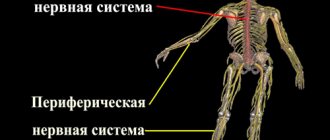
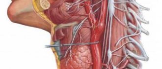
Sympathetic (shown in blue) and parasympathetic (shown in red) divisions of the autonomic nervous system.
This term has other meanings, see ANS.
Autonomic nervous system
(from lat. vegetatio - excitement, from lat. vegetativus - plant),
ANS
,
autonomic nervous system
,
ganglion nervous system
(from lat. ganglion - nerve ganglion), visceral nervous system (from lat. viscera - insides), organ nervous system , celiac nervous system,
systema nervosum autonomicum
(PNA) is part of the body’s nervous system, a complex of central and peripheral cellular structures that regulate the functional level of the body, necessary for the adequate response of all its systems.
The autonomic nervous system is a section of the nervous system that regulates the activity of internal organs, endocrine and exocrine glands, blood and lymphatic vessels[1]. Plays a leading role in maintaining the constancy of the internal environment of the body and in the adaptive reactions of all vertebrates.
Anatomically and functionally, the autonomic nervous system is divided into sympathetic, parasympathetic and metasympathetic. The sympathetic and parasympathetic centers are under the control of the cerebral cortex and hypothalamic centers [2].
The sympathetic and parasympathetic divisions have central and peripheral parts. The central part is formed by the bodies of neurons lying in the spinal cord and brain. These clusters of nerve cells are called vegetative nuclei. Fibers extending from the nuclei, autonomic ganglia lying outside the central nervous system, and nerve plexuses in the walls of internal organs form the peripheral part of the autonomic nervous system.
The sympathetic nuclei are located in the spinal cord. The nerve fibers extending from it end outside the spinal cord in the sympathetic ganglia, from which the nerve fibers originate. These fibers are suitable for all organs.
The parasympathetic nuclei lie in the midbrain and medulla oblongata and in the sacral part of the spinal cord. Nerve fibers from the nuclei of the medulla oblongata are part of the vagus nerves. From the nuclei of the sacral part, nerve fibers go to the intestines and excretory organs.
The metasympathetic nervous system is represented by nerve plexuses and small ganglia in the walls of the digestive tract, bladder, heart and some other organs.
The activity of the autonomic nervous system does not depend on the will of a person. This means that under normal conditions a person cannot force his heart to beat less often or his stomach muscles not to contract. However, it is possible to achieve conscious influence on many parameters controlled by the ANS using special training methods - for example, using biofeedback methods.
The sympathetic nervous system enhances metabolism, increases the excitability of most tissues, and mobilizes the body's forces for vigorous activity. The parasympathetic system helps restore spent energy reserves and regulates the functioning of the body during sleep.
The organs of circulation, respiration, digestion, excretion, reproduction, as well as metabolism and growth are under the control of the autonomous system. In fact, the efferent section of the ANS carries out nervous regulation of the functions of all organs and tissues, except for skeletal muscles, which are controlled by the somatic nervous system.
Unlike the somatic nervous system, the motor effector neuron in the autonomic nervous system is located in the periphery, and the spinal cord only indirectly controls its impulses.
Terms autonomous system
,
visceral system
,
sympathetic nervous system
are ambiguous. Currently, only part of the visceral efferent fibers is called sympathetic. However, different authors use the term "sympathetic" in different ways:
- in the narrow sense, as described in the sentence above;
- as a synonym for the term "autonomous";
- as the name of the entire visceral (“autonomic”) [3] nervous system - both afferent and efferent.
Terminological confusion also arises when the entire visceral system (both afferent and efferent) is called autonomous.
The classification of parts of the vertebrate visceral nervous system, given in the manual [4] by A. Romer and T. Parsons, is as follows:
Visceral nervous system:
- afferent;
- efferent: special gill;
- autonomic: sympathetic;
- parasympathetic.
Content
- 1 Morphology 1.1 Central and peripheral sections 1.1.1 Central section
- 1.1.2 Peripheral department
- 2.1 General significance of autonomic regulation
The role of the autonomic nervous system
The ANS provides regulation of the activity of internal organs, which include smooth muscle and glandular tissue. Such organs include all organs of the digestive, respiratory, urinary, reproductive systems, heart and blood vessels (blood and lymphatic), and endocrine glands. The ANS also takes part in the work of skeletal muscles, regulating metabolism in the muscles.
The role of the ANS is to maintain a certain level of functioning of organs, to strengthen or weaken their specific activity depending on the needs of the body. In this regard, the ANS has two parts (sympathetic and parasympathetic), which have opposite effects on the organs.
Morphology
The distinction of the autonomous (autonomic) nervous system is due to certain features of its structure. These features include the following:
- focal localization of vegetative nuclei in the central nervous system;
- accumulation of bodies of effector neurons in the form of nodes (ganglia) as part of the autonomic plexuses;
- two-neuronality of the nerve pathway from the autonomic nucleus in the central nervous system to the innervated organ.
The fibers of the autonomic nervous system do not emerge segmentally, as in the somatic nervous system, but from three limited areas of the brain spaced apart from each other: cranial, sternolumbar and sacral.
The autonomic nervous system is divided into sympathetic, parasympathetic and metasympathetic parts. In the sympathetic part, the processes of spinal neurons are shorter, while the ganglion ones are longer. In the parasympathetic system, on the contrary, the processes of spinal cells are longer, those of ganglion cells are shorter. Sympathetic fibers innervate all organs without exception, while the area of innervation of parasympathetic fibers is more limited.
Central and peripheral sections
The autonomic (autonomic) nervous system is divided according to topographical characteristics into central and peripheral sections.
Central department
- parasympathetic nuclei of the 3rd, 7th, 9th and 10th pairs of cranial nerves lying in the brain stem (craniobulbar region).
- sympathetic nuclei located in the lateral horns of the thoracolumbar spinal cord; nuclei located in the gray matter of the three sacral segments (sacral section)[5][6];
Peripheral department
- autonomic (autonomic) nerves, branches and nerve fibers emerging from the brain and spinal cord;
- vegetative (autonomous, visceral) plexuses;
- nodes (ganglia) of the autonomic (autonomous, visceral) plexuses;
- sympathetic trunk (right and left) with its nodes (ganglia), internodal and connecting branches and sympathetic nerves;
- terminal nodes (ganglia) of the parasympathetic part of the autonomic nervous system.
Sympathetic, parasympathetic and metasympathetic divisions
Based on the topography of the autonomic nuclei and nodes, differences in the length of the axons of the first and second neurons of the efferent pathway, as well as the characteristics of the function, the autonomic nervous system is divided into sympathetic, parasympathetic and metasympathetic.
Location of ganglia and structure of pathways
Neurons
nuclei of the central part of the autonomic nervous system are the first efferent neurons on the way from the central nervous system (spinal cord and brain) to the innervated organ. The nerve fibers formed by the processes of these neurons are called prenodal (preganglionic) fibers, since they go to the nodes of the peripheral part of the autonomic nervous system and end with synapses on the cells of these nodes. Preganglionic fibers have a myelin sheath, which makes them whitish in color. They leave the brain as part of the roots of the corresponding cranial nerves and the anterior roots of the spinal nerves.
Vegetative nodes
(ganglia): are part of the sympathetic trunks (present in most vertebrates, except cyclostomes and cartilaginous fish), large vegetative plexuses of the abdominal cavity and pelvis, located in the head and in the thickness or near the organs of the digestive and respiratory systems, as well as the genitourinary system, which are innervated by the autonomic nervous system. The nodes of the peripheral part of the autonomic nervous system contain the bodies of second (effector) neurons lying on the way to the innervated organs. The processes of these second neurons of the efferent pathway, carrying nerve impulses from the autonomic ganglia to the working organs (smooth muscles, glands, tissues), are post-nodular (postganglionic) nerve fibers. Due to the absence of the myelin sheath, they are gray in color. Postganglionic fibers of the autonomic nervous system are mostly thin (most often their diameter does not exceed 7 µm) and do not have a myelin sheath. Therefore, excitation spreads slowly through them, and the nerves of the autonomic nervous system are characterized by a longer refractory period and greater chronaxis.
Reflex arc
The structure of the reflex arcs of the autonomic part differs from the structure of the reflex arcs of the somatic part of the nervous system. In the reflex arc of the autonomic part of the nervous system, the efferent link consists not of one neuron, but of two, one of which is located outside the central nervous system. In general, a simple autonomic reflex arc is represented by three neurons.
The first link of the reflex arc is a sensory neuron, the body of which is located in the spinal ganglia and in the sensory ganglia of the cranial nerves. The peripheral process of such a neuron, which has a sensitive ending - a receptor, originates in organs and tissues. The central process, as part of the dorsal roots of the spinal nerves or sensory roots of the cranial nerves, is directed to the corresponding nuclei in the spinal cord or brain.
The second link of the reflex arc is efferent, since it carries impulses from the spinal cord or brain to the working organ. This efferent pathway of the autonomic reflex arc is represented by two neurons. The first of these neurons, the second in a simple autonomic reflex arc, is located in the autonomic nuclei of the central nervous system. It can be called intercalary, since it is located between the sensitive (afferent) link of the reflex arc and the second (efferent) neuron of the efferent pathway.
The effector neuron is the third neuron of the autonomic reflex arc. The bodies of effector (third) neurons lie in the peripheral nodes of the autonomic nervous system (sympathetic trunk, autonomic ganglia of cranial nerves, nodes of extraorgan and intraorgan autonomic plexuses). The processes of these neurons are directed to organs and tissues as part of organ autonomic or mixed nerves. Postganglionic nerve fibers end on smooth muscles, glands and other tissues with the corresponding terminal nerve apparatus.
Structure of the somatic nervous system
The animal nervous system has a simple structure; it is controlled by neurons, on the work of which activities and functions are based:
- sensory (spinal) neurons - deliver impulses to the central nervous system;
- motor (cranial) neurons - deliver information from the brain to muscle tissue.
Neurons are located throughout the body from the center to important receptors and muscles. Their bodies are located in the central nervous system, and their axons extend to the skin, muscle tissue, and sensory organs. The muscles located on the left are under the control of the right hemisphere of the brain, and the muscles on the right are controlled by the left side. In addition to supplying nerves, it also influences interaction with muscles. The somatic nervous system includes reflex arcs designed to control unconscious actions and reflexes. With their help, the motor work of muscles is controlled without signals from the brain.
- Burpee - what kind of exercise is it? Techniques for weight loss and Burpees method for beginners
- How to lose weight with Bonn soup
- Do-it-yourself elastic bands made from satin ribbons: master class with photos and videos
Cranial nerves
The somatic nervous system includes 12 pairs of cranial nerves that carry information to and from the brain stem:
- olfactory;
- visual;
- oculomotor;
- block;
- trigeminal;
- diverting;
- facial;
- auditory;
- glossopharyngeal;
- wandering;
- additional;
- sublingual.
Almost all of them innervate the area of the head and neck, that is, the sensory organs, muscle tissue inside the skull, and include motor and secretory cells of the brain, where nuclear clusters of neurons are formed. Individual cranial nerves (for example, the optic nerve) are built only from sensory fibers. The vagus nerve innervates the heart, gastrointestinal tract, lungs and is responsible for their activity. The sensory fiber bodies are located near the brain, and the motor fiber bodies are located inside it.
Spinal nerves
Other structures with somatic innervation are the 31 pairs of spinal nerves with numerous branches to supply areas below the neck. Each spinal nerve is formed by the connection of the posterior and anterior (sensitive and motor) roots, the fusion of their fibers. The posterior ones supply the skin, muscles of the dorsal region, areas of the coccyx, sacrum, the anterior ones - the skin, muscle tissue of the arms, legs and the front part of the body.
Physiology
General importance of autonomic regulation
The autonomic nervous system adapts the functioning of internal organs to environmental changes. The ANS ensures homeostasis (constancy of the internal environment of the body). The ANS is also involved in many behavioral acts carried out under the control of the brain, influencing not only physical, but also mental activity of a person.
The role of the sympathetic and parasympathetic divisions
The sympathetic nervous system is activated during stress reactions. It is characterized by a generalized effect, with sympathetic fibers innervating the vast majority of organs.
It is known that parasympathetic stimulation of some organs has an inhibitory effect, while others have an excitatory effect. In most cases, the action of the parasympathetic and sympathetic systems is opposite.
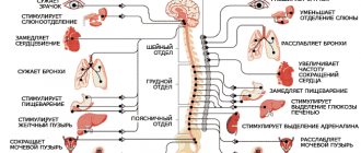
The influence of the sympathetic and parasympathetic departments on individual organs
Influence of the sympathetic department:
- On the heart - increases the frequency and strength of heart contractions.
- On the arteries—[7]dilates the arteries.
- On the intestines - inhibits intestinal motility and the production of digestive enzymes.
- On the salivary glands - inhibits salivation.
- On the bladder - relaxes the bladder.
- On the bronchi and breathing - expands the bronchi and bronchioles, enhances ventilation of the lungs.
- On the pupil - dilates the pupils.
Influence of the parasympathetic department:
- On the heart - reduces the frequency and strength of heart contractions.
- On the arteries - does not affect most organs, causes dilation of the arteries of the genital organs and brain, narrowing of the coronary arteries and arteries of the lungs.
- On the intestines - enhances intestinal motility and stimulates the production of digestive enzymes.
- On the salivary glands - stimulates salivation.
- On the bladder - contracts the bladder.
- On the bronchi and breathing - narrows the bronchi and bronchioles, reduces ventilation of the lungs.
- On the pupil - constricts the pupils.
Neurotransmitters and cellular receptors
The sympathetic and parasympathetic departments have different, in some cases opposite, effects on various organs and tissues, and also cross-influence each other. The different effects of these sections on the same cells are associated with the specificity of the neurotransmitters they secrete and with the specificity of the receptors present on the presynaptic and postsynaptic membranes of neurons of the autonomic system and their target cells.
Preganglionic neurons of both parts of the autonomic system secrete acetylcholine as the main neurotransmitter, which acts on nicotinic acetylcholine receptors on the postsynaptic membrane of postganglionic (effector) neurons. Postganglionic neurons of the sympathetic department, as a rule, secrete norepinephrine as a transmitter, which acts on adrenergic receptors of target cells. On the target cells of sympathetic neurons, beta-1 and alpha-1 adrenergic receptors are mainly concentrated on the postsynaptic membranes (this means that in vivo
they are mainly affected by norepinephrine), and al-2 and beta-2 receptors are on extrasynaptic areas of the membrane (they are mainly affected by blood adrenaline). Only some postganglionic neurons of the sympathetic division (for example, those acting on the sweat glands) release acetylcholine.
Postganglionic neurons of the parasympathetic division release acetylcholine, which acts on muscarinic receptors on target cells.
On the presynaptic membrane of postganglionic neurons of the sympathetic division, two types of adrenergic receptors predominate: alpha-2 and beta-2 adrenergic receptors. In addition, the membrane of these neurons contains receptors for purine and pyrimidine nucleotides (P2X ATP receptors, etc.), nicotinic and muscarinic cholinergic receptors, neuropeptide and prostaglandin receptors, and opioid receptors [8].
When norepinephrine or blood adrenaline acts on alpha-2 adrenoreceptors, the intracellular concentration of Ca2+ ions drops, and the release of norepinephrine at the synapses is blocked. A negative feedback loop occurs. Alpha-2 receptors are more sensitive to norepinephrine than to epinephrine.
When norepinephrine and epinephrine act on beta-2 adrenergic receptors, the release of norepinephrine usually increases. This effect is observed during normal interaction with the Gs protein, during which the intracellular concentration of cAMP increases. Beta two receptors are more sensitive to adrenaline. As adrenaline is released from the adrenal medulla under the influence of norepinephrine from the sympathetic nerves, a positive feedback loop occurs.
However, in some cases, activation of beta-2 receptors can block the release of norepinephrine. It has been shown that this may be a consequence of the interaction of beta-2 receptors with Gi/o proteins and their binding (sequestering) of Gs proteins, which, in turn, prevents the interaction of Gs proteins with other receptors [1].
When acetylcholine acts on muscarinic receptors of sympathetic neurons, the release of norepinephrine in their synapses is blocked, and when it acts on nicotinic receptors, it is stimulated. Because muscarinic receptors predominate on the presynaptic membranes of sympathetic neurons, activation of the parasympathetic nerves typically reduces the level of norepinephrine released from the sympathetic nerves.
Alpha-2 adrenergic receptors predominate on the presynaptic membranes of postganglionic neurons of the parasympathetic department. When norepinephrine acts on them, the release of acetylcholine is blocked. Thus, the sympathetic and parasympathetic nerves mutually inhibit each other.
CNS and its activities
The central nervous system is the main part of the total nervous systems of the human body, consisting of neurons that have axons and glia.
The central nervous system consists of two main organs:
- brain;
- spinal cord.
The main job of the central nervous system is to provide reflexes of varying complexity. It combines information coming from all organs of the body. Bone shells protect the major organs.
The brain is located inside the cranium, the spinal cord is protected by the vertebrae. Each of them has three more shells: hard, arachnoid and vascular.
It should be noted that the central nervous system differs from the peripheral nervous system at the microscopic level. They differ in tissues and neurons.
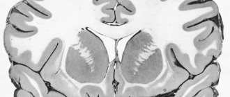
The organs of the central nervous system contain gray and white matter. White represents axons and oligodendrocytes, gray represents neurons and unmyelinated fibers.
Both substances contain glia. Their content in white is higher than in gray.
The medulla oblongata, middle cord, intermediate cord, spinal cord and cerebellum are responsible for processes occurring throughout the body. Subcortical formations and the cerebral cortex are responsible for connections between the external environment and the body.
The peripheral nervous system provides communication between the central nervous system and organs and tissues; it also includes cranial, spinal and intervertebral nerves and nodes.
Two-way signal transmission is carried out. The central nervous system receives excitatory impulses from peripheral receptors, they are transmitted by afferent nerves, and from the central nervous system signals are transmitted to target tissues.
Afferent and efferent cells interact with each other, and a two-neuron reflex arc is formed. In this case, the brain controls the functions performed by the spinal cord and regulates spinal reflexes.
Along the entire length of the spinal cord there are pathways that transmit nerve impulses, formed by white matter and connecting the spinal cord with the brain. They provide reflex and conductive functions.
They contain descending and ascending paths. Nerve signals, through ascending pathways, enter the brain, and from there, through descending pathways, they reach the spinal cord.
Development in embryogenesis
- Development of the peripheral (somatic) and autonomic nervous system.
The peripheral (somatic) and autonomic nervous system develops from the outer germ layer - the ectoderm. Cranial and spinal nerves in the fetus are formed very early (5-6 weeks). Myelination of nerve fibers occurs later (for the vestibular nerve - 4 months; for most nerves - at 6-7 months).
Spinal and peripheral autonomic ganglia are formed simultaneously with the development of the spinal cord. The starting material for them is the cellular elements of the ganglion plate, its neuroblasts and glioblasts, from which the cellular elements of the spinal ganglia are formed. Some of them are shifted to the periphery to the localization of the autonomic nerve ganglia
Functional division of the nervous system
In functional terms, the nervous system is divided into somatic and autonomic.
The somatic nervous system perceives irritations from the external environment and regulates the functioning of skeletal muscles, i.e. responsible for the movements of the body and its movement in space. The autonomic nervous system (ANS) regulates the functions of all internal organs, glands and blood vessels, and its activity is practically independent of human consciousness, which is why it is also called autonomous. The nervous system is a huge collection of neurons (nerve cells), consisting of a body and processes. With the help of processes, neurons connect with each other and with innervated organs. Any information from the external environment or from the body and internal organs is transmitted along chains of neurons to the nerve centers of the central nervous system in the form of a nerve impulse.
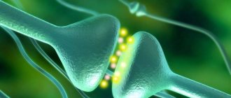
After analysis in the nerve centers, the corresponding commands are also sent along chains of neurons to the working organs to carry out the necessary action, for example, contraction of skeletal muscles or increased production of juices by the digestive glands. The transmission of a nerve impulse from one neuron to another or to an organ occurs at synapses (translated from Greek.
Notes
- Brief medical encyclopedia.
- Biologist's Dictionary. (unavailable link)
- For example, in the book “Human Physiology / Ed. V. M. Pokrovsky, G. F. Korotko. - M.: Medicine, 1997. - T. 1 - 448 p.; T. 2 - 368 pp.”
- Romer A., Parsons T. Anatomy of vertebrates. - T. 2. - P. 260.
- I. Espinosa-Medina, O. Saha, F. Boismoreau, Z. Chettouh, F. Rossi.
The sacral autonomic outflow is sympathetic (English) // Science. — 2016-11-18. - Vol. 354, iss. 6314. - P. 893–897. — ISSN 1095-9203 0036-8075, 1095-9203. — DOI:10.1126/science.aah5454. - The sacral division of the autonomic nervous system is sympathetic. Retrieved February 21, 2020.
- Brin V.B. et al. Fundamentals of human physiology in 2 volumes. Textbook for higher educational institutions. Ed. B. I. Tkachenko. St. Petersburg, 1994. T.1 - 567 p. T.2 - 413 p.
- Stefan Boehm, Sigismund Huck. Receptors controlling transmitter release from sympathetic neurons in vitro // Progress in Neurobiology. — Volume 51, Issue 3, February 1997, Pages 225-242.
- G. Ross, C. Ross, D. Ross. Entomology. - M.: Mir, 1985. - P.109.
Well-functioning mechanism of the autonomic nervous system
The division of the brain into functional elements is described rather conventionally, since it is a complex, well-functioning mechanism. The ANS, on the one hand, coordinates the activity of its structures, and on the other, is influenced by the cortex.
The visceral system is responsible for performing many tasks. Higher nerve centers are responsible for the coordination of the ANS.
The neuron is the main structural unit of the ANS. The path along which the impulse signals travel is called a reflex arc. Neurons are necessary for conducting impulses from the spinal cord and brain to somatic organs, glands and smooth muscle tissue.
An interesting fact is that the heart muscle is represented by striated tissue, but also contracts involuntarily.
The ANS is divided into the sympathetic and parasympathetic subsystems (SNS and PNS, respectively). They differ in the specificity of innervation and the nature of the reaction to substances that affect the ANS, but at the same time they closely interact with each other - both functionally and anatomically. Sympathy is stimulated by adrenaline, parasympathetic by acetylcholine. The former is inhibited by ergotamine, the latter by atropine.
The tasks of the autonomous system include the regulation of all internal processes occurring in the body: the work of somatic organs, blood vessels, glands, muscles, and sensory organs.
The ANS maintains the stability of the human internal environment and the implementation of such vital functions as breathing, blood circulation, digestion, temperature regulation, metabolic processes, excretion, reproduction and others.
The ganglionic system participates in adaptation-trophic processes, that is, it regulates metabolism according to external conditions.
Thus, vegetative functions are as follows:
- support of homeostasis (constancy of the environment);
- adaptation of organs to various exogenous conditions (for example, in the cold, heat transfer decreases and heat production increases);
- vegetative implementation of human mental and physical activity.
Suprasegmental
It includes the hypothalamus, the reticular formation (waking and falling asleep), the visceral brain (behavioral reactions and emotions).
The hypothalamus is a small layer of brain matter. It has thirty-two pairs of nuclei that are responsible for neuroendocrine regulation and homeostasis. The hypothalamic region interacts with the cerebrospinal fluid circulation system because it is located next to the third ventricle and the subarachnoid space.
In this area of the brain there is no glial layer between neurons and capillaries, which is why the hypothalamus immediately responds to changes in the chemical composition of the blood.
The hypothalamus interacts with the organs of the endocrine system by sending oxytocin and vasopressin, as well as releasing factors, to the pituitary gland. The visceral brain (psycho-emotional background during hormonal changes) and the cerebral cortex are connected to the hypothalamus.
So, the work of this important area is dependent on the cortex and subcortical structures. The hypothalamus is the highest center of the ANS, which regulates various types of metabolism, immune processes, and maintains environmental stability.
Segmental
Its elements are localized in the spinal segments and basal ganglia. This includes the SMN and PNS. Sympathy includes the Yakubovich nucleus (regulation of the muscles of the eye, constriction of the pupil), the nuclei of the ninth and tenth pairs of cranial nerves (the act of swallowing, providing nerve impulses to the cardiovascular and respiratory systems, gastrointestinal tract).
The parasympathetic system includes centers located in the sacral spinal cord (innervation of the genital and urinary organs, rectal area). Fibers emanate from the centers of this system and reach the target organs. This is how each specific organ is regulated.
Thus, sympathetic irritation manifests itself everywhere - in different parts of the body. Acetylcholine is involved in sympathetic regulation, and adrenaline is involved in the periphery. Both subsystems interact with each other, but not always antagonistically (sweat glands are innervated only sympathetically).
Peripheral
It is represented by fibers entering the peripheral nerves and ending in organs and vessels. Particular attention is paid to the autonomic neuroregulation of the digestive system - an autonomous formation that regulates peristalsis, secretory function, etc.
Autonomic fibers, unlike the somatic system, lack a myelin sheath. Because of this, the speed of impulse transmission through them is 10 times less.
All organs are under the influence of these subsystems, except the sweat glands, blood vessels and the inner layer of the adrenal glands, which are innervated only sympathetically.
The parasympathetic structure is considered more ancient. It helps create stability in the functioning of organs and conditions for the formation of an energy reserve. The sympathetic department changes these states depending on the function performed.
Both departments interact closely. When certain conditions occur, one of them is activated, and the second is temporarily inhibited. If the tone of the parasympathetic department predominates, parasympathotonia occurs, and the tone of the sympathetic department - sympathotonia. The former is characterized by a state of sleep, the latter by heightened emotional reactions (anger, fear, etc.).
Command centers
Command centers are localized in the cortex, hypothalamus, brain stem and lateral spinal horns.
Peripheral sympathetic fibers arise from the lateral horns. The sympathetic trunk extends along the spinal column and unites twenty-four pairs of sympathetic nodes:
- three cervical;
- twelve breasts;
- five lumbar;
- four sacral.
The cells of the cervical ganglion form the nerve plexus of the carotid artery, the cells of the lower ganglion form the superior cardiac nerve. The thoracic nodes provide innervation to the aorta, bronchopulmonary system, and abdominal organs, while the lumbar nodes provide innervation to organs in the pelvis.
In the midbrain there is a mesencephalic section in which the nuclei of the cranial nerves are concentrated: the third pair is the Yakubovich nucleus (mydriasis), the central posterior nucleus (innervation of the ciliary muscle).
The medulla oblongata is also called the bulbar region, the nerve fibers of which are responsible for the processes of salivation.
Also here is the vegetative nucleus, which innervates the heart, bronchi, gastrointestinal tract and other organs.
Nerve cells of the sacral level innervate the genitourinary organs and the rectal gastrointestinal tract.
In addition to the listed structures, the fundamental system, the so-called “base” of the ANS, is distinguished - this is the hypothalamic-pituitary system, the cerebral cortex and the striatum. The hypothalamus is a kind of “conductor” that regulates all underlying structures and controls the functioning of the endocrine glands.
VNS Center
The leading regulatory link is the hypothalamus. Its nuclei communicate with the cortex of the telencephalon and the underlying parts of the brainstem.
Role of the hypothalamus:
- close relationship with all elements of the brain and spinal cord;
- implementation of neuroreflex and neurohumoral functions.
The hypothalamus is penetrated by a large number of vessels through which protein molecules penetrate well. Thus, this is a rather vulnerable area - against the background of any diseases of the central nervous system or organic damage, the work of the hypothalamus is easily disrupted.
The hypothalamic region regulates falling asleep and waking up, many metabolic processes, hormonal levels, the functioning of the heart and other organs.
The brain is formed from the anterior wide part of the brain tube. Its posterior end transforms into the spinal cord as the fetus develops.
At the initial stage of formation, three brain vesicles are born with the help of constrictions:
- rhomboid – closer to the spinal cord;
- average;
- front.
Next, the medulla oblongata and hindbrain are formed from the rhomboid region. The forebrain is divided into telencephalon and diencephalon. All structural elements of the brain are formed from these bubbles.
The canal located inside the anterior part of the brain tube, as it develops, changes its shape, size and is modified into the cavity - the ventricles of the human brain.
Highlight:
- lateral ventricles - cavities of the telencephalon;
- 3rd ventricle – represented by the cavity of the diencephalon;
- cerebral aqueduct – midbrain cavity;
- The 4th ventricle is the cavity of the hindbrain and medulla oblongata.
All ventricles are filled with cerebrospinal fluid.
ANS dysfunctions
When the VNS malfunctions, a variety of disorders are observed. Most pathological processes do not entail the loss of one or another function, but increased nervous excitability.
Problems in some parts of the ANS can spread to others. The specificity and severity of symptoms depend on the level affected.
Damage to the cortex leads to autonomic, psychoemotional disorders, and tissue nutritional disorders.
When the limbic system is irritated, vegetative-visceral attacks appear (gastrointestinal, cardiovascular, etc.). Psycho-vegetative and emotional disorders develop: depression, anxiety, etc.
When the hypothalamic area is damaged (neoplasms, inflammation, toxic effects, injury, circulatory disorders), vegetative-trophic (sleep disorders, thermoregulatory function, stomach ulcers) and endocrine disorders develop.
Damage to the nodes of the sympathetic trunk leads to impaired sweating, hyperemia of the cervico-facial area, hoarseness or loss of voice, etc.
Dysfunction of the peripheral parts of the ANS often causes sympathalgia (painful sensations of various localizations). Patients complain of a burning or pressing nature of the pain, and there is often a tendency to spread.
Conditions may develop in which the functions of various organs are disrupted due to the activation of one part of the ANS and the inhibition of another. Parasympathotonia is accompanied by asthma, urticaria, runny nose, sympathotonia is accompanied by migraine, transient hypertension, and panic attacks.
Additional features
The SNS can suffer even against the background of a mental disorder. For example, under severe psycho-emotional stress, a person suddenly begins to limp, experience severe trembling, and cannot clench or unclench his fingers. These symptoms are not related to physical health, the problem is psychological in nature. When a competent specialist finds out its source and helps the patient eliminate the problem, physical difficulties will go away.
The nervous system of the body is complex and multifaceted. Each of the departments has its own purpose, and the role of the somatic nervous system is extremely important. It is she who controls motor processes and allows people to feel comfortable in the world around them.
Therefore, it is necessary to monitor your health and avoid stress on the nervous system, because they can lead to irreversible processes and severe pathologies.
Functions of SNA
This system takes over the functions of controlling the human body. The following departments are responsible:
- Motor. Exercises control over the actions of the striated muscles of the body.
- Sensory. Responsible for receiving and processing information coming from the external world to internal systems through the senses.
The functioning of the SNS largely depends on the quality of protection of hard bone tissue and the supply of nutrients to all parts of the brain. These factors allow the somatic system to operate normally, and the person gains the ability to analyze information and learn, has the ability to make purposeful movements and navigate in space, and can also see, hear, smell, feel touch, and taste different tastes.
The importance of the autonomic nervous system
The activity of most internal organs is regulated by both parts of the ANS, which, as already noted, have different, sometimes opposite, effects due to the action of mediators.
The main transmitter of the sympathetic part of the ANS is norepinephrine, and the parasympathetic part is acetylcholine. The sympathetic part of the ANS mainly provides activation of trophic functions (increased metabolic processes, breathing, cardiac activity), and the parasympathetic part provides their inhibition (decreased heart rate, decreased respiratory movements, bowel movements, bladder emptying, etc.).
Irritation of the parasympathetic nerves leads to constriction of the pupils, bronchi, arteries of the heart, slowing and weakening of the heartbeat, but increased intestinal motility and opening of the sphincters, increased secretion of the glands, and dilation of peripheral vessels.
In general, the sympathetic portion of the ANS is associated with the body's fight-or-flight response, which increases the delivery of oxygen and nutrients to the muscles and heart, causing them to contract more. The predominance of activity of the parasympathetic part of the ANS causes reactions such as “rest and recovery,” which leads to the accumulation of vitality by the body. Normally, body functions are ensured by the coordinated action of both parts of the ANS, which is controlled by the brain.
Why does the body need two systems?
The central nervous system is divided into two main components: autonomic and somatic. What is the difference between these departments and why does the body need both of these departments?
The work of the somatic nervous system is subject to the will of a person, while the autonomic system is responsible for unconscious work. The main task of this department is to ensure the normal functioning of smooth muscles, acting in an independent mode.
A person cannot change the rhythm of the heart muscle at will, force the pupil to narrow or dilate, etc. The Armed Forces are responsible for these actions and many others.
It, interacting with internal organs, monitors their activity and contributes to the normal functioning of the body.
- The somatic system has a high speed of impulse transmission. Thanks to this, a person can quickly react to danger, correctly assess the current situation, play sports, hunt, etc.
- The vegetative system functions much more slowly, but its tasks are different. It ensures vital functions by controlling the processes of respiration, blood circulation, distribution of nutrients, and excretion.
Due to division, the load on the brain is distributed evenly. If a person had to independently control all the processes of the body, it would be difficult and the brain simply could not stand it. Nature has created segments, each with its own function.


