THE MOST EFFECTIVE AND SAFE WAY TO STOP PROGRESSIVE MYOPIA (myopia) IN CHILDREN AND ADULTS IS ORTHOCERATOLOGY (NIGHT, OK LENSES). SINCE 2013, CORNEOREFRACTIVE THERAPY HAS BEEN INCLUDED IN THE LIST OF MANDATORY RECOMMENDATIONS AS THE SAFEST WAY TO INHER THE MYOPIC PROCESS
Deterioration in visual function occurs at any age. They are temporary or permanent. In most cases, refractive errors can be prevented or corrected. Let's look at the main causes of visual impairment.
Soderazhine
- Physiology of the visual process
- Causes of visual impairment
- Symptoms of the disorder
- Temporary loss of vision
- Types of violations
- Correction of visual impairments
- Prevention of visual impairment
- What to do if you notice violations
Physiology of the visual process
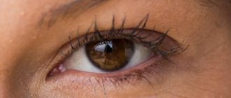
In order for a person to perceive visual information, rays of light pass through his visual apparatus, which are refracted in the lens at a certain angle. Thanks to this, the image is projected onto the retina, from which the optic nerve transmits information to the brain, which processes the image and is responsible for its perception.
The retina contains nerve receptors - cones and rods; they are responsible for color perception. During the daytime, vision is provided by cones, which come in three types. They help us perceive the primary colors of the spectrum: blue, red and green. Thanks to rods, a person sees at dusk and at night. They are designed to perceive black and gray shades.
The optic nerve sends impulses received by the rods and cones to the brain. As a result, a person understands what he saw.
The ciliary muscle also takes part in the functioning of the visual apparatus, which reduces and increases the thickness of the lens. The extraocular muscles are responsible for moving the eyeball and turning it in different directions.
Diagnostics
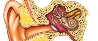
You can determine the reasons why everything is blurry in your eyes using an MRI or CT scan of the brain, X-ray examination of the spine, and laboratory tests. Additionally, the patient visits an ophthalmologist. This specialist undergoes a standard examination: they alternately close the left and right eyes, and recognize the letters that the doctor points to. Other types of diagnostics depend on the characteristics of the clinical case.
Causes of visual impairment
The causes of visual impairment can be congenital or acquired. By impairment, doctors mean not only a decrease in visual acuity, but also image distortion, the presence of dark spots before the eyes, poor visibility at night, and incorrect perception of colors.
Congenital
Congenital causes of poor vision lie in abnormal development of the fetus, complications during pregnancy and childbirth. The most common of them:
- genetic mutations in the fetus;
- consumption of alcoholic beverages, drugs, and certain types of medications while pregnant;
- injury during pregnancy;
- prematurity (vision problems are diagnosed in a third of premature babies);
- pathological formation of organs in a child;
- endocrine diseases in the mother;
- exposure to poisons or toxic substances on a pregnant woman;
- infections suffered by a pregnant woman (rubella, toxoplasmosis, syphilis, etc.).
Often, pathologies of the visual organs are caused by heredity, but it is not the disease itself that is transmitted, but a strong tendency to it. If both parents have the disease, then the child will have it with a probability of 70-90%.
Prevention of such deviations is impossible, but knowing about the pathologies of the parents, you can identify them in the child as early as possible and begin treatment.
Purchased
Visual impairments acquired during life are considered acquired. Most pathologies occur after 40-45 years, when age-related changes in the visual apparatus begin.
The most common causes of complete or partial vision loss are:
- infectious, parasitic, fungal and other eye diseases;
- age-related changes (clouding of the lens, deviation from normal parameters of the eyeball, etc.);
- stressful conditions resulting in deterioration of vision;
- diseases of the circulatory system that provoke circulatory disorders in the organs of vision;
- poor nutrition, lack of vitamins and nutrients necessary for the full functioning of the visual system;
- mechanical eye injuries (bruises, burns, foreign body);
- work associated with constant eye strain, poor lighting of the workplace;
- previous head injuries with cerebral hemorrhage, brain diseases in which the visual center is involved;
- heart attacks and strokes;
- excessive consumption of alcoholic beverages, drugs, caffeine;
- constant use of hormonal drugs, antibiotics, incorrect dosage of medications, or their use without a doctor’s prescription;
- neglect of visual hygiene.
Preventive measures will help to avoid many factors that cause deterioration in visual function.
Why does dizziness occur during pregnancy?
A consultation with an ophthalmologist is one of the mandatory consultations for a pregnant woman. If a problem is detected, the doctor prescribes the necessary treatment. It is especially dangerous if degenerative processes occur in the eye, the retina is damaged, or there are tears or detachments.
Consultations are carried out due to the fact that pregnancy is often accompanied by the appearance of spots, circles, light, blurred vision occurs, silhouettes are distorted, and become blurry. Strabismus is often observed.
Ophthalmologists advise resorting to a cesarean section for myopia of more than 6 diopters. Below this threshold, natural delivery is completely acceptable. However, the indications for cesarean section include pathological processes that occur with myopia.
Symptoms of the disorder
If any symptoms, even minor ones, appear, you should pay attention to them, including if they are periodic. These include:
- Deterioration of vision. The person begins to have difficulty seeing objects near or far, or objects become irregular in shape.
- Double vision. Objects may begin to double after eye or head injuries, but this symptom can also be a sign of serious illness.
- Dizziness and headaches. This sign may indicate both neurological problems and eye diseases. If it is combined with deterioration of vision, then pathology of the visual apparatus is likely.
- Itching, burning, feeling of “sand”. This indicates problems with the mucous membrane.
- Painful sensations. Pain in the eyeball is often caused by infectious or inflammatory diseases, increased intraocular pressure, pathologies of the lens and retina.
- Dry eyes. The reasons may be different: insufficient secretion of tear fluid, diseases of the eyelids, in which the eyelids cannot close completely.
- Impaired pupil reaction. The pupil does not constrict under the influence of light rays and does not expand in the dark. This condition can occur due to head and eye injuries, or the use of drugs or medications.
Why is MS dangerous for vision?
A common ophthalmological manifestation of the disease is inflammation of the optic nerve, which is experienced by more than half of patients. In medicine it is called retrobulbar neuritis. Often this symptom occurs several years before other complaints appear. At the same time, one should not draw premature conclusions and certainly associate neuritis with MS, since it may have other causes. Inflammation can be suspected in case of severe loss of vision, dyschromatopsia, or pain when moving the eyes. This condition lasts no more than 3 months, after which an improvement in well-being follows. However, in case of relapse, optic nerve atrophy may occur. Therapy for this disease is extremely complex and requires the intervention of more than one doctor.
Temporary loss of vision
Sometimes vision impairment is temporary, but the reasons do not lie in eye disease. Symptoms appear mainly in the evenings, and by the morning they disappear without a trace. Sometimes a person sees worse for several weeks, and then the vision is restored on its own.
It is believed that visual function may deteriorate for the following reasons:
- constant lack of sleep, poor sleep quality;
- constant stress, severe nervous shock;
- severe visual stress, overwork at work.
When overtired and lacking sleep, it becomes difficult for a person to focus on images, and the brain processes information slowly. The picture becomes fuzzy, floats and undresses.
Under stress, the pupil dilates, so image quality deteriorates. Also, due to increased breathing, the oxygen content in the blood increases, so “spots” and “stars” flash before the eyes.
In severe emotional states, visual impairment may persist for weeks or even months. Additionally, headaches and eyelid twitching appear. These problems require treatment because they can cause serious health problems.
Causes and symptoms of the disease
As a result of the disease, the myelin sheath of nerve fibers is affected and plaques are formed, which are commonly called sclerotic. It affects the brain and spinal cord. The optic nerve is also affected. It is worth noting that experts do not have a consensus on the causes of the development of the disease. Thus, it is believed that multiple sclerosis is caused by a whole complex of factors, such as:
- genetic;
- viral;
- allergic;
- endocrine;
- geographical.
The symptoms of the disease vary from person to person. Patients may complain of symptoms such as lack of coordination of movements, muscle flaccidity, numbness, sudden paralysis and loss of balance. However, most often patients are brought to consult a doctor by visual impairment due to multiple sclerosis. This manifests itself in blurred images, pain and double images.
Types of violations
There are more than two hundred eye pathologies, but some of them are quite rare. Let's look at the most common types of visual impairment.
Myopia (myopia)
Myopia is a refractive error in which light rays refract early and therefore do not reach the retina. The image is focused in front of the retina.
Patients with myopia can only see objects at a short distance. With a severe degree of the disorder, a person literally cannot see beyond his nose.
The causes of myopia lie in the fact that the eyeball seems to stretch in length, that is, its anteroposterior size increases. In rare cases, myopia is caused by spasm of the ciliary muscle, a change in the structure of the cornea.
Farsightedness (hypermetropia)
Hypermetropia is a visual impairment in which rays are focused behind the retina. A person sees objects located in the distance well, but the picture near them blurs and becomes unclear.
The main cause of hypermetropia is shortening the anteroposterior size of the eyeball. Other reasons include a decrease in the elasticity of the lens.
The concept of hypermetropia also includes presbyopia. This disorder occurs in old age due to the natural aging of the body, so age-related farsightedness cannot be prevented, you can only delay its occurrence.
Astigmatism
Astigmatism is a defect in which rays are refracted in different directions, so the image is projected behind and in front of the retina. Only some of the rays hit the retina. The patient sees periodically clearly or blurred, and the distance to the object does not matter. Many people with astigmatism perceive images with distorted shapes and blurred outlines.
Astigmatism appears due to the curvature of the shape of the cornea, lens, and other parts of the eyeball.
Strabismus (strabismus)
With strabismus, the eyes do not look symmetrically, but in different directions. This makes it impossible to focus on one image.
Causes of strabismus:
- there are pathologies of the extraocular muscles;
- the eyeballs are positioned incorrectly;
- visual acuity decreases in only one eye;
- there are problems with accommodation;
- the person has undergone injury or eye surgery;
- tumors of the visual organs and brain appeared;
- the central nervous system was affected.
Strabismus has a classification depending on the nature of the deviation: convergent, divergent, vertical and mixed form.
Amblyopia
Amblyopia is characterized by the presence of one “lazy” eye that does not function properly, that is, it is not involved in the visual process. The brain cannot combine two images into one, so monocular vision develops. If the problem is not addressed, visual functions will increasingly deteriorate.
Most often, amblyopia occurs due to untreated strabismus. It can also be caused by pathologies of the fundus and cornea.
Anisometropia
With anisometropia, the refractive power of the eyes varies significantly. In this case, one eye may have 100% vision, while the other may have significant impairments (myopia, farsightedness). Also, both eyes may have anomalies, but the visual acuity in each of them will vary significantly (from 2 or more diopters). With such a colossal difference in refraction, the brain cannot perceive information, and a failure in perception occurs. Therefore, the nervous system gives preference to only one eye, or nerve impulses are read in turn from each eye.
Prerequisites for the occurrence of anisometropia: abnormal structure of the visual organs, surgical operations on the eyeballs, ophthalmological diseases.
Colorblindness
With color blindness, a person does not distinguish colors correctly; he does not perceive red, green or blue. In severe cases, the patient sees everything in black and white. This type of disorder is called achromasia.
The cause of the pathology lies in defects in the cones: there are either few of them or there are not enough coloring pigments in certain types.
Usually this anomaly is congenital and cannot be treated. It is extremely rare that color blindness occurs due to inflammatory diseases, damage to the optic nerve, and age-related degeneration of the visual apparatus.
Arterial pressure
Darkening and dots before the eyes occur during a sharp surge in pressure. This happens in healthy people if:
- Make sudden movements;
- Stay upside down for a long time;
- Quickly change body position.
The blurry vision is explained by a sharp increase in pressure, arterial and ocular. You should measure your blood pressure and lie quietly for a couple of minutes. During this time, the pressure will normalize and vision will be restored.
If rest does not help, the cause of blurred vision may be cardiovascular pathologies:
- Arterial hypertension;
- Hypotension;
- Vasospasm;
- Dysfunction of the autonomic nervous system;
- Anemia.
Glucose level
Diabetes mellitus is one of the causes of decreased vision. The risk of developing eye diseases occurs during periods of high blood sugar. The patient's vision becomes cloudy due to swelling of the lens.
To restore visual function, it is necessary to normalize glucose levels.
People with diabetes should visit an eye doctor regularly. Sometimes such patients have blurred vision due to concomitant diseases:
- Retinopathy (progressive damage to the retina);
- Intraocular pressure;
- Cataract.
These pathologies are dangerous due to decreased or loss of vision.
To prevent the development of complications, people with diabetes are required to monitor the following indicators:
- Blood sugar;
- Cholesterol;
- Arterial pressure.
The problem occurs in people who engage in sports and heavy physical labor. A decrease in blood sugar after exercise is accompanied by dizziness and the appearance of black spots before the eyes.
Visual function is completely restored, additional symptoms disappear after rest and a carbohydrate snack.
Correction of visual impairments
Most disorders can be successfully treated if you consult a doctor in time. In this case, there are no negative consequences.
The ELITE PLUS clinic uses only progressive methods for eliminating violations:
- Night lenses. This is a modern way to correct myopia, astigmatism, and farsightedness. Night lenses not only help you see better, but also have a therapeutic effect.
- Hardware treatment. Courses of hardware therapy relieve spasm of accommodation, improve blood supply to the eyeball, eliminate visual strain, and help cure myopia (including false), strabismus, amblyopia, etc. without surgery. This treatment method is well suited for children, since the sessions are held in a playful way.
It is especially effective to use the techniques in childhood, since it is possible to completely eliminate refractive errors. The sooner treatment is started, the faster and easier it is for a person to get rid of the disease.
Dizziness and eye pain: why they occur and what to do with such symptoms
Often the cause of dizziness, nausea, and lack of coordination is heart disease. Moreover, they can rather be attributed to the consequences of the general weakening of the patient’s body. After all, the heart is the main worker of the human body.
And it is impossible to eliminate dizziness without treating the underlying disease.
Heart rhythm disturbances
Dizziness often accompanies heart rhythm disturbances (arrhythmia), especially bradycardia (decreased rhythm) and extrasystole (failure to contract the rhythm of the heart or its individual parts).
The person feels weak, tired and dizzy. Sometimes mild nausea is felt, but there is no vomiting.
Cardiomyopathy
This is a whole group of ailments in which, for various reasons, pathological changes in heart tissue occur. As a result, it does not perform its functions well enough, which leads to an imbalance of the entire human body. Including weakness and dizziness.
Heart defects
This is a negative change (congenital or acquired) in the structure of the heart or large vessels, which results in a defect in one or more heart valves.
In this case, blood circulation is carried out insufficiently efficiently. Therefore, dizziness, nausea, and lack of coordination occur, the cause of which lies in the poor supply of oxygen to the brain.
Orthostatic collapse
Sharp darkening of the eyes and even fainting are possible with orthostatic collapse due to a drop in blood pressure. This occurs when a person quickly changes the position of a person’s body from horizontal to vertical, or when they remain in a standing position for a long time.
Anemia
Dizziness is one of the common symptoms of anemia, in which the supply of hemoglobin to the organs is impaired. It does not have any pronounced symptoms and lasts until the cause is eliminated, intensifying with physical exertion or blood loss.
Hypoglycemia as a cause of dizziness and loss of coordination
Unpleasant symptoms may occur due to a drop in blood glucose levels. Therefore, you should be careful about eating in a timely manner, especially during stress and increased physical activity.
People prone to hypoglycemia need to strictly monitor their diet and meal times. Meals should be divided into smaller portions and taken every 3 hours.
Very often, dizziness, nausea, and lack of coordination, the cause of which does not lie in a disease of the body, are the result of improper human behavior.
For example, such moments include:
- poor nutrition (fasting or violation of diet rules);
- "seasickness";
- sudden change in weather;
- drinking alcohol.
Diet violations
Diets are a complex process that should be carried out under the supervision of a specialist. But often people try to adhere to various dietary restrictions without consulting a doctor.
If you make mistakes in your diet (ill-thought-out vegetarianism, complete refusal of carbohydrates, etc.), manifestations of hypoglycemia, anemia, or simply hungry dizziness are possible. If you completely give up salt, your blood pressure may drop, which can also cause dizziness.
Starvation
Fasting is always accompanied by a decrease in blood glucose levels. The brain does not receive enough nutrition. And the person feels dizzy. Small but frequent (at least 6 times a day) consumption of a minimum amount of food will help to avoid this.
Motion sickness
“Seasickness” is weakness and dizziness, as well as nausea and vomiting during a long trip by train, car, ship, plane flight or excessive rides on attractions.
In these situations, the human body may not be able to cope with signals arriving through various channels. Children and only about 1% of adults are more susceptible to motion sickness. Special medications help relieve discomfort.
Change in atmospheric pressure
Any chronic diseases, especially those of a vascular nature, as well as weakening of the human body after illness and in the presence of age-related changes, can cause increased sensitivity to changes in weather conditions.
In this case, mild dizziness or lightheadedness, migraines, which can be relieved with short-term medications, are possible.
Prevention of visual impairment
Preventive measures to help avoid vision loss:
- Proper organization of the workplace. The monitor should be moved 50-60 cm away from the eyes. The lighting should fall from the left. The chair should have back support and not be too high so that your feet reach the floor.
- A complete diet. The menu should contain products with substances beneficial to the eyes. These include: blueberries, citrus fruits, persimmons, eggs, seafood, nuts, honey, pumpkin, liver.
- Vitamin complexes. It is better to take vitamins and minerals to improve visual abilities on the recommendation of an ophthalmologist. They should contain vitamins A, B, C, E, lutein, zeaxanthin, zinc, selenium, copper.
- Eye drops. Medicinal drops should be used as recommended by a doctor. You can use “artificial tears” yourself to prevent excessive dryness.
- Gymnastics for the eyes. Exercises are done in the mornings and evenings, as well as during the working day during breaks when working at the computer. Breaks are required every 40 minutes.
Fundus of the eye in arterial hypertension
Have you been struggling with HYPERTENSION for many years without success?
Head of the Institute: “You will be amazed at how easy it is to cure hypertension by taking it every day...
Read more "
The supply of useful substances to the retina is carried out using blood vessels located in the fundus of the eye. The development of arterial hypertension leads to increased intraocular pressure. This is fraught with a decrease in visual acuity, pressing pain in the area of the superciliary arches, and a significant decrease in performance. Many people attribute migraines and “floaters before their eyes” to fatigue, lack of sleep or prolonged work at the computer. The fundus of the eye in hypertension may be damaged due to vascular spasm. There are cases when vision deteriorates directly during a hypertensive crisis, and then is restored back.
Causes of fundus changes
Arterial hypertension is an insidious disease that can be asymptomatic and is accidentally discovered only during a routine medical examination. Signs of fundus changes in hypertension resemble vascular inflammation caused by glaucoma, which is a local pathology.
Normal intraocular pressure is 12–22 mmHg. Art. If, in addition to changes in blood pressure, there are no other symptoms of glaucoma, we are talking about hypertension.
The development of hypertension can be provoked by:
- bad habits (alcohol, smoking, drugs);
- abuse of coffee and other tonics;
- overweight, unhealthy diet, physical inactivity;
- old age, genetic predisposition, chronic stress;
- improperly functioning cardiovascular, endocrine and nervous systems.
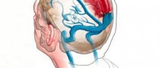
Fundus examination for hypertension is included in the list of mandatory preventive procedures, since its malignant course leads to damage to internal organs. Together with the vessels located in the retina, the cerebral arteries suffer, which is fraught with an attack of hemorrhagic stroke.
With the help of a visual analyzer, more than 80% of information about the world around us is learned. Visual impairment due to hypertension is one of the serious complications of the disease. High blood pressure is accompanied by spasm of blood vessels, tension in their walls, and thickening of the blood, which can lead to retinal infarction, the formation of microthrombi, and hemorrhage.
Classification of vascular pathologies of the retina
With the help of ophthalmoscopy, even minor changes in the fundus of the eye are diagnosed in hypertension. Based on the nature of inflammation of the retinal vessels, the ophthalmologist determines the etiology of the disease in order to predict its further course and select the appropriate treatment. Sometimes contrast methods, such as angiography, are allowed. Eye pain accompanied by lacrimation may be of allergic origin, so it is important to differentiate the two conditions through therapeutic and ophthalmological examinations.
Among the fundus lesions associated with a persistent increase in blood pressure are:
- Hypertensive angiopathy.
- Hypertensive angiosclerosis.
- Hypertensive retinopathy.
- Hypertensive neuroretinopathy.
These pathologies differ in the localization of inflammation, the size of the affected area and the level of vision loss. Damage to the optic nerve is very dangerous, since it is used to conduct nerve impulses from the receptors of the retina to the occipital lobe of the brain, where visually received information is processed. Changes in the eyes with hypertension gradually progress, which is fraught with negative consequences.
The above stages of development of retinal vascular lesions can transform into one another. First, inflammation of the eye arteries and veins occurs; they cannot withstand the excessive load caused by increased pressure in the body. Compensatory mechanisms are depleted, resulting in tissue sclerosis. The malignant course of the disease leads to generalized damage to the retina along with the optic nerve.
Signs of increased intraocular pressure
With cardiovascular diseases, performance and concentration levels are significantly reduced. The visual analyzer plays an important role in various activities. Hypertension and glaucoma negatively affect the condition of the retina.
The first symptoms of damage to the ocular vessels are:
- redness of the protein membrane;
- rapid development of fatigue during reading, prolonged work at the computer;
- a person sees poorly in the twilight;
- the field of view becomes smaller, the picture seems to blur;
- pressing pain in the temporal region;
- sunlight causes unpleasant sensations, “floaters appear before the eyes.”
People who naturally have very good vision begin to be frightened by the rapid development of symptoms of arterial hypertension. Today, there are various treatment methods, which include surgical correction, therapy with vitamins and minerals. Before starting to fight eye angiopathy, it is worth achieving normalization of blood pressure in the whole body.
Clinical picture of changes in the fundus in hypertension
The degree of vascular damage depends on the stage of the disease. At first, it may resemble fatigue caused by excessive load on the visual analyzer. As the symptoms progress, they intensify and do not disappear even after proper rest. People run to buy drops for conjunctivitis, put on safety glasses, try to avoid prolonged work at the computer, without realizing the true nature of the visual impairment. Unfortunately, many patients turn to the doctor when the disease has already significantly affected the level of vision.
The following periods are distinguished in the development of ocular hypertension:
- Retinal angiopathy occurs from hypertension in a mild stage, which is accompanied by a short-term increase in blood pressure. Symptoms of the disease, such as headaches, “jumping midges” before the eyes, redness of the sclera, may disappear over time and then reappear. A slight dilation of the veins, together with spasm of the arteries, causes hyperemia of the fundus.
- Hypertensive angiosclerosis. Pathological changes in the ocular vessels acquire an organic character. Discomfort and redness are accompanied by hardening of the arterial walls, which leads to the “copper wire symptom” (the vessels of the fundus become yellow-red). Over time, it develops into the “silver wire symptom,” characterized by a white tint. At the site of crossing of the vessels, compression of the ophthalmic vein is observed, which causes the Salus-Hun symptom.
- Generalized retinopathy. Pathological changes from the vessels spread directly to the retina, causing its swelling, the appearance of white and yellowish spots, and figures in the form of a ring or a star form around the visual spot. At this stage of the disease, visual impairment is pronounced due to a decrease in its acuity.
- Involvement of the optic nerve in the inflammatory process is neuroretinopathy. Its disc swells, and over time the entire retina becomes swollen. The permeability of blood vessels increases significantly, and their plasmatic cutting occurs.
At the last stage of development of ocular hypertension, an irreversible decrease in visual acuity occurs. Only timely treatment will help a patient with high blood pressure maintain the function of the visual analyzer and avoid dangerous complications.