Epilepsia is a fairly common neurological disease that causes seizures. Seizures usually involve disturbances in consciousness, sensory and motor functions, emotions and behavior.
The clinical picture manifests itself in the form of generalized convulsive attacks or in small forms, for example, absence seizures, talking or walking during sleep, twitching.
Epilepsia is treatable, but before determining treatment options, an accurate diagnosis must be made. The fact is that a single case of a seizure is not enough to establish the presence of a disease; there must be two or more of them.
Electroencephalography is one of the effective methods for examining patients. The disease has more than 50 forms, so it is extremely important to identify them in order to choose the right treatment method.
Since the pathology is expressed by the discharge of neurons in the brain, using electroencephalography it becomes possible to identify indicators characteristic of each form.
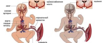
The causes of the disease are different and depend on age. So, in adults it can be brain tumors, severe head injuries, a stroke, or alcohol addiction. The causes of childhood illness can be oxygen starvation during difficult childbirth, infections suffered by the mother during pregnancy - toxoplasmosis, herpes, rubella, cytomegaly and others.
In adolescence, the disease more often manifests itself due to hereditary predisposition. If children are born to people who are closely related, there is a high probability that they will be susceptible to this disease.
Based on the causes of occurrence, the disease is divided into three groups:
- Symptomatic, when it is possible to identify structural defects in the brain.
- Ideopathic – the presence of a genetic predisposition.
- Cryptogenic – the cause cannot be determined.
It is to determine the hereditary factor that it is necessary to conduct genetic research. They will show the presence of a predisposition. In addition, when treatment is started, other tests for epilepsy are taken to determine the concentration in the body of the substances contained in the drugs. This allows you to regulate the dosage of medications. To identify side effects after taking AEDs, a biochemical blood test is performed and an MRI is prescribed.
What is EEG: the essence of the method
An EEG or electroencephalogram is an effective diagnostic method for patients with signs of epilepsy and other various brain injuries. This method is often prescribed to patients who do not need it at all.
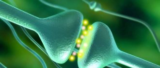
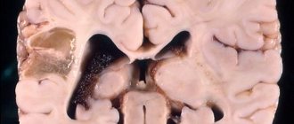
The essence of the technique is that it records electrical signals sent by neurons - nerve cells of the brain. In fact, many pathologies are manifested by severe disturbances in the electrical activity of the brain. Most often this is epilepsy, during which a group of neurons exhibits serious activity, and structural changes in the brain are detected: tumors, cysts, consequences of stroke and hemorrhages.
This technique is considered to be as accurate as possible, and all because it can fully show the entire clinical picture of the disease:
- how far the inflammation has spread and its level;
- what changes have occurred in the vessels;
- early signs of epilepsy;
- neoplasms and stages of their development;
- how much brain function is impaired due to an illness affecting the nervous system;
- what are the consequences of a stroke, hemorrhage or surgery.
According to ICD-10, epilepsy is assigned code G40; a detailed description of the EEG for this pathology allows you to track what changes have occurred in the brain, especially if this study was not carried out for the first time. Thus, the doctor has the opportunity to monitor brain activity during treatment and adjust it at any time. Almost always, after diagnosis, the doctor can accurately determine where exactly the source of excitation is localized.
Characteristic
An indicator of epileptic activity on the EEG is recorded in the form of certain waves, or peaks of an acute nature.
Their difference lies in their high amplitude; these are typical graph elements and patterns. They differ depending on the specific form of epilepsy. When the disease manifests itself in the form of foci, then such changes are characterized by:
- Rolandic epilepsy. In this situation, a wave of sharp peaks is visible. They are collectively called the “Rolandic complex.” They are most often formed in the area of the central temporal leads, face, lips, tongue;
- The focus of nocturnal or frontal epilepsy is localized in the frontal lobes.
Generalized epilepsy, when diagnosed on an EEG, manifests itself in the form of a whole complex of waves throughout the entire region of the brain.
Subclinical patterns are distinguished - these are cases of epilepsy when there are no symptoms as such. This condition is rare and occurs in a small number of patients.
With the help of the procedure, the doctor, in combination with the main methods of treatment, will see the complete clinical picture and make a diagnosis based on this. A neurologist or epileptologist takes into account the patient’s complaints and, if the diagnosis is confirmed, is able to determine the exact form of development of the disease.
Sometimes repeated procedures are performed to confirm a previously made diagnosis. The body is examined using functional tests and loads. The procedure continues for several hours or a day. All results are recorded using video monitoring.
Note! The patient should not self-diagnose. This is done exclusively by the doctor. Self-medication in this case is unacceptable.
When should an EEG be performed?
This diagnostic method is used for various speech, mental and neurological disorders. As a preventive measure, EEG can be prescribed to people who are taking a driver's license exam, as well as to obtain permission to store and carry weapons. Positive results can exclude the presence of schizophrenia and other mental disorders. EEG shows epilepsy, and also provides other data, so this technique is used for medicinal purposes:
- after surgery that could affect the functioning of brain cells;
- when identifying and determining the location of tumor and cystic formations;
- with traumatic brain injuries obtained in various ways;
- to confirm or deny the presence of epilepsy;
- if the patient experiences convulsions, numbness of the limbs and fainting;
- with chronic hypertension and circadian rhythm disturbances;
- if the child has developmental delay.
An EEG of the brain can reveal not only disturbances in the functioning of brain tissue, but also the severity and depth of their damage, and the location of the source of the disease. In some cases, regular monitoring may be recommended, in other words, several studies at short intervals, which will help not only to identify signs of epilepsy on the EEG, but also to determine further treatment, which can be adjusted.
By studying the processes of activity of the nervous system, it is possible to prevent another attack. For patients in a coma or under long-term general anesthesia, this type of diagnosis is mandatory, because it will help determine the performance and vital activity of brain tissue.
The concept of “epileptic activity”
This term is used in two cases:
- Registration of epileptiform phenomena on the EEG during a seizure (psychomotor seizure pattern or ongoing polyspike). The activity may not contain epileptic seizure patterns.
- In the case of a clear activity schedule. Can be recorded outside of an attack.
Hereditary EEG patterns may be associated with epileptic seizures. Some specific combinations have different epileptic syndromes.
The presence of epileptiform activity and epileptic seizure patterns on the EEG, high-amplitude bursts of activity (more than 150 μV) are important signs of the presence of epilepsy.
Value of the study
Epilepsy code according to ICD-10 G40, EEG in this disease helps to identify and record foci of neuronal activity. The main aspects of the application of this technique are:
- determination of the form of the disease;
- opportunities to track development dynamics;
- monitor improvements in the patient's condition;
- selection of the correct therapy and dosage of drugs.
The main value of diagnosis is that all pathological changes can be detected between seizures.
If anomalies are present, then the equipment records peaks and waves, as well as graph elements specific to its definition. Therefore, as soon as bursts of activity, peaks and waves appear on the EEG, this already indicates the presence of a pathological condition, but this is not enough for an accurate diagnosis. Similar changes are often observed in cases of malignant neoplasm, after a stroke, sleep disorder, and encephalopathy. That is why additional studies are being carried out.
Each form of the disease has its own characteristic waves. With rolandic, their greater concentration is observed in the central temporal lobes, with nocturnal - in the frontal part.
How to prepare for an EEG?
After 12 o'clock at night before the diagnosis, the patient should avoid drinking drinks containing caffeine. Hair should be washed and dried, and oils, lotions and aerosols should not be applied to it. There are no other requirements, but the child needs to be prepared more carefully to get the right results.
An epileptic attack in a child can begin at any moment and parents are not always prepared for it. Therefore, if even the slightest deviations are noticed, you need to seek help and undergo an examination. An EEG will help make an accurate diagnosis, but you should prepare for the study:
- Take a good look at the child's head. If wounds and scratches are found, they should be reported to the doctor. Electrodes should not be attached to damaged areas of the skin.
- Feed the baby. The study is carried out on a full stomach, this is the only way to get clear results. But you can’t give sweets with chocolate. Infants are fed before the procedure in a medical facility. In this case, the baby will fall asleep peacefully and sleep peacefully during the examination.
Stop taking medications, but if the child receives them on an ongoing basis, then doctors warn about this. Children of school and preschool age need to be explained what they will be doing; only the right psychological attitude will help to avoid excessive emotionality. The child can take toys with him, but not electronic ones.
Remove all foreign objects, such as elastic bands and hairpins, from the head; hair should be loose. If an EEG for epilepsy is not done for the first time, then be sure to take the previous transcript with you. If a child is sick, no diagnosis is carried out; a full recovery is expected.
Features of diagnostics
An EEG of the brain is a painless procedure that will not harm either an adult or a child. During diagnosis, the patient sits comfortably in a chair, electrodes are placed on his head, but initially measurements are taken in three positions: head circumference, the distance between the bridge of the nose and the protrusion of the occipital bone, from one ear to the other through the crown. Only after this can you determine exactly where to attach the electrodes. The attachment point is degreased with alcohol, after which a gel is applied and the sensor is installed. In some cases, special helmets or caps may be used.
What does an EEG look like for epilepsy? Yes, in different ways, even in a healthy person you can sometimes detect waves and peaks of activity, which is mainly due to his individual characteristics.
In children who suffer from neuroses and psychopathy, as well as those with an aggressive character, the study reveals activity, but there are no clinical signs. But most young patients with such data are eventually diagnosed with epilepsy.
With widespread seizures, activity is observed in all areas, and when it is a focal form, then only in certain areas. It is not always possible to detect signs of illness in people who abuse alcohol. The following may cause activity in such patients: eye movement, swallowing, touching sensors, contraction of head muscles, heartbeat, pulsation of blood vessels.
The patient's age, taking pills for epilepsy or other diseases, time of the last seizure, visual impairment, abnormal shape of the skull - all this can affect the EEG results. Therefore, the study is carried out taking into account all related factors.
How long does the study take?
A routine examination is a routine EEG or diagnosis of a paroxysmal state. The duration of the study depends on which area is being tested and what functional tests are used. On average, the procedure takes about half an hour. During this time, the specialist manages to:
- perform rhythmic photostimulation using different frequencies;
- check for hyperventilation;
- carry out a load in the form of blinking;
- detect hidden changes.
If the data obtained is not enough, then in addition to the EEG for epilepsy, the specialist can use a more in-depth and effective examination:
- EEG of night sleep.
- EEG with deprivation.
- Long-term EEG.
The duration of these techniques can take from 20 minutes to 15 hours.
3.4. Epilepsy with seizures provoked by specific factors
Epilepsy with seizures provoked by specific factors is characterized by partial and partial-complex seizures, which are regularly reproduced by some direct influence. A large group consists of reflex attacks.
Haptogenic seizures are caused by thermal or tactile stimulation of a certain area of the body surface, usually projected into the zone of epileptogenic focus in the cortex when it has a destructive focal lesion.
Photogenic seizures are caused by flickering light and manifest as minor, myoclonic, and grand mal seizures.
Audiogenic seizures are caused by sudden sounds, certain melodies [Ictal..., 2003] and are manifested by temporal psychomotor, grand mal, myoclonic or tonic seizures.
Startle seizures are triggered by a sudden startling stimulus and manifest as myoclonic or brief tonic seizures.
EEG rhythms
During an EGG, the device detects four main types of rhythms:
- Alpha waves are the main element in the diagnosis of a healthy adult patient and are recorded in 90% of people. These waves have a frequency of around 13 hertz per second and are fundamental during wakefulness, when the patient simply lies with his eyes closed. The maximum activity of alpha waves is observed in the area of the back of the head and crown.
- Beta, like alpha waves, are considered normal manifestations in the body of a healthy adult. But the number of their oscillations reaches 35 hertz per second, they are recorded mainly over the frontal part. The beta rhythm appears if the sensory organs are irritated: by touching the patient, when stimulated by light or sound.
- Delta waves with a frequency of up to 3 hertz during decoding of an EEG for epilepsy can mean normal in a baby up to one year old. The indicator partially persists for up to 7 years. In adults, they are recorded during sleep.
- The theta rhythm with a frequency of up to 7 oscillations per second is normally found in children from one to 6 years of age, gradually being replaced by alpha waves as they grow older. In adults they are observed during sleep.
Analysis transcript
Before answering the question of what to do with epilepsy, you need to accurately decipher the EEG data. Research data is displayed on a monitor or on paper in the form of graphic curves that only an experienced specialist can decipher. The analysis and conclusion of an EEG for epilepsy is issued by a neurophysiologist, who, when decoding, takes into account the patient’s age, his complaints, the clinical picture of disorders in the body and many other factors, such as heredity.
Key points of decoding:
- It is determined which of the rhythms is the main one, predominant in the patient.
- The symmetry of the electrical potentials of nerve cells, which are recorded from the left and right hemispheres of the brain, is carefully studied.
- Pathological changes, such as delta and theta waves in an adult patient while awake, are carefully analyzed.
- The regularity and amplitude of the rhythms are checked.
- Paroxysmal activity is revealed when sharp waves, peaks and sky-waves are detected on the curve.
- If there are no pathological changes in the background EEG, then additional functional tests are performed, such as hyperventilation or photostimulation, repeated recording of electrical potentials and decoding.
Types of procedure
To make an accurate diagnosis, a set of studies is prescribed, which includes repeated electroencephalography and long-term monitoring from 12 to 24 hours.
The process does not cause pain and is tolerated calmly. To take readings, electrodes are attached to the head of the person being examined, which record the activity of neurons and send them to an electroencephalograph, computer or flash card. Here the information is processed by comparing the obtained data with normal values. At the same time, the device records peaks and waves, reflecting them on paper in the form of a curve, which is deciphered by a specialist. The following types of procedure are used today:
- "Routine." Its purpose is to take readings about the biopotential of the brain.
- With stimulation of activity, for example, through hyperventilation, when the patient is asked to breathe quickly and forcefully. Photostimulation, in which the subject is exposed to flashing LEDs. And other types - reading, listening to music, etc.
- An encephalogram may be performed with a larger number of electrodes than usual.
- During night sleep.
- With deprivation.
- Long-term monitoring takes several hours or even days. In this case, the patient behaves as usual and can perform his usual housework.
- VEEG involves recording throughout the entire electroencephalography process for further analysis.
Different activity rhythms are recorded, e.g. Alpha, Beta, Mu, Theta and Delta, which differ in frequency measured in hertz. The readings are interpreted in accordance with age standards.
What changes are observed on the EEG in epilepsy?
During an epileptic seizure, EEG recording allows recording high-amplitude activity in the form of peaks and sharp waves. Outside of an attack, convulsive activity in the brain may not manifest itself in any way, therefore, to provoke epileptic activity, various tests are used. Patients often experience paroxysmal activity in the form of high-voltage theta and delta waves. For long-term EEG recording, it is allowed to use video monitoring, when the study is carried out over a long period of time, in some cases up to 8 hours, and subsequently a specialist deciphers it.
EEG allows not only to determine the location of the source of the disease, but also to recognize its type. If, nevertheless, the diagnosis is confirmed and bright and unlike anything else changes can be seen on the graph, then the description of the EEG for epilepsy will contain the following information:
- waves with sharp angles, rising and falling sharply;
- pronounced slow waves with sharp angles;
- a sharp increase in amplitude by several units;
- during testing for hyperventilation, narrowing and severe spasm of blood vessels appears;
- During photostimulation, an unusual reaction to the test is observed.
If there is a suspicion that this is indeed epilepsy, then during the control study the tests are carried out in a gentle mode, and all because the load can cause another epileptic attack in the patient.
When the diagnosis is definitely established, many people wonder what to do with epilepsy, how to help such a patient.
Basis of epileptiform activity
At the basis of epileptiform activity at the cellular level, a paroxysmal displacement of the membrane occurs, which causes a burst of action potentials. They are followed by a long period of hyperpolarization.
This action occurs regardless of what kind of epileptiform activity is recorded, focal or generalized.
Each of these patterns can also be observed in phenotypically healthy people. The presence of these patterns is not a clear basis for diagnosing epilepsy, but does indicate the possibility of a genetic predisposition.
In some patients, epileptiform activity is recorded only during sleep. It can be triggered by certain stressful situations or the behavior of the person himself.
To clearly determine the pathology, you can provoke an attack with special stimuli. If the patient is subjected to rhythmic light stimulation during sleep, it is possible to detect the presence of epileptiform discharges and patterns of epileptic seizures.
To generate epileptiform activity, it is necessary to involve a huge number of nerve cells – neurons.
There are 2 types of neurons that play an important role in this process:
- 1 type of neurons – “epileptic” neurons. PD flashes are issued autonomously;
- Type 2 – surrounding neurons. They are under afferent control, but can be involved in the process.
There are some exceptions to severe epileptic activity that occurs without seizures but reaches the level of status epilepticus.
- Landau-Kleffner syndrome;
- ESES;
- various non-convulsive epileptic encephalopathies.
What other diagnoses does EEG reveal?
After undergoing an examination, neurologists often diagnose children and adults not only with epilepsy, but also with other ailments. Among the common diseases are the following:
- neoplasm in the brain of various etiologies, the cause of which is not clear;
- traumatic brain injury;
- an inflammatory process that simultaneously affects the membranes of the brain and the medulla; the cause of this condition can be an infection;
- abnormal accumulation of fluid in the structures of the brain, often this pathology is congenital, perhaps the pregnant woman did not undergo mandatory screenings, or this disease developed as a result of an injury that the baby received during childbirth;
- a chronic disease that affects the mental and nervous state with characteristic attacks - epilepsy, provoking factors can be heredity, birth injuries, infections, improper behavior of the mother during pregnancy;
- hemorrhage in the brain due to rupture of blood vessels, a condition that can be provoked by high blood pressure, head injuries, blockage of blood vessels with cholesterol plaques;
- cerebral palsy;
- sleepwalking, somnambulism.
Many diseases can be detected, therefore, if suddenly the EEG does not show epilepsy, but there are seizures, the doctor prescribes other types of studies. This is the only way to exclude other pathologies and make a more accurate diagnosis.