Cerebellar atrophy is a degenerative disease of the small brain that progresses over time. Develops with vascular disorders, some metabolic disorders, infections, tumors of the nervous system. Atrophic processes lead to impaired coordination of movements, speech, and paralysis.
Functions and structure of the cerebellum
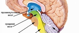
The human brain has a complex structure and consists of several sections. One of them is the cerebellum, which is also called the small brain. This department performs a wide range of functions necessary to maintain the performance of the entire body.
The main function of the described part of the brain is motor coordination and maintaining skeletal muscle tone. Due to the work of the cerebellum, it is possible to coordinate the work of individual muscle groups, which is necessary for performing any everyday movements.
In addition, the cerebellum is directly involved in the reflex activity of the body. Through nerve connections, it is connected to receptors in different parts of the human body. When exposed to a certain stimulus, a nerve impulse is transmitted to the cerebellum, after which a response is formed in the cerebral cortex.
The ability to conduct nerve signals is possible due to the presence of special nerve fibers in the cerebellum. The development of atrophy has a direct impact on these tissues, as a result of which the disease is accompanied by various movement disorders.
The cerebellum is supplied with blood through three groups of arteries: anterior, superior and posterior. Their function is to provide an uninterrupted supply of oxygen and nutrients. In addition, certain components in the blood provide local immunity.
The cerebellum is one of the main parts of the brain responsible for motor coordination and many reflex movements.
Causes of atrophy
In general, atrophic processes in the brain, and in particular in the cerebellum, can be triggered by a large number of reasons. These include various diseases, exposure to pathogenic factors, and genetic predisposition.
With atrophy, the affected organ does not receive the required amount of nutrients and oxygen. Because of this, irreversible processes develop associated with the cessation of the normal functioning of the organ, a decrease in its size, and general exhaustion.
Possible causes of cerebellar atrophy include the following:
- Meningitis. With this disease, an inflammatory process develops in various parts of the brain. Meningitis is an infectious disease that, depending on its form, is caused by bacteria or viruses. Cerebellar atrophy due to the disease can develop due to prolonged exposure to blood vessels, the direct influence of bacteria, and blood poisoning.
- Tumors. A risk factor is the presence of tumors in the patient in the posterior part of the cranial fossa. As the tumor grows, pressure on the cerebellum and the brain regions located in close proximity increases. Because of this, blood flow to the organ may be disrupted, which subsequently provokes atrophic changes.
- Hyperthermia. One of the causes of cerebellar damage is prolonged exposure to high temperature. This may be due to increased body temperature due to some illness or heat stroke.
- Vascular diseases. Often, cerebellar atrophy occurs against the background of cerebral atherosclerosis. The pathology is associated with a decrease in the patency of blood vessels, depletion of their walls and a decrease in tone caused by focal deposits. Against the background of atherosclerosis, oxygen deficiency develops and the flow of substances worsens, which, in turn, causes atrophic changes.
- Complications after a stroke. Stroke is a sudden disruption of blood circulation in the brain caused by hemorrhages and cranial hematomas. Due to a lack of blood in the affected tissue areas, they die. Cerebellar atrophy acts as a consequence of this process.
The diseases described above have a direct impact on the functioning of the cerebellum, causing irreversible changes in it. The danger of atrophy of any parts of the brain lies in the fact that they consist mainly of nerve tissue, which practically does not recover even after long-term complex treatment.
Cerebellar atrophy can be caused by the following factors:
- Constant drinking of alcohol.
- Diseases of the endocrine system.
- Traumatic brain injuries.
- Hereditary predisposition.
- Chronic intoxication.
- Long-term use of certain drugs.
Thus, cerebellar atrophy is a condition associated with an acute deficiency of oxygen and nutrients, which can be provoked by diseases and a wide range of harmful factors.
Cerebellar ataxia in humans. How to treat?
In cases where ataxia manifests itself as a secondary disease, treatment should begin with eliminating the underlying disease. Infectious and inflammatory processes require antibacterial and antiviral therapy. If the etiology of the pathology lies in vascular disorders, normalization of blood circulation will be required. For this purpose, drugs from the category of anticoagulants, thrombolytics, vasodilators, and angioprotectors are prescribed.
It will not be possible to completely get rid of the hereditary form of the disease. With this type of disease, the only help will be metabolic therapy - the use of B vitamins, preparations based on ginkgo biloba, nootropics.
A mandatory part of the treatment process is massage and physical therapy. A properly selected set of exercises will normalize the condition of muscle tissue, restore tone, and coordinate movements.
Types of cerebellar atrophy
The form of the disease depends on a number of aspects, among which the most significant are the cause of the lesion and its location. Atrophic processes can occur unevenly and are more expressed in certain parts of the cerebellum. This also affects the clinical picture of the pathology, which is why it is often individual for each individual patient.
Main types:
Cerebellar vermis atrophy is the most common form of the disease. The cerebellar vermis is responsible for conducting information signals between different parts of the brain and individual parts of the body. Due to the lesion, vestibular disorders occur, manifested in imbalances and coordination of movements.
Diffuse atrophy. The development of atrophic processes in the cerebellum often occurs in parallel with similar changes in other brain regions. The simultaneous lack of oxygen in the nerve tissues of the brain is called diffuse atrophy. In the overwhelming majority of cases, atrophy of several brain regions occurs against the background of age-related changes. The most common manifestations of this pathology are Alzheimer's and Parkinson's diseases.
Atrophic processes of the cerebellar cortex. Atrophy of the tissues of the cerebellar cortex, as a rule, is a consequence of damage to other parts of the organ. The pathological process most often passes from the upper part of the cerebellar vermis, increasing the area of atrophic lesion. Subsequently, atrophy may extend to the cerebellar olives.
Determining the form of the disease is one of the important criteria for selecting a treatment method. However, quite often it is impossible to make an accurate diagnosis, even when performing a comprehensive hardware examination.
In general, there are different types of cerebellar atrophy, the distinctive feature of which is the location of the lesion and the nature of the symptoms.
Cognitive impairment
The cerebellum is involved in such aspects of language as verb selection, the perception of temporal differences between phonemes, word formation and fluency, and verbal working memory. Executive functions modulated by the cerebellum include set switching, multitasking, and attention. The degree to which the cerebellum is involved in executive functions may depend on motor demands, automaticity, and the speed required to perform the task at hand.
Cognitive deficits after cerebellar strokes develop in 35-100% of patients. The deficits themselves are heterogeneous and may include impairments in executive functions, language, praxis, memory, and visuospatial coordination. In many cases, the disturbances are mild and the patient's condition improves over time. More extensive lesions are associated with more severe cognitive deficits, and deficits associated with posterior inferior cerebellar artery infarction tend to be milder than cognitive dysfunction associated with superior or anterior inferior cerebellar artery infarction. Excision of tumors along the midline of the posterior cranial fossa in children results in posterior fossa syndrome in 13% of cases. This syndrome is characterized by mutism, oropharyngeal dyspraxia, impaired initiation of voluntary movements, oculomotor apraxia, incontinence, emotional lability, as well as personality changes in the child with the manifestation of an autism-like syndrome. Although the condition of most children improves, in some children the deficiency does not completely resolve. Excision of tumors of the right hemisphere of the cerebellum can lead to impairment of speech and auditory sequential memory, and removal of tumors of the left hemisphere of the cerebellum can lead to impairment of visual sequential memory. In general, deficits in executive functions in cerebellar lesions are more pronounced than deficits in memory. It is important to note that executive dysfunction in degenerative cerebellar lesions may be present even in patients with normal results on general intelligence assessment and bedside cognitive testing.
Clinical picture
The nature of symptoms in cerebellar atrophy manifests itself in different ways. Signs of the disease often differ in intensity and severity, which directly depends on the form and cause of the pathology, the individual physiological and age characteristics of the patient, and possible concomitant disorders.
The following symptoms are characteristic of cerebellar atrophy:
- Motor disorders. The cerebellum is one of the organs that ensures normal human motor activity. Atrophy causes symptoms that occur both during movement and at rest. These include loss of balance, deterioration in motor coordination, drunken gait syndrome, and deterioration in hand motor skills.
- Ophthalmoplegia. This pathological condition is associated with damage to the nerve tissues responsible for transmitting signals to the eye muscles. Such a violation is usually temporary.
- Decreased mental activity. Impairment of the passage of nerve impulses caused by atrophy of the cerebellum affects the functioning of the entire brain. Due to the pathological process, the patient’s memory and ability to think logically and analytically deteriorate. Speech disorders are also observed - confusion or slowness of speech.
- Reflex activity disorders. Due to damage to the cerebellum, many patients experience areflexia. With such a disorder, the patient may have no reaction to any stimulus that, in the absence of pathology, causes a reflex. The development of areflexia is associated with a violation of the signal patency in the nerve tissues, as a result of which the previously formed reflex chain is broken.
The symptoms and manifestations of cerebellar atrophy described above are considered the most common. However, in some cases, damage to the brain may be practically invisible.
The clinical picture is sometimes supplemented by the following manifestations:
- Nausea and regular vomiting.
- Headache.
- Involuntary urination.
- Trembling in the limbs, eyelids.
- Slurred speech.
- Increased intracranial pressure.
Thus, a patient with cerebellar atrophy may experience various symptoms, the nature of which depends on the form and stage of the disease.
Symptoms
The pathology is characterized by pronounced polymorphism of clinical signs and the absence of pathognomonic (specific, unambiguous) symptoms. Often the manifestations of the disease are similar to those of symptomatic forms (develop as a result of primary pathologies) of cerebellar atrophy. In 29% of cases, the symptoms are identical to those of multiple system atrophy. Symptoms of cerebellar atrophy:
- Head pain, dizziness.
- Nausea accompanied by bouts of vomiting.
- Ataxia (impaired coordination during contraction of a muscle group), unsteadiness of gait, instability in the vertical position of the body.
- Fine motor disorder.
- Scanned, slow speech.
- Visual dysfunction, most often nystagmus.
- Deterioration of cognitive abilities.
Symptoms are often observed: pyramidal syndrome (a combination of central paresis and paralysis with increased muscle tone and increased tendon reflexes), bulbar disorders, and sensitivity disorders. The progression of the pathology leads to disruption of the pelvic organs (urinary incontinence), the development of dementia and chorea (choric hyperkinesis), which is manifested by erratic, irregular, jerky movements.
The initial stage of the disease is often manifested by unsteadiness and instability of gait. Primary symptoms are mild. The pathology usually progresses slowly. Several years may pass before significant loss of motor coordination occurs. Later on, motor disturbances include speech disorder and postural tremor (trembling of the limbs when performing voluntary movements). Patients in the Romberg position (standing, feet together, straight arms extended forward) experience instability.
Typically, an expansion of the support base (widely spaced legs) when walking, difficulty moving when turning, weakening of the knee reflexes. The disease tends to have a late onset (40-70 years). In the differential diagnosis of cortical cerebellar atrophy and the olivo-ponto-cerebellar form, the presence of a specific symptom - a sensitivity disorder, not typical for OPCA - is of great importance.
In infants, congenital hypoplasia (underdevelopment) of the cerebellum, which is associated with gene mutations, is more often diagnosed. The pathology is manifested by nystagmus and oculomotor disorders, dysarthria, mental retardation, hydrocephalic and pyramidal syndrome. Parallel development of optic nerve atrophy and deafness is possible.
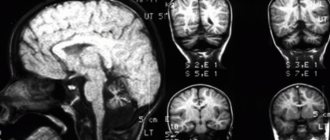
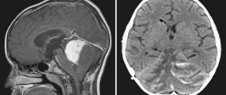
CT and MRI studies are considered the most informative. Neuroimaging reveals changes in the structural structure of the tissue - deepening and widening of the grooves, an increase in the volume of the subarachnoid space, the size of the large tank located in the occipital area.
Diagnostic methods
Many methods and tools are used to detect cerebellar atrophy. In addition to directly confirming the presence of atrophic processes, the purpose of diagnosis is to determine the form of the disease, detect concomitant pathologies, possible complications, and make predictions regarding treatment methods.
To carry out diagnostic procedures, the patient must seek the help of a neurologist. You should visit a medical facility if any manifestations of atrophy occur, since timely assistance provided significantly reduces the likelihood of serious consequences for the patient’s health.
Basic diagnostic methods:
- Examination and questioning of the patient is the primary diagnostic method, which is aimed at identifying complaints and signs of the disease. During the examination, the neurologist checks the patient’s nervous reactions, notes possible motor and speech disorders and other symptoms. In addition, anamnesis is studied - the history of diseases that could act as a provoking factor for atrophy.
- MRI is considered the most reliable diagnostic method, as it can detect even minor atrophic changes. Using this method, the exact location and area of damage to the cerebellum, as well as possible concomitant changes in other parts of the brain, are determined.
- Computed tomography is also a very reliable diagnostic method, allowing you to confirm the diagnosis and obtain additional information about the nature of the disease. Usually prescribed in cases where MRI is contraindicated for some reason.
- Ultrasound examination. This method is used to diagnose extensive brain damage caused by stroke, trauma, and age-related changes. Ultrasound examination allows you to identify areas of atrophy and, similarly to other hardware methods, determine the stage of the disease.
Diagnosis of cerebellar atrophy is carried out using various hardware and non-hardware methods when early signs of the disease appear.
Which doctor should I contact?
If the slightest signs of pathology appear, you should urgently visit a neurologist; only timely treatment will avoid serious complications and irreversible consequences that significantly worsen the quality of life.
Diagnosis and complex therapy at an early stage of pathology will improve blood circulation and eliminate hypoxia of nerve tissue. This will stop the atrophic process. Timely medical care will reduce the risk of complications and pathological changes in other areas of the GM.
Neurons that undergo atrophy cannot be restored.
Therapy
Unfortunately, there are no special methods aimed at eliminating cerebellar atrophy. This is due to the fact that medicinal, physiotherapeutic or surgical methods of therapy are not able to restore nerve tissue damaged due to circulatory disorders and oxygen starvation. Therapeutic measures are reduced to eliminating pathological manifestations, reducing negative consequences for other parts of the brain and the whole body, and preventing complications.
With careful diagnosis, the cause of the disease is determined. Eliminating it allows for positive changes in the patient’s condition, especially if treatment began at an early stage.
The following medications can be used to relieve symptoms:
- "Teralen."
- "Alimemazine."
- "Levomepromazine."
- "Thioridazine."
- "Sonapax".
The action of such drugs is aimed at eliminating psychotic disorders caused by pathological processes in the cerebellum. In particular, medications are used for manic-depressive states, neuroses, panic attacks, increased anxiety, and sleep problems.
Depending on the medication, administration can be carried out orally (when using tablets), intravenously and intramuscularly (if using appropriate solutions). The optimal method of administration, dosage and duration of the therapeutic course are prescribed by a neurologist individually, in accordance with the diagnosis.
During therapy, it is extremely important to provide the patient with careful care. Because of this, many experts recommend carrying out the initial stages of treatment at home. At the same time, self-treatment and the use of non-traditional folk methods are strictly prohibited, as they can cause even greater harm.
The patient should regularly undergo repeated examinations and examinations by a neurologist. The main purpose of secondary diagnostics is to monitor the effectiveness of treatment, provide recommendations to the patient, and adjust drug dosages.
Thus, cerebellar atrophy is not amenable to direct therapeutic intervention, which is why treatment is symptomatic.
Undoubtedly, cerebellar atrophy is a very serious pathological condition, accompanied by deterioration of functions and tissue death of this part of the brain. Due to the lack of special treatment methods and the high likelihood of complications, you should pay attention to any potential signs of the disease and promptly visit a neurologist.
Treatment of pathology
As we mentioned, the main method of treatment is surgery. This is a radical excision of the formation. But if it grows into the fourth ventricle, a complex anatomical structure, then this makes it difficult to remove the cerebellar tumor. Then, to restore normal liquor circulation, the maximum possible volume of pathogenic tissue is cut out.
Surgeries for a cerebellar tumor also involve partial resection of the foramen of the occipital bones, the first cervical vertebra. These manipulations help reduce the pressure of the formation on the brain stem.
To reduce hydrocephalus, with its sharp development, shunting measures, ventricular external drainage, and puncture of the cerebral ventricles are also indicated.
After removal of the tumor, its tissue is sent for histological analysis to determine malignancy and stage of development.
Additionally, the patient is prescribed chemotherapy and radiation therapy, sedatives, antiemetics, and painkillers.