Optic neuritis
(optic neuritis) - inflammatory damage to the optic nerve. This disease also includes nerve damage in demyelinating diseases. Within the framework of optical neuritis, intra- and retrobulbar neuritis are distinguished, which differ significantly in the ophthalmoscopic picture. Common symptoms are: decreased vision and the appearance of scotomas; In some forms, pain in the eye is possible. Ophthalmoscopy plays a primary role in diagnosis. Treatment is based on a combination of anti-edematous, anti-inflammatory, desensitizing, antibacterial or antiviral, immunocorrective, detoxification and metabolic therapy.
What contributes to the appearance of the disease
Retrobulbar neuritis is a neurological pathology in which an inflammatory process develops in nerve fibers. The disease is characterized by a gradual weakening of visual function. Due to the characteristic location of the inflammatory process, consultations with a neurologist and ophthalmologist are required.
To understand the features of the causes of development and symptoms of neuritis, let us turn to the anatomy of the optic nerve. This nerve runs through the intraocular and retrobulbar space, where it connects with other fibers and, forming a new structure, reaches the corresponding parts of the brain.
Retrobulbar neuritis causes complications.
In the absence of adequate and timely treatment, the disease provokes atrophy of the optic nerve, which can lead to blindness and disability.
Etiology and pathogenesis of optic neuritis
Among the factors that provoke optic neuritis, the most common are inflammatory processes of the orbit (periostitis, phlegmon), eyeball (iridocyclitis, retinitis, keratitis, panophthalmitis) and brain (arachnoiditis, meningitis, encephalitis); infectious processes in the nasopharynx (ethmoiditis, sinusitis, frontal sinusitis, chronic tonsillitis, tonsillitis, pharyngitis). Common infections can lead to the development of optic neuritis: tuberculosis, malaria, typhus, brucellosis, ARVI, diphtheria, gonorrhea, etc. Other causes include alcoholism, head injury, complicated pregnancy, systemic diseases (gout, collagenosis), blood diseases, diabetes mellitus, autoimmune disorders. Optic neuritis often manifests itself in multiple sclerosis.
The inflammatory process (neuritis) can develop both in the optic nerve sheath and in its trunk. In this case, inflammatory edema and infiltration lead to compression of the optic fibers with their subsequent degeneration, which is the cause of decreased visual acuity. After acute inflammation subsides, some fibers can restore their function, which is clinically manifested by improved vision. Severe optic neuritis often leads to the disintegration of nerve fibers and the growth of glial tissue in their place. Optic nerve atrophy develops with an irreversible decrease in visual acuity.
In multiple sclerosis, neuritis is based on the process of demyelination of nerve fibers - the destruction of their myelin sheath. Although demyelination is not an inflammatory process, in the medical literature and in practice demyelinating lesion n. opticus are classified as retrobulbar neuritis, since their clinical symptoms are identical.
Forms of neuritis
The optic nerve consists of a central and peripheral part. Based on the zone of localization of the inflammatory process, retrobulbar neuritis is divided into the following types:
- Axial. With this form of the disease, the central part of the nerve is affected, which is why visual acuity can sharply decrease. Also, with axial neuropathy, the appearance of blind spots (scotomas) in the visible zone is noted.
- Peripheral. The inflammatory process in the peripheral part causes the formation of exudate, which causes the patient to experience pain. Visual acuity is maintained, but the field of visibility of objects is narrowed.
- Transversal. The inflammatory process spreads along the entire length of the optic nerve. Transversal neuropathy causes partial or complete blindness.
Depending on the stage of development of the disease, chronic and acute retrobulbar neuritis are distinguished. The latter is characterized by a rapid decrease in visual acuity and occurs mainly in young people. The chronic form is characterized by a slow increase in the intensity of symptoms.
Optic neuritis
The optic nerve (n. opticus) consists of processes (axons) of retinal neurons. The latter perceive the image and transmit information about it in the form of nerve impulses traveling along axons to the cerebral visual centers. Each optic nerve consists of more than 1 million axons. It begins with the optic disc, located on the retina and accessible to ophthalmological examination. The part n located inside the orbit. opticus is called intrabulbar (intraorbital). After leaving the orbit, the optic nerve passes into the cranial cavity, this part is called retrobulbar. In the area of the sella turcica, the optic nerves cross (chiasma), where they partially exchange their fibers. The optic nerves end in the visual centers of the midbrain and diencephalon.
Throughout its entire length, the optic nerve is enveloped in membranes that are closely connected with nearby structures of the orbit and brain, as well as with the cerebral membranes. This causes the frequent occurrence of optic neuritis in inflammatory diseases of the orbit, brain and its membranes.
Reasons for the development of the disease
The reasons for the development of pathology are varied. One of the causes of eye inflammation in retrobulbar neuritis is the activity of cytomegalovirus. This pathogen provokes an abnormal reaction of the immune system and it attacks its own cells. As a result of an autoimmune response, nerve fibers become inflamed.
The second most common cause of the disease is considered to be infectious pathologies:
- sinusitis;
- otitis;
- uveitis;
- iridocyclitis;
- chorioretinitis;
- tonsillitis.
Less commonly, the optic nerve is affected due to the spread of infection throughout the body from distant foci of inflammation.
The appearance of neuritis is associated with the course of multiple sclerosis and brain tumors. In multiple sclerosis, demyelination of the optic nerve tissue occurs, which leads to inflammation of local fibers, which is why symptoms of retrobulbar disease are often diagnosed in people with MS. Neoplasms compress blood vessels, nerve fibers experience an acute deficiency of micronutrients, and inflammation of local tissues occurs.
Possible causes of retrobulbar optic neuritis include the course of systemic pathologies of an infectious nature:
- tuberculosis;
- neurosyphilis;
- Lyme disease.
In some cases, inflammation of the optic nerve is possible due to AIDS or toxic damage to the body caused by the penetration of methyl alcohol, poisons, chemical compounds, and iodine-containing drugs.
The possibility of damage to the structures of the organs of vision due to renal failure and metabolic disorders cannot be ruled out.
Treatment of neuromyelitis optica
A patient with symptoms of optic neuritis requires emergency hospitalization.
Before determining the etiology of the disease, various drugs relieve inflammation, reduce the increased sensitivity of the body, initiate metabolic processes in the tissues of the central nervous system, and strengthen the immune system.
The patient is given intramuscular antibiotics, excluding gentamicin, neomycin, streptomycin and other drugs that cause side effects on the optic nerve. Parenteral hormonal drugs are prescribed - dexamethasone and prednisolone.
After clarifying the etiology of neuritis, they proceed to treatment of the underlying disease - sinusitis, herpes, tuberculosis, etc. The course of emergency treatment includes gastric lavage, injections, incl. taking special alcohol solutions orally.
Symptoms
At the initial stage of the disease, deformation of the optic nerve head occurs, which manifests itself in the form of hyperemia.
Basically, the inflammatory process occurs in one eye. Bilateral lesions are rarely diagnosed.
With retrobulbar neuritis, symptoms manifest as deterioration in visual acuity over several days (diagnosed in 60% of patients). If the disease has become chronic, eye function deteriorates gradually over 1-2 weeks.
The development of the disease is indicated by the following signs:
- a veil before the eyes;
- objects become blurry;
- sudden "flashes of light";
- prolonged adaptation of vision at dusk (hemeralopia).
A decrease in the quality of vision is caused by the appearance of dark spots in front of the eyes and a narrowing of the field of vision. Most patients have a color perception disorder (the patient does not distinguish colors). This condition is usually detected only through an ophthalmological examination.
More than half of patients complain of constant or temporary pain behind the eyeball. With retrobulbar optic neuritis, the intensity of this symptom varies depending on the characteristics of the development of the inflammatory process. The pain syndrome intensifies when moving the eyes and more often occurs when the peripheral part of the nerve fibers is damaged.
The intensity of the manifestation of general symptoms depends both on concomitant pathologies and on the patient’s condition. Stress, physical fatigue and a number of other factors provoke a decrease in visual acuity.
With the development of neuritis due to toxic damage, the nature of the clinical picture changes: attacks of nausea with vomiting are noted. The toxic form does not cause fundus deformation. During the examination, a weakened reaction to light and pupil dilation are diagnosed.
Symptoms of intrabulbar neuritis
Typically acute onset of visual disturbances. Their severity and character depend on the degree of damage to the diameter of the optic nerve. With a total process, visual acuity drops to the point of complete blindness (amaurosis). With partial vision, visual acuity can be maintained even at 1.0. However, spots appear in the field of vision - paracentral or central scotomas, having an arched or rounded shape; There is a decrease in color perception and dark adaptation, a low level of lability of the optic nerve and a critical frequency of flicker fusion.
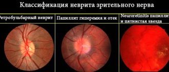
From the first days of neuritis, a pathognomonic picture of changes in the optic disc is revealed: hyperemia, blurred boundaries, exudative swelling, moderate vasodilation, the presence of streak-like hemorrhages in the tissue of the disc and peridiscal region. If the exudate fills the vascular funnel and imbibes the adjacent layers of the vitreous, then the fundus is not clearly visualized. Unlike congestive discs associated with intracranial hypertension and hydrocephalus, with optic neuritis there is no pronounced protrusion (prominence) of the disc, the changes are usually unilateral.
The acute period lasts from 3 to 5 weeks. Then the hyperemia and swelling of the disc gradually disappear, the hemorrhages resolve, and the boundaries of the disc again acquire clear outlines. In more rare cases, with severe optic neuritis, atrophy of n. opticus. In this case, ophthalmoscopy reveals a pale disc with thread-like narrowed vessels and clear boundaries.
Diagnostic measures
Diagnostics involves conducting a comprehensive examination of the patient, which includes:
- Visometry. The method allows you to diagnose decreased visual acuity.
- Assessment of color perception. The examination is carried out using a special table.
- Perimetry. Helps identify narrowing of the visual field and scotoma.
- Ophthalmoscopy. As part of this procedure, the condition of the fundus and nerve fibers is assessed, hemorrhages and hyperemia are determined.
In order to exclude concomitant pathologies, the patient's neurological status is assessed. To determine the cause of the development of retrobulbar neuritis, MRI of the brain and cerebrospinal fluid examination are used. The first method helps to identify tumors, the second - infectious infection.
Visometry
Perimetry
If multiple sclerosis is suspected, other procedures are prescribed.
Diagnosis of optic neuritis
Since optic neuritis is an interdisciplinary pathology, its diagnosis often requires the joint participation of specialists in the field of neurology and ophthalmology. In typical cases, to verify the diagnosis, a consultation with an ophthalmologist is sufficient, during which the patient’s complaints, visual acuity test data, perimetry and ophthalmoscopy results are compared.
The most important task is to differentiate disc changes in optic neuritis from congestive disc. This is especially true for mild neuritis with minimal disturbances in visual function and when neuritis is combined with disc swelling. In such cases, the identification of foci of exudation and minor hemorrhages in the disc tissue indicates neuritis. Fluorescein angiography of the fundus helps to distinguish between these conditions. To exclude a stagnant disc in complex cases, a consultation with a neurologist, echo-encephalography, or lumbar puncture may be required.
In order to determine the etiology of optic neuritis, it is possible to conduct an MRI of the brain, blood culture for sterility, PCR studies, ELISA, RPR test, consultation with an infectious disease specialist, rheumatologist, immunologist, etc.
How to treat retrobulbar neuritis?
Treatment tactics for the disease are developed based on the characteristics of the causes of the disease. Full recovery of the patient is possible provided that therapy is carried out at the initial stage of development of the inflammatory process. If a brain tumor is detected, surgery is prescribed to remove the tumor and restore damaged nerve fibers.
Traditional therapy
When retrobulbar optic neuritis is detected, treatment begins with desensitization and dehydration of the body. It is also recommended to take medications that accelerate the flow of nutrients to tissues and stimulate the functioning of the immune system.
The effectiveness of treatment methods for retrobulbar neuritis directly depends on the success of the diagnosis. If during the examination of the patient it was not possible to identify the strain of the pathogenic agent (a common case), broad-spectrum antibiotics are used in therapy, however, the use of ototoxic drugs such as Gentamicin and Streptomycin is contraindicated. These medications negatively affect the condition of the optic nerve.
When the body is infected with a virus, antiviral drugs are used. Due to the fact that a common cause of inflammation of the optic nerve is the activity of cytomegalovirus, Acyclovir and other systemic antiherpetic drugs are used in the treatment of the disease. With such damage to the body, synthetic interferon inducers are additionally prescribed.
Regardless of the causes of the disease, anti-inflammatory therapy is carried out, in which glucocorticosteroids are prescribed.
Dexamethasone is mainly used for retrobulbar neuritis. In advanced cases, it is recommended to take corticosteroids.
At the same time, decongestant therapy is used, which involves taking diuretics (Furosemide, Acetazolamide) and antihistamines (Desloratadine, Chloropyramine).
Since the course of retrobulbar neuritis causes damage to the optic nerve, to restore it, the doctor prescribes neuroprotective treatment using the following drugs:
- vitamins B and C;
- "Pentoxifylline";
- "Nicotinamide".
If damage to the optic nerve is caused by poisoning of the body, severe inflammatory pathologies or neuroinfections, detoxification is carried out. For this purpose, intravenous administration of saline solutions and Dextran is prescribed.
Drug treatment is combined with physiotherapeutic procedures: magnetotherapy, laser stimulation. The positive effect of the measures taken becomes noticeable after a few months.
In advanced cases, to restore visual functions, a surgical operation is performed in which the sheath of the affected nerve is opened. This reduces the level of pressure on the inflamed fibers.
The course of the disease in multiple sclerosis causes serious complications, so treatment of both diseases is carried out in a hospital setting. In multiple sclerosis, self-healing of the optic nerve is possible without outside intervention. To speed up this process, doctors use B vitamins and immunoglobulin.
Treatment with folk remedies
Retrobulbar neuritis cannot be treated with traditional medicine, but their use helps reduce the intensity of symptoms and speed up the patient’s recovery.
Treatment with aloe leaves, which must be crushed and mixed with 50 grams, shows good results. flowers of field cornflower and eyebright. Also add 200 grams to the composition. honey and 700 ml of red wine. This mixture is kept in a water bath for 40 minutes. It is recommended to take the medicine half an hour before meals, one tablespoon.
The second effective remedy against inflammation of the optic nerve is prepared from a collection of fragrant rue and green cones (mixed in a ratio of 1 to 5). The components are brewed in boiling water with lemon and granulated sugar. The mixture simmers for half an hour in a water bath. After preparation, the product is taken 3 times a day 30 minutes before meals.
A positive effect is achieved through regular consumption of herbal teas. They strengthen general immunity, increasing the body's resistance to infections and viruses, improve vascular trophism and nutrition of eye cells.
Drug treatment
The main goal of treatment is to relieve inflammation and the causes of its occurrence.
Therapy for retrobulbar neuritis involves the use of several groups of drugs. These include:
- antibiotics;
- antihistamines;
- antiviral;
When diagnosing neuritis, the use of penicillin and broad-spectrum antibiotics is indicated. The most commonly prescribed drug is streptomycin. The dosage and duration of treatment is determined by the attending physician.
Taking antihistamines and sulfonamide drugs is also indicated. For mild cases, local hormonal therapy using eye drops is prescribed. But in severe cases, general treatment is necessary.
Prognosis and prevention
The prognosis for inflammation of the optic nerve is determined depending on the severity of the case. With timely treatment of acute retrobulbar neuritis, complete restoration of vision is possible.
The chronic form of the disease cannot be cured. This pathology is characterized by alternating periods of remission and relapse; the patient’s visual acuity gradually decreases until the complete loss of the ability to see with one or both eyes. This is explained by the fact that the chronic course of the inflammatory process is accompanied by atrophy of nerve fibers, the place of which is taken by connective tissue.
To reduce the likelihood of developing retrobulbar neuritis, it is recommended to begin treatment of inflammatory diseases in a timely manner.
This is especially true in cases where the pathological process occurs in close proximity to the optic nerve (in the brain, eyes, nasopharynx). It is also recommended to limit your intake of alcoholic beverages, stop smoking and consult a doctor as soon as persistent vision loss is detected.
It is impossible to completely exclude the possibility of nerve inflammation. But if you follow these recommendations, you can prevent the early development of neuritis or the appearance of complicated forms of the disease.
Forecast
Optic neuritis has a favorable prognosis. In most cases, patients manage to restore their previous visual acuity a year after the first signs of the problem appeared.
If the pathological process arose as a result of multiple sclerosis or neuromyelitis optica, then the prognosis for its complete elimination will be less favorable. Exacerbations of the disease in such situations occur constantly.
To prevent the problem from occurring, it is important to eat a balanced diet and avoid consuming unhealthy foods - canned food, smoked foods and sweets. In addition, diseases should be treated in a timely manner so that they do not become chronic and cause complications in the future. Measures to prevent optic neuritis also include:
- avoiding situations that could lead to traumatic brain injury;
- to give up smoking;
- regular physical activity.
Optic neuritis may in some cases disappear on its own without special treatment. However, when the first signs of pathology appear, you must consult an ophthalmologist: only a doctor will be able to correctly assess the possible risks and prescribe a suitable treatment regimen. Otherwise, the problem can cause complete atrophy of the optic nerve.
Retrobulbar neuritis: causes of the disease, main symptoms, treatment and prevention
An inflammatory process that affects the area of the second pair of cranial nerves, which is located between the optic chiasm and the orbit.
The patient complains of blurred vision, loss and limitation of the visual field, pain during movements of the eyeballs. To establish and confirm the diagnosis, the doctor analyzes the clinical manifestations, studies the medical history, conducts an ophthalmological examination, and then refers the patient to a consultation with a neurologist and additional examinations.
As part of the diagnosis, magnetic resonance imaging, ultrasound, lumbar puncture followed by cerebrospinal fluid examination, polymerase chain reaction, enzyme immunoassay, retinal angiography and Doppler ultrasound can be performed. Therapeutic tactics are determined after identifying the cause of the disease.
The drug regimen may include antibacterial and antiviral agents, glucocorticosteroids, diuretics, antihistamines and vitamins. For tumors, surgery is possible. The prognosis is favorable. Axial, peripheral and transversal forms of neuritis are classified.
Causes of retrobulbar neuritis
The disease can develop with cytomegalovirus infection, herpes, infectious mononucleosis, sinusitis, otitis, chronic tonsillitis, pyelonephritis, cystitis, uveitis, iridocyclitis, chorioretinitis.
Secondary retrobulbar neuritis is characterized by formation against the background of multiple sclerosis, brain neoplasia, neuroAIDS, neurosyphilis, tuberculous meningitis, and Lyme disease.
In addition, the pathology can be provoked by toxic damage to the optic nerve by pesticides, methyl alcohol, iodine preparations and some pharmaceuticals. Endogenous toxicosis can also be caused by dysmetabolic diseases, pregnancy, and renal failure.
Symptoms of retrobulbar neuritis
The pathology is manifested by a significant decrease in vision within 1-2 days in acute cases, or 14 days in chronic cases. Patients see objects blurry and may complain of blurred vision. Sometimes flashes of light suddenly appear in your field of vision.
With a gradual onset, hemeralopia develops - poor visual adaptation in the dark. The disease is also characterized by tunnel vision and dark spots before the eyes. During an ophthalmological examination, the doctor may detect dyschromatopsia.
Most patients experience pain that intensifies when the eyeball moves. Sometimes headaches occur. In the secondary form of the disease, neurological symptoms are noted.
The disease can be complicated by descending atrophy of the optic nerve, which manifests itself as a persistent decrease in vision. With total atrophy, partial or complete blindness develops.
Diagnosis of retrobulbar neuritis
The pathology is differentiated from neuromyelitis optica, intrabulbar lesions and ischemic neuropathy of the second pair of cranial nerves.
To establish and confirm the diagnosis, the doctor analyzes the clinical manifestations, studies the medical history, checks visual acuity, determines color vision, performs perimetry, ophthalmoscopy, and then refers the patient to a consultation with a neurologist and additional examinations.
As part of the diagnosis, magnetic resonance imaging, ultrasound, lumbar puncture followed by cerebrospinal fluid examination, polymerase chain reaction, enzyme immunoassay, retinal angiography and Doppler ultrasound can be performed.
Treatment of retrobulbar neuritis
Therapeutic tactics depend on the cause of the disease. For neoplasia, surgical intervention is performed, for multiple sclerosis, immunocorrective treatment is performed, and for neurofincia, etiotropic therapy is performed. As a rule, the patient is prescribed antibacterial or antiviral drugs.
To relieve the inflammatory process, glucocorticosteroids are administered retrobulbarly. To reduce swelling, diuretics and antihistamines are used. The drugs of choice are usually acetazolamide, furosemide, chloropyramine and desloratadine. The medication regimen is supplemented with B vitamins, nicotinamide, ascorbic acid and pentoxifylline.
In addition, the patient is recommended physical therapy.
Prevention of retrobulbar neuritis
Specific methods of prevention have not been developed. It is necessary to promptly seek medical help in order to identify and treat diseases that can provoke inflammation of the optic nerve in the early stages.
Source: //www.obozrevatel.com/health/bolezni/retrobulbarnyij-nevrit.htm