Neurological damage in syphilis - myth or reality?
In the article we will consider damage to the central nervous system in syphilis - its cause, pathogenesis, symptoms, a number of pathologies with which it is necessary to differentiate it, diagnostic measures that are aimed not only at confirming, but also at assessing the progression of the disease. We will analyze the therapeutic tactics used for this pathology, as well as the prognosis with and without specific treatment.
Neurosyphilis is one of the most complex processes to which syphilis can progress. Involves damage to the central nervous system.
Depending on the duration of the process, at first the most common forms of neurosyphilis are considered to involve the cerebrospinal fluid, meninges and vascular system of the brain. Later, damage occurs directly to the tissue itself - the parenchyma of the brain and spinal cord.
How the disease develops
Treponema pallidum penetrates the nervous system by hematogenous and lymphogenous routes in the early stages of untreated syphilis. They affect the membranes, vessels and membranes of the roots and peripheral nerves. Over time, these structures lose the ability to hold treponema pallidum and neutralize them, and then the bacteria penetrate the substance (parenchyma) of the brain and spinal cord, causing the development of a number of diseases.
In the first years from the onset of infection, the patient may develop a latent (asymptomatic) form of neurosyphilis, when the patient does not have any neurological disorders, but lymphocytic pleocytosis and increased protein content are noted in the cerebrospinal fluid.
In the primary (rarely) and secondary (more often) periods of syphilis, the development of syphilitic meningitis is recorded. The main symptom complex called neurosyphilis develops in the tertiary period of syphilis.
- In the first five years of the disease, early syphilis of the nervous system develops, which is characterized by the development of inflammatory changes in the mesenchyme - blood vessels and meninges.
- Late neurosyphilis develops in later stages of the disease - 10 - 25 or more years from the moment of primary infection. Following the mesenchyme, the parenchyma begins to be affected - nerve cells, fibers and glia.
Modern neurosyphilis occurs with minimal severity of symptoms and is characterized by a milder course and less change in the cerebrospinal fluid. Complaints that come to the fore include weakness, lethargy, insomnia, and decreased performance. The longer the infectious process, the more often the symptoms and clinical manifestations of neurosyphilis are recorded.
Rice. 2. The photo shows manifestations of tertiary syphilis - gumma. During this period, late neurosyphilis develops.
Therapeutic approach
The treatment regimen for neurosyphilis varies depending on the severity of the course, the allergic characteristics of the body and its comorbid conditions.
Standard treatment protocols follow the following recommendations:
- water-soluble crystalline penicillin G;
or
- procaine penicillin G in combination with probenecid.
Patients who are sensitive to penicillin antibiotics can also be treated according to the above scheme, but after a course of desensitization. Treatment of neurosyphilis with Ceftriaxone is used in cases of mild allergic reaction to penicillins in the absence of “cross-reaction”. An alternative treatment option is Doxycycline.
Important! Instructions for using an antibacterial drug must be prescribed by a doctor and strictly followed by the patient.
In conclusion, it is important to note that timely diagnosis of syphilis and treatment by a specialist will help to avoid such complex conditions as damage to the central nervous system by Treponema pallidum. Health requires close attention and self-care.
Stages of neurosyphilis
Stage I. Latent (asymptomatic) syphilitic meningitis.
Stage II. Damage to the meninges (meningeal symptom complex). Damage to the soft and hard membranes of the brain: acute syphilitic meningitis, basal meningitis, local damage to the membrane of the brain. Damage to the soft and hard membranes of the spinal cord, its substance and spinal roots - syphilitic meningoradiculitis and meningomyelitis.
Stage III. Vascular lesions (secondary and tertiary periods of syphilis). More often there is simultaneous damage to the soft meninges and blood vessels of the brain - meningovascular syphilis.
Stage IV. Late neurosyphilis (tertiary period of syphilis). There are late latent syphilitic meningitis, late vascular and diffuse meningovascular syphilis, tabes dorsalis, progressive paralysis, taboparalysis, gumma cerebri.
Rice. 3. Nietzsche, V. Lenin and Al Capone suffered from neurosyphilis.
Asymptomatic meningitis
Asymptomatic (latent) meningitis is registered in 10 - 15% of cases in patients with primary syphilis, in 20 - 50% in patients with secondary and latent early syphilis. In most cases, symptoms of meningitis cannot be identified. Previously, latent meningitis was called “syphilitic neurasthenia”, since the symptoms of neurasthenia came to the fore - severe fatigue, exhaustion, decreased mood, absent-mindedness, forgetfulness, indifference, irritability, decreased performance. Sometimes patients are bothered by persistent headaches, attacks of dizziness, a feeling of stupefaction, and difficulty concentrating. Meningeal symptoms are rare. Serological reactions of the cerebrospinal fluid (Wassermann reaction and RIF) are positive, pleocytosis (increased lymphocytes and polynuclear cells) of more than 5 cells per 1 mm3 and an increased amount of protein are noted - more than 0.46 g/l.
In early forms of syphilis, asymptomatic meningitis is one of its manifestations, like chancre or secondary syphilides. But in late forms of syphilis, asymptomatic meningitis requires active treatment, as neurosyphilis develops against its background.
Only with neurosyphilis are changes in the cerebrospinal fluid observed in the absence of clinical symptoms.
Rice. 4. Damage to the oculomotor nerve (photo on the left) and pupillary disorders (anisocoria) in the photo on the right with neurosyphilis.
Treatment
Therapy is carried out exclusively inpatient. The patient is administered a drug with a high concentration of penicillin. The course lasts at least two weeks. Doctors often prescribe complex therapy using several medications at once. funds. Standard scheme:
- penicillin;
- probenecid;
- ceftriaxone.
Medicines are administered intravenously, and penicillin is also injected into the spinal canal. After two weeks, a check is made to see if the virus has been eliminated; if not, the course is extended.
On the first day of treatment, headaches may intensify, so the patient is also prescribed corticosteroid and anti-inflammatory medications. facilities.
At a later stage, bismuth and arsenic are used, which, as you can imagine, are very toxic.
Damage to the meninges
In the second stage of neurosyphilis, the soft and hard membranes of the brain and spinal cord are affected.
Syphilis of the meninges
Acute syphilitic meningitis
Acute syphilitic meningitis is rare. The disease manifests itself in the first years after infection. Body temperature rarely rises. Sometimes the oculomotor, visual, auditory and facial nerves are involved in the pathological process, and hydrocephalus develops.
Meningoneuritic form of syphilitic meningitis (basal meningitis)
This form of neurosyphilis is more common than acute meningitis. The disease is acute. The clinical picture of the disease consists of symptoms of meningitis and neuritis. The nerves that originate at the base of the brain become inflamed. Headache, worse at night, dizziness, nausea and vomiting are the main symptoms of basal meningitis. The mental status of patients is disturbed. Excitability, depression, irritability are noted, and an anxious mood appears.
When the abducens, oculomotor and vestibular-cochlear nerves are damaged, facial asymmetry and drooping of the eyelid (ptosis) are noted, the nasolabial fold is smoothed, the tongue deviates from the midline (deviation), drooping of the soft palate is noted, and bone conduction decreases. Damage to the optic nerve is manifested by deterioration of central vision and narrowing of the fields. Sometimes inflammation affects the pituitary gland area. When the convex surface of the brain is affected, the disease proceeds as vascular syphilis or progressive paralysis. In the cerebrospinal fluid, protein is 0.6 - 0.7%, cytosis is from 40 to 60 cells per mm3.
Rice. 5. Damage to the oculomotor nerve in neurosyphilis - ptosis (drooping eyelids).
Syphilis of the dura mater of the brain
The cause of the disease is either a complication of the bone process or a primary lesion of the dura mater.
Rice. 6. Damage to the oculomotor nerve in neurosyphilis.
Syphilis of the spinal cord membranes
Syphilis of the soft membranes of the spinal cord
The disease is diffuse or focal in nature. The pathological process is most often localized in the thoracic spinal cord. The disease manifests itself as paresthesia and radicular pain.
Acute syphilitic inflammation of the soft membranes of the spinal cord
The disease occurs with pain in the spine and paresthesia. Skin and tendon reflexes increase, and contractures of the limbs are noted. Due to pain, the patient takes a forced position.
Chronic syphilitic inflammation of the soft membranes of the spinal cord
The disease is registered more often than acute. The membranes of the brain thicken, often along the entire length, less often in limited areas.
When the meninges and spinal nerve roots are simultaneously involved in the process, syphilitic meningoradiculitis develops. The main symptoms of the disease are symptoms of root irritation. The clinical picture depends on the localization of the pathological process.
When the substance of the spinal cord, membranes and spinal roots is involved in the process, syphilitic meningomyelitis develops. More often, peripheral parts of the spinal cord are involved in the pathological process. Spastic paraparesis develops, tendon reflexes increase, and all types of sensitivity are impaired. Sphincter disorders are an early and persistent symptom of the disease.
Syphilis of the dura mater of the spinal cord
The symptom complex was first described by Charcot and Geoffroy. The first stage of the disease is characterized by a symptom complex of root irritation. The patient experiences pain in the back of the head, neck, and in the area of the median and ulnar nerves. In the second stage of the disease, loss of sensitivity is noted, flaccid paralysis, paresis and muscle atrophy develop. In the third stage, symptoms of spinal cord compression appear: sensory disturbances, spastic paralysis, trophic disorders, often including bedsores. Sometimes spontaneous hemorrhages occur on the inner surface of the dura mater, accompanied by radicular and spinal phenomena such as strokes.
Rice. 7. MRI of a patient with neurosyphilis. The subarachnoid space is expanded. The meninges are thickened.
Damage to cerebral vessels
In the third stage of neurosyphilis, damage to small or large vessels is noted. The clinical picture of the disease depends on the location, number of affected vessels and their size. In neurosyphilis, vascular damage is often combined with damage to the meninges. In this case, focal symptoms are combined with general cerebral symptoms. Syphilitic arteritis is recorded both in the brain and in the spinal cord. The vessels at the base of the brain are most often affected.
Damage to large vessels is complicated by strokes, small ones - by general disorders of brain function, paresis and damage to the cranial nerves.
With vascular syphilis of the spinal cord, the pathological process affects the venous system. Paresis, sensitivity disorders and sphinter functions develop slowly. Lesions of the spinal cord vessels are manifested by symptoms that depend on the location of the pathological process.
Young age, normal blood pressure, “scattered” neurological symptoms, positive serological reactions are the hallmarks of vascular syphilis.
The prognosis of the disease is favorable. Specific treatment leads to complete cure.
Rice. 8. Damage to large vessels in neurosyphilis is complicated by strokes.
Therapy
Treatment of neurosyphilis is mainly carried out with the antibiotic Penicillin.
Treatment of neurosyphilis is mainly carried out with the antibiotic Penicillin. The treatment regimen is drawn up by the attending physician on an individual basis; the dosage of the drug directly depends on the degree of damage to the body. The most effective therapy is considered to be using intravenous administration of sodium benzylpenicillin salt. Injections or droppers are given 6 times a day, the course of treatment is 14 days.
When the doctor recommends administering the drug intramuscularly, benzylpenicillin novocaine salt can be used plus probenecid taken orally 4 times a day, the course of treatment is 14 days. The drug Probenecid stimulates the complete absorption of the antibiotic Penicillin by soft tissues.
After completing the course described above, therapy continues, the patient is given an injection with benzathine benzylpenicillin once a week, the course is 3 weeks. At the beginning of therapy, the infected person’s health deteriorates significantly: fever, headache and muscle pain, attacks of tachycardia, surges in blood pressure. In such cases, corticosteroids and non-steroidal anti-inflammatory drugs are additionally prescribed.
If the patient is found to be intolerant to penicillin, it is replaced with Ceftriaxone, Chloramphenicol. The effectiveness of a particular drug is assessed by improving the patient’s health and cerebrospinal fluid indicators. A repeat examination is carried out immediately after the initial course of penicillin administration; a puncture and cerebrospinal fluid are taken. After that, every 6 months for two years. If test results show no improvement, antibiotics are administered again.
Signs and symptoms of late neurosyphilis
Late forms of syphilis have become increasingly rare in many countries around the world in recent decades. This is facilitated by the widespread use of antibacterial drugs, improved diagnostics and therapy. Among patients with neurosyphilis, tabes dorsalis and progressive paralysis are becoming less common. The incidence of meningovascular syphilis is increasing. Late forms of neurosyphilis often develop in patients who have been insufficiently treated or not treated for early syphilis. The development of the disease is facilitated by a decrease in immunity, which is negatively affected by physical and mental trauma, intoxication, allergies, etc.
The following forms of late neurosyphilis are distinguished:
- late hidden (latent) syphilitic meningitis,
- late diffuse meningovascular syphilis,
- vascular syphilis (syphilis of the brain vessels),
- tabes dorsalis,
- progressive paralysis,
- taboparalysis,
- gumma brain.
Late latent syphilitic meningitis
The disease occurs 5 or more years after infection. Quite difficult to treat. Against this background, other manifestations of neurosyphilis are formed. Often patients do not show any complaints; some patients experience headache, dizziness, tinnitus and hearing loss. When examining the fundus, changes are revealed in the form of hyperemia of the optic nerve nipple and papillitis. An increased content of cellular elements and protein is noted in the liquor. Wasserman's reaction is positive.
Late diffuse meningovascular syphilis
Dizziness, headaches, epileptiform seizures, hemiparesis, speech and memory disorders are the main symptoms of the disease. Damage to cerebral vessels is complicated by the development of strokes and thrombosis. A small amount of protein and cellular elements is detected in the cerebrospinal fluid.
Rice. 9. Late neurosyphilis. MRI of a patient with mental disorders.
Tabes dorsalis
Tabes dorsalis is becoming less and less common over the years. Vascular forms of late neurosyphilis are more common. The disease is diagnosed in 70% of cases 20 or more years after infection. The dorsal roots, dorsal columns and membranes of the spinal cord are affected. The specific process is most often localized in the lumbar and cervical spine. The inflammatory process eventually leads to the destruction of nerve tissue. Degenerative changes are localized in the dorsal roots in the areas of their entry into the spinal cord and the posterior cords of the spinal cord.
The disease in its development goes through three stages, which successively replace each other: neuralgic, ataxic and paralytic.
Pain is an early symptom of tabes dorsalis
Pain from tabes dorsalis occurs suddenly, has the character of a lumbago, spreads quickly and disappears just as quickly. Pain during tabes dorsalis is an early symptom of the disease that requires serious treatment. In 90% of patients, severe pain crises (tabetic crises) are recorded, the cause of which is damage to the autonomic nodes. In 15% of patients, visceral crises are recorded, characterized by dagger-like pain, often in the epigastrium, always accompanied by nausea and vomiting. The pain may resemble an attack of angina, hepatic or renal colic. In some patients, the pain is of a circling, compressive nature.
Paresthesia
Paresthesia is an important sign of sensory impairment in tabes dorsalis. Patients experience numbness and burning in the Hitzig area (3rd - 4th thoracic vertebrae), in the areas of the medial surfaces of the forearms and lateral surfaces of the legs, and there is pain when the Achilles tendon and ulnar nerve are compressed (Abadi and Bernadsky's symptom). “Cold” paresthesia appears in the area of the feet, legs and lower back. Tingling and numbness appear in the legs.
Tendon reflexes
Already in the early stages, patients with tabes dorsalis experience a decrease, and over time, a complete loss of tendon reflexes. First, the knee reflexes disappear, and then the Achilles. The disease is characterized by the preservation of skin reflexes throughout the disease. Hypotonia of the muscles of the lower extremities is noted, which is why when standing and walking the legs hyperextend at the knee joints.
Damage to cranial nerves
Paresis of the cranial nerves leads to ptosis, strabismus, tongue deviation (deviation from the midline) and facial asymmetry.
Pupillary disorders appear: the shape (irregular with uneven edges) and size of the pupils change (anisocoria), their dilation (mydriasis) or narrowing (myiasis) is noted, there is no reaction of the pupils to light with preserved accommodation and convergence (Argyll-Robertson symptom), pupils Both eyes are different in size (anisocoria).
Atrophy of the optic nerves with tabes dorsalis is one of the early symptoms. As the disease progresses over a short period of time, complete blindness develops. If the disease is stationary, then vision decreases to a certain level. The rate of vision loss is rapid; both eyes are affected. With ophthalmoscopy, the pallor of the optic nerve nipple and its clear delineation are determined. Over time, the nipple acquires a grayish-blue tint. Dark spots appear on the fundus of the eye.
Damage to the auditory nerves is also an early symptom of tabes dorsalis. At the same time, bone conduction is reduced, but air conduction is preserved.
Rice. 10. Pupillary disorders in tabes dorsalis: the pupils of both eyes are deformed and differ in size.
Rice. 11. Pupillary disorders in tabes dorsalis: the pupils are narrow and deformed, do not respond to light (Argyll-Robertson symptom).
Pelvic organ dysfunction
At the onset of sexual dysfunction in men, priapism (excessive arousal) is observed. As degenerative changes in the spinal centers increase, excitation decreases until impotence develops. Urinary retention and constipation are replaced by urinary and fecal incontinence.
Movement coordination disorders
“Stamping” gait is a characteristic clinical sign of the disease. The gait becomes unsteady, the patient spreads his legs wide and hits the floor with them when walking.
70% of patients experience instability in the Romberg position. The finger-nose and heel-knee tests are violated. The paralytic stage of tabes dorsalis is characterized by increased disturbances in gait and coordination of movements. There is an inability of patients to move independently, loss of professional and everyday skills. Ataxia and severe hypotension are the main reason why patients become bedridden.
Trophic disorders
With tabes dorsalis, trophic disorders are recorded. Bone degeneration is the most characteristic of these. The disease causes pathological fragility of bones in the absence of severe pain, brittle nail plates, dry skin, hair and teeth loss, bone atrophy, and ulcers appear on the feet. In rare cases, joints are affected. More often - the knees, less often - the spine and hip joints. Dislocations, subluxations, fractures, displacement of articular surfaces lead to severe deformation of the joints. In this case, the pain syndrome is mild.
Rice. 12. Myelopathy and arthropathy in a patient with neurosyphilis.
Taboparalysis
Taboparalysis is spoken of when progressive paralysis develops against the background of tabes dorsalis. Decreased memory for immediate events, intelligence, ability to count, write and read fluently are the first signs of taboparalysis. Mental degradation of the individual increases slowly. In patients with tabes dorsalis, a demented form of progressive paralysis is more often recorded, which is characterized by the patient’s loss of interest in others, the rapid onset of apathy, dullness and progressive dementia.
With tabes dorsalis, positive serological reactions are recorded only in 50 - 75% of patients. In 50% of cases, changes in the cerebrospinal fluid are observed: protein - up to 0.550/00, cytosis - up to 30 per 1 mm3, positive Wasserman reactions and globulin reactions.
Rice. 13. Trophic disorders with tabes dorsalis - ulcers on the foot.
Progressive paralysis
Progressive paralysis is a chronic frontotemporal meningoencephalitis with a progressive decline in cortical functions. The disease is sometimes called paralytic dementia. The disease manifests itself 20 to 30 years after infection, as a rule, in patients who were not treated or insufficiently treated during early syphilis. The disease is characterized by complete collapse of personality, degradation, progressive dementia, various forms of delusions, hallucinations and cachexia. With progressive paralysis, neurological symptoms are recorded: pupillary and motor disorders, paresthesia, epileptiform seizures and anisoreflexia.
Patients with progressive paralysis are treated in psychiatric hospitals. Timely initiation of specific treatment improves the prognosis of the disease.
Rice. 14. V.I. Lenin suffered from neurosyphilis. Progressive paralysis is a late stage of neurosyphilis.
Gumma brain
The convex surface of the hemispheres and the area of the base of the brain are the main places of localization of gummas (late syphilides). The gumma begins to develop in the pia mater. Next, the process involves the area of the dura mater. Gummas can be single or multiple. Multiple small gummas merging, resembling a tumor.
Located at the base of the skull, gummas compress the cranial nerves. Intracranial pressure increases. Spinal cord gummas manifest as paresthesia and radicular pain. Over time, movement disorders arise and the function of the pelvic organs is disrupted. Symptoms of complete transverse spinal cord lesions develop very quickly.
Rice. 15. The photo shows the gumma of the brain.
Erased, atypical, low-symptomatic and seronegative forms are the main forms of manifestation of modern neurosyphilis.
Forecast
The best prevention of late forms of neurosyphilis is complete treatment of early forms, as well as a study of cerebrospinal fluid, which is carried out to assess the effectiveness of therapy. It is also used once every six months for 2 years after the onset of neurosyphilis in case of recurrence of the disease. If a pathogen is identified, the treatment course with antibiotics is repeated.
We invite you to familiarize yourself with: Traditional methods of treating prostatitis and prostate adenoma
With timely and effective treatment of neurosyphilis, the prognosis in most cases is favorable. After therapy, patients are advised to undergo a comprehensive medical examination by doctors such as an ophthalmologist, therapist and otolaryngologist to resolve the issue of deregistering the patient.
Attention! This article is posted for informational purposes only and under no circumstances constitutes scientific material or medical advice and should not serve as a substitute for an in-person consultation with a professional physician. For diagnostics, diagnosis and treatment, contact qualified doctors!
Almost all individuals know what Syphilis is, but have no idea how serious this disease is. It is imperative to follow preventive measures. A healthy person is advised to avoid casual sex and observe hygiene rules in all public places. When visiting a medical facility, ask for a medical record.
After infection and successful treatment, the patient is not released from the diagnosis. He must undergo periodic examinations, consult with a neurologist, venereologist, mandatory preventive measures are necessary. Once a year, the infected person must undergo a cerebrospinal fluid test. Only if all of the above is observed, the prognosis will be positive.
Diagnosis of neurosyphilis
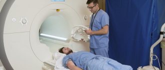
Positive serological reactions, characteristic neurological syndromes and changes in the cerebrospinal fluid (cytosis more than 8 - 10 in 1 mm3, protein more than 0.4 g/l and positive serological reactions) are the main criteria for the diagnosis of neurosyphilis. Computed tomography, magnetic resonance imaging, and positron emission tomography help in differential diagnosis.
Rice. 16. Lumbar puncture for neurosyphilis is a mandatory diagnostic procedure.


