Content
- 1 Pathogenesis
- 2 Types of dislocation syndromes 2.1 Displacement of the cerebral hemispheres under the falciform process
- 2.2 Temporotentorial and cerebellartentorial herniation
- 2.3 Herniation of the cerebellar tonsils into the foramen magnum
- 2.4 Displacement of the cerebral pons through the foramen of the tentorium cerebellum
- 2.5 Filling the middle and side tanks of the bridge
- 2.6 Displacement of the posterior part of the corpus callosum in the dorsal direction into the cistern of the same name
Pathogenesis
The brain does not occupy the entire volume of the cranium. Between it and the arachnoid membrane there is a subarachnoid space. In some sections it expands and forms the so-called subarachnoid cisterns.
When pressure increases in a certain part of the brain and cranium (the appearance of an area of distension), processes of displacement of parts of the brain occur within the subarachnoid space. Thus, in acute pathological processes of different etiologies, the same anatomical structures with stereotypical clinical manifestations are involved in dislocation syndromes. In other words, the clinical picture of acute dislocation syndrome does not depend on the etiology of the process. The difference in clinical manifestations in different patients depends on the rate of its development, location and volume.
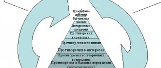
The fact is that dislocation syndromes are essentially internal hernias of the brain, that is, invagination of its parts into the holes and crevices formed by the bones and dura mater. There are 3 degrees of brain dislocation: protrusion, herniation and pinching. There are lateral and axial (along the axis of the brain stem) dislocations of the brain.
Drawing a parallel with hernias, it should be remembered that a sharp disruption of the patient’s vital functions arises not from the fact of the existence of a hernia, but from its strangulation. Infringement represents a protrusion, which is accompanied by cessation of blood flow due to compression of blood vessels.
Determining the shape of the dislocation
The presence of this disorder in the human body can be determined by the following signs. These include:
- Severe pain in the head.
- Nausea, vomiting.
- Deterioration of vision. The decline may occur intermittently.
- Cramps.
- Semi-conscious state or complete loss of consciousness.
All symptoms associated with dislocation are called dislocation syndrome. Similar signs also appear in humans with a brain tumor. A doctor can make an accurate diagnosis.
Types of dislocation syndromes
Most medical literature describes 4 types of dislocation syndromes (1-4), which are of greatest importance in clinical practice, as they can be accompanied by a significant deterioration in the patient’s condition. At the same time, the Great Medical Encyclopedia identifies 8 dislocation syndromes. Some sources [1][2] also describe external dislocation (number 9), which is a protrusion of parts of the brain through a traumatic defect of the skull.
- Displacement of the cerebral hemispheres under the falciform process
- Temporotentorial displacement
- Cerebellar-tentorial displacement
- Displacement of the cerebellar tonsils in the foramen magnum of the occipital bone
- Displacement of the cerebral pons through the foramen of the tentorium cerebellum
- Filling the middle and side tanks of the bridge
- Displacement of the posterior part of the corpus callosum in the dorsal direction into the cistern of the same name
- Displacement of the gyri of the frontal lobe into the chiasm cistern
- External brain dislocation
Displacement of the cerebral hemispheres under the falciform process
When the brain is compressed on one side by an acute volumetric process, with an increase in cerebrospinal fluid pressure in one lateral ventricle due to acute occlusive hydrocephalus, a difference in cerebrospinal fluid pressure develops between the right and left lateral ventricles. In this case, conditions are created for the dislocation of the cerebral hemisphere in the transverse direction into the gap between the falx cerebrum and the corpus callosum. The wedged area is mainly the cingulate gyrus and its adjacent areas. Thus, the cingulate gyrus fills the cistern of the corpus callosum. The cingulate gyrus, bulging in the opposite direction, displaces the anterior cerebral arteries, pressing them against the falciform process. The lateral and third ventricles are deformed. Perifocal edema can generalize due to slowing of blood circulation, as a result of compression of arteries and veins. All this leads to insufficient supply of nutrients and oxygen to the brain tissue, which leads to the development of even greater edema-swelling of the brain and to even greater dislocation. These processes can lead to blockage of liquor outflow. The cerebrospinal fluid produced in the lateral ventricles, having no outflow tract, creates an area of distension in the supratentorial space with the development of temporotentorial herniation, which leads to compression of the midbrain.
Temporotentorial and cerebellartentorial herniation
These 2 dislocation syndromes can be combined due to the commonality of the affected structures and the correspondingly developing clinic. They differ in the location (sub- or supratentorial) of the area of distension. Temporotentorial herniation
It is a protrusion into the tentorial foramen (Bishat’s fissure) of a section of the temporal lobe of the brain and, first of all, the medial sections of the hippocampal gyrus, partly the anterior part of the lingual gyrus and the isthmus of the vaulted gyrus.
Wedged between the free edge of the cerebellar tentorium and the oral parts of the brainstem, the area of the temporal lobe can occupy different positions in relation to the trunk: anterior - when located in front of the trunk; anterolateral - when located anterior to the trunk and along its anterior quadrant, posterolateral, posterior. Intrusion of brain areas into the tentorial foramen can occur not only from the cerebral hemispheres, but also in the opposite direction, that is, from the posterior cranial fossa. In such cases, a portion of the cerebellum protrudes between the free edge of the tentorium and the quadrigeminal region. This herniation is called cerebellentorial
. It occurs during pathological processes in the posterior cranial fossa. The occurrence of temporo-tentorial and cerebellar-tentorial herniations is inevitably associated with a displacement of the trunk in the opposite direction, which depends on the size and characteristics of the herniation.
The ipsilateral oculomotor nerve is compressed - the pupil dilates after a short-term miosis.
Lateral displacement of the homolateral parahippocampal gyrus causes the midbrain to move across the midline toward the opposite edge of the tentorium. This small change in midbrain configuration can lower the patient's level of consciousness and cause targeted arousal.
Eventually, the globus pallidus, internal capsule, and thalamus opticum on the homolateral side move caudally, and the parahippocampal gyrus protrudes beyond the edge of the cerebellar tentorium into the subtentorial space. The mamillary bodies are wedged into the narrowed interpeduncular fossa. Under these conditions, the midbrain experiences intense pressure, which causes the development of coma. The midbrain may be pressed against the opposite edge of the tentorium so tightly that the descending motor fibers in the compressed cerebral peduncle are damaged. The hemiplegia caused by this type of herniation is not contralateral, but homolateral in relation to the volumetric process - Kernohan's cerebral peduncle syndrome
. Due to compression, hemorrhages occur in the inferotemporal and occipital regions of the brain, midbrain, which can often lead to irreversible consequences and death of the patient
Herniation of the cerebellar tonsils into the foramen magnum
The tonsils of the cerebellum, shifting into the foramen magnum, sometimes reach the level of the second cervical vertebra. Wedged between the caudal part of the medulla oblongata, the adjacent part of the spinal cord and the occipito-cervical ring, they tightly envelop and compress this part of the trunk from the dorsal surface.
In this case, cerebral circulation disorders occur, inevitably leading to hypoxia and to an even greater increase in edema-swelling of the brain. Thus, a vicious circle is closed, which leads to the progression of compression of the brain stem, which leads to disruption of the function of the vital centers of blood circulation and respiration embedded in them and, as a consequence, to the death of the patient.
Displacement of the cerebral pons through the foramen of the tentorium cerebellum
The pons is displaced in the oral direction into the interpeduncular cistern.
Filling the middle and side tanks of the bridge
It occurs as a result of pressing the brain bridge against the slope of the base of the skull.
Displacement of the posterior part of the corpus callosum in the dorsal direction into the cistern of the same name
Occurs with closed hydrocephalus of the brain, which can also occur with the above dislocation syndromes. Due to increased pressure in the ventricles, the corpus callosum is displaced into the cistern of the same name. The free edge of the falx forms a notch on the corpus callosum. Since this dislocation syndrome occurs against the background of deep disturbances in the vital functions of the body, it is rarely described in the literature devoted to this problem.
CSF circulation disorders
Brain dislocations are dangerous not only by the development of cerebrovascular accidents. There is also displacement and compression of the cerebrospinal fluid-containing spaces, both as a whole and in their individual parts, in particular the cerebral aqueduct, the fourth ventricle, etc. Compression leads to disruption of the cerebrospinal fluid circulation. The normal state of the cerebrospinal fluid pathways is disrupted due to their narrowing at the level of the tentorial foramen or foramen magnum, which leads to the development of hydrocephalus. Naturally, if the outflow of cerebrospinal fluid from the ventricles is disrupted, a pressure gradient occurs between the ventricles and the spinal sac. This pressure difference leads to even greater displacements of the brain stem in the caudal direction, to the development of new dislocation syndromes.
Indications for echoencephalography
There are a number of conditions and diseases that echoencephalography has been demonstrated to detect and identify. So, the procedure is carried out when:
- headache;
- frequent dizziness (loss of consciousness and balance);
- head injuries;
- diffuse and local cerebral edema;
- intracranial hematomas;
- abscesses;
- brain tumors;
- intracranial hypertension;
- hydrocephalus;
- inflammatory diseases of the brain;
- other cerebral diseases;
- cerebral ischemia;
- stroke;
- concussion and bruises of the brain;
- vertebrobasilar insufficiency;
- vegetative-vascular dystonia;
- disorders of cerebral blood flow;
- tinnitus;
- neck injuries;
- Parkinson's disease;
- pituitary adenoma.
Clinic
In parallel with focal neurological disorders associated with an acute pathological focus, during dislocation, general cerebral phenomena also develop due to increasing intracranial hypertension. The clinical picture of dislocation syndrome and damage to the brain stem can develop against the background of local manifestations of the main acute pathological process. And only at the very latest stages of the disease, with the development of a deep or extreme coma, clinical manifestations of the main acute pathological process may not be observed and only severe bilateral damage to the brain stem and other general cerebral pathological signs are revealed.
When the cerebral hemispheres are dislocated under the falx cerebri
clinically, symptoms of disorders of higher nervous activity and mental processes appear first. Clinically, first of all (if the patient’s consciousness is preserved) mental disorders appear, sometimes with hallucinations or delirium-like states. Psychomotor agitation often occurs, which can occur with both preserved and lost consciousness. Subsequently, adynamia, akinesia appear, and an oneiric state occurs. A serious symptom with this type of herniation is epileptiform convulsions (due to the involvement of the cingulate gyrus in the process). Usually their occurrence is accompanied or preceded by a deepening disturbance of consciousness, up to coma.
With temporo-tentorial and cerebellar-tentorial herniations
oculomotor disturbances appear (horizontal, rotatory, vertical nystagmus, discrepancy of the eyeballs, Hertwig-Magendie symptom, partial or complete paresis of upward or sideways gaze or complete immobility of the eyeballs, weakened or absent reaction of the pupils to light, etc.). Unilateral pyramidal insufficiency, which occurs during an acute space-occupying process (Kernohan's cerebral peduncle syndrome), becomes bilateral. This fact indicates the involvement of the second leg of the brain in the pathological process. The clinical picture of these types of dislocations depends on which arteries to which areas of the brain are compressed in the tentorial foramen.
Compression of the vessels leading to the basal ganglia of the extrapyramidal system leads to dysregulation of muscle tone, first in the form of increased extensor tone and the appearance of cogwheel symptoms. Then hormetonia develops, muscle rigidity and, as a result, muscle atony - evidence of separation of the midbrain from the overlying parts. Diencephalic disorders are detected in the form of tachycardia, which replaces bradycardia. The ECG records various forms of arrhythmia, infarction-like ischemia and other disorders that are functional in nature, secondary to cerebral pathology and disappear without a trace when the acute period is eliminated. There is also tachypnea, hyperthermia, in some patients up to 39-40°C, hyperemia and greasiness of the skin, etc. The consciousness of patients is depressed, up to coma of varying degrees of severity.
Displacement of the cerebellar tonsils into the foramen magnum
clinically is the most severe. In this case, vital centers suffer, primarily the centers of respiration and circulation. Respiratory disturbances of central origin of varying degrees occur, which depend on the rate of development, degree and duration of the herniation. These disorders are detected in tachypnea (at the beginning of the process), which then develops into deeper forms of pathology, up to Cheyne-Stokes, Biot breathing, terminal types and apnea. In parallel with the breathing disorder, the depth of the disturbance of consciousness increases, and anabolic disorders develop. Swallowing is severely impaired, the pharyngeal reflex decreases or disappears—bulbar syndrome develops. Vascular tone decreases. Arterial hypotension develops.
Clinical manifestations of brain dislocation in the acute period of severe traumatic brain injury
The results of surgical treatment of 168 patients with dislocation syndrome in acute severe traumatic brain injury were treated in the National Hospital. The analysis of the clinical manifestations and treatment of dislocation of the brain in patients with severe traumatic brain injury in the acute period. The dynamic observation of dislocation syndrome clinic patients examined. Outcomes depend both on the primary and secondary factors of brain damage.
Key words: Severe craniocerebral trauma, dislocation syndrome, a horizontal and axial dislocation, surgical treatment.
Relevance of the problem : In victims with severe traumatic brain injury (STBI), intracranial hypertension and dislocation manifestation of the brain structure are an important link in the pathogenesis of this nosology, and is the main cause of mortality [3, 9, 20].
According to research by L. B. Likhterman (1999), mortality in severe TBI in the Russian Federation is at least 68–70%, and, according to S. V. Astrakov (2007), from 38 to 80%.
Dislocation syndrome (DS) is a common complication of emergency neurosurgical diseases. As reserve cerebrospinal fluid spaces are exhausted, various intracranial pressure gradients may arise. This leads to displacement of brain structures, deformation and infringement of the brain [3,6]. Dislocation syndrome with STBI develops in a short period of time, resulting in an immediate threat to the patient’s life requiring emergency medical care [4, 8].
The clinical picture of acute dislocation syndrome does not depend on the etiology of the process. The clinical manifestation of brain dislocation in different patients depends on the rate of development of the dislocation, localization and volume of the pathological process of the brain [1, 6, 18].
The critical level of increase in intracranial pressure is considered to be 20–25 mmHg. Art., although clinical experience demonstrates the absence of a close connection between the absolute value of intracranial pressure and the development of brain dislocation. However, in the presence of a volumetric process of temporal localization, dislocation and herniation can occur even with intracranial pressure less than 20 mm Hg. Art. [13]. Intracranial pressure level is more than 20–25 mmHg. Art. in most cases, it requires the appointment of adequate anti-edematous therapy, and in some cases, surgical decompression.
Treatment of brain injuries accompanied by dislocation syndromes is an extremely complex, at the same time unsolved to date [2] and controversial problem.
According to most retrospective and prospective non-randomized clinical studies, hemicraniectomy with dural plastic surgery reduces associated mortality from 80 to 30% [15, 16, 19]. Some studies indicate that mortality may be further reduced if hemicraniectomy is performed within the first 24 hours after stroke [14].
All neurosurgeons write about the severe course of dislocation syndrome and the danger to the patient’s life. However, recently little attention has been paid to the treatment, including surgery, of this formidable complication. And only a few reports describe attempts to eliminate dislocations [5,10,11,12,18].
Purpose of the study: To analyze the clinical manifestations of brain dislocation in the acute period of severe traumatic brain injury, depending on the location, form and degree of manifestation of brain dislocation and to develop principles for eliminating brain dislocation.
Research objectives:
- To determine the location and types of the source of damage in the acute period of TBI and, depending on the severity of the brain contusion, to establish the types and forms of dislocation syndrome.
- To study the clinical manifestation of dislocation syndrome with various forms and degrees of brain dislocation based on neuroimaging examination methods.
- To analyze comparative results in surgical and conservative treatment of brain dislocations.
Materials and methods: The results of clinical and instrumental examination methods, as well as surgical treatment of 168 patients with severe traumatic brain injury (STBI), who were hospitalized in the neurosurgery clinic of the National Hospital of the Ministry of Health of the Kyrgyz Republic, aged from 21 to 61 years, were studied. , on average 40–41 years old. 138 (82.1%) patients were men, 30 (17.9%) patients were women.
To assess the degree of dislocation syndrome (DS), the following signs were taken into account: the level of depression of consciousness [7], general cerebral and focal symptoms, respiratory rate, pulse, blood pressure. Instrumental examination included CT and (or) MRI of the brain. Consciousness of patients was assessed using the Glasgow Coma Outcome Scale [17]. In most cases (76%), the operation was performed by decompressive craniotomy with removal of the lesion and elimination of dislocation; in other cases, resection craniotomy or osteoplastic craniotomy.
In most cases, patients 137 (81.5%) were transported by ambulance after injury from 30 minutes to 12–14 hours, on average 6–7 hours. In the remaining cases (18.5%), patients were admitted to the nearest medical institutions, and after first aid was provided, they were subsequently transferred to a neurosurgical clinic.
There were 112 patients with closed craniocerebral injury, 56 patients with open craniocerebral injury, of which 7 cases had penetrating TBI. Various types of fractures of the vault and base of the skull were found in 97 patients.
CT examination was performed in 84 patients, and MRI in 65 patients. 135 patients underwent surgical treatment, 33 patients received conservative treatment.
Results and discussion : Subdural hematoma (SDH) was found in 42 patients, epidural hematoma (EDH) in 19 patients, a focus of contusion with hemorrhagic impregnation and the formation of intracerebral hematoma in 24 patients, sub- and epidural hematoma in 12 patients, in 22 cases focus of contusion with hemorrhage and epi-, subdural hematomas, multiple foci of contusion with crushing of the brain substance with hemorrhage in 8 patients. In 57 (33.9%) patients there was bilateral brain damage and in 111 (66.1%) cases a unilateral focal lesion was established (on the left in 48 patients and on the right in 63 patients).
CT and MRI examinations of the brain revealed horizontal and axial types of displacement (dislocation) of brain structures. Horizontal displacement (under the falx of the brain) was found in 49 patients, axial displacement (at the level of the tentorial foramen or foramen magnum) in 16 patients, horizontal and axial dislocations were found in 94 patients. Thus, the following forms of dislocation have been established: 1) cingular (under falx) dislocation in 49 cases; 2) temporotentorial dislocation, 14 cases; 3) temporotentorial and foramenal dislocations were found in 2 patients. 4) cingulate and temporal-tentorial dislocation were found in 78 patients. 5) cingulate, temporotentorial and foraminal dislocations in 16 patients.
Horizontal - transverse types of displacement were identified in 111 (86.6%) of 127 patients with unilateral lesions, and in 35 (85.3%) of 41 patients with bilateral lesions.
In unilateral traumatic lesions out of 127 patients, axial dislocation was observed in 75(59%) cases, in 34(46.6%) patients temporotentorial dislocation up to 4mm, in 31(41.3%) patients more than 4mm. With bilateral focal lesions, out of 41 patients, temporotentorial dislocation was observed in 29 patients, temporotentorial and foramenal dislocations were found in 2 patients. Bilateral temporotentorial dislocation was found in 3 patients.
According to our data, the degree of horizontal type of brain dislocation depended on the location of the lesion. When the lesion was localized in the parietotemporal region, out of 19 patients, in 5 (26.3%) cases, the displacement of the median structures was 10 mm or more, in the fronto-parietotemporal region, out of 31 patients, 14 (45.1%) cases, as well as with extensive traumatic damage to one hemisphere of the brain out of 17 patients in 6 (35.3%) cases. When the lesions were localized in the frontal lobe of the brain, out of 14 patients, horizontal dislocation in 6 (42.6%) patients was 4 mm, in 5 (35.7 %) patients up to 8 mm. In case of traumatic lesion of the parietal region, out of 7 patients, in 3 patients the horizontal dislocation reached 4 mm and in 1 patient up to 8 mm. With bilateral lesions out of 41 patients, a horizontal transverse dislocation with a unilateral accent of displacement was detected, but the size of the displacement was not expressed, in 12 (29.2%) patients up to 4 mm and in 15 (36.5%) up to 8 mm.
Table 1
Causes of development of dislocation syndrome ( n -168)
| Types of dislocation | Cingular dislocation | Axial dislocation | ||||||
| With one side | On both sides | |||||||
| Dimensions (mm) mixing Types of outbreak defeats | Up to 4mm | Up to 8mm | Up to 12mm | Up to 17–18mm | Up to 4mm | More than 4mm | Up to 4mm | More than 4mm |
| 5 | 4 | 6 | — | 5 | 4 | — | — |
| 8 | 9 | 15 | 1 | 2 | 9 | 1 | — |
| 6 | 8 | 5 | 1 | 7 | 5 | 1 | 1 |
| 4 | 11 | 12 | 7 | 13 | 11 | — | — |
| 2 | 1 | 5 | — | 2 | 4 | 1 | 0 |
| 12 | 15 | 8 | — | 7 | 7 | 8 | 11 |
EDH-epidural hematoma; SDH-subdural hematoma; OUiRGM VMH - foci of contusion and crushing of the bare brain, intracerebral hematoma; MOUiRGMG - multiple foci of contusion and crushing of the brain with hematomas; Combined - VMG + SDH or + EDH in one hemisphere; DMOUiRGVG - bilateral multiple foci of contusion and crushing of the brain.
Satisfactory - moderate residual effects and recovery;
Unsatisfactory - gross residual effects or vegetative state;
Axial types of dislocation were often observed in bilateral lesions out of 41 patients in 33 (80.4%) cases: bilateral temporotentorial herniation was found in 19 (46.3%) patients, 8 cases up to 4 mm and in 11 patients more than 4 mm, unilateral temporotentorial herniation. tentorial herniation in 14 (34.1%) patients, of which up to 4 mm in 7 and more than 4 mm in 7 patients. With a unilateral lesion, frequent temporotentorial dislocation was observed when the lesion was localized in the parietotemporal region out of 19 patients in 15 (78.9%) cases, temporal localization of the lesion out of 6 patients in 4 (66.6%) patients, fronto-parieto- temporal localization out of 31 patients in 21 (67.7%) patients. With extensive localization of the lesion, out of 17 patients, 14 (82.3%) cases, in the frontotemporal region, out of 12 patients, 8 (66.6%) cases.
Cingular and temporotentorial forms of dislocation were often observed with combined lesions, in 6 (70%) cases out of 8 patients, and in 24 (69%) cases with multiple lesions with hematomas out of 34 patients.
The clinical picture of 49 (29.1%) patients with the cingulate form of dislocation was marked by general cerebral symptoms, focal symptoms were detected in 31 (63.2%) patients, mild brainstem symptoms in the form of pupil dilation, absence of photoreaction and decreased corneal reflexes in 9( 18.4%) patients.
Of the 78 (46.4%) patients with cingulate and temporotentorial dislocation: in 66 (84.6%) patients, along with general cerebral symptoms, focal disorders in the form of pyramidal insufficiency dominated, in 41 (52.6%) patients the lungs stem symptoms, 14 (17.9%) patients had severe brainstem symptoms with impaired breathing, cardiac activity, decreased and lack of reactions to painful stimuli.
With temporotentorial dislocation, out of 14 (8.3%) patients: focal symptoms were observed in 9 (64.2%) patients, 3 patients had mild brainstem symptoms and 4 patients had severe brainstem symptoms.
Of 16 (20.5%) patients with cingulate, temporotentorial and foramenal dislocation: focal symptoms were detected in 7 (43.7%) patients, mild brainstem symptoms in 4 patients and severe brainstem symptoms in 6 (37.5%) patients symptoms.
In 2 patients with temporotentorial and foramenal dislocation, the condition was extremely severe with severe brainstem symptoms.
When analyzing the results of conservative treatment, we identified patients admitted at the stage of compensation according to GCS 12–14 points, displacement of the midline structures of the brain not exceeding 5 mm and the volume of the hematoma not exceeding 50 cm3. showed good results. Of the 33 patients, 13 (39.1%) were discharged with recovery (on the Karnofsky scale 70–90%), 9 (27.3%) patients were in unsatisfactory condition (on the Karnofsky scale 50–60 points). In extremely serious condition, the admitted patients had a GCS score of 3–5 points, with multiple foci of brain damage, with horizontal dislocation of more than 10 mm and axial dislocation of 2–4 mm, with severe vital disturbances, 10 (43.5%) patients, despite the intensive 6 patients died within 48 hours of therapy and 4 patients died before the 4th day.
Surgical treatment was performed in 135 patients using three methods: decompressive craniotomy (DCT) - 51 (37.8%) patients, osteoplastic craniotomy (OPCT) - 49 (36.3%) and resection craniotomy (RTC) - 35 ( 25.9%) to patients with removal of hematomas and foci of contusion and crush injuries with elimination of dislocation syndrome. Of the 135 patients operated on, 69 (51.1%) patients were discharged in satisfactory condition (on the Karnovsky scale 80–90 points) with recovery, 25 (18.5%) patients were in unsatisfactory condition (on the Karnovsky scale 50–60 points), not Despite intensive therapy, 39 (28.9%) patients died.
The outcomes depending on the location of the lesion were as follows. Multiple lesions with hemorrhage in the brain out of 8 patients, death was in 5 (62.5%) patients, 2 (25%) patients were discharged in unsatisfactory condition according to the Karnofsky scale 60–60 points, 1 (12.5%) the patient was discharged in satisfactory condition. With bilateral lesions, out of 41 patients, death occurred in 17 (41.4%) patients, unsatisfactory condition on the Karnovsky scale of 60–70 points in 12 (29.3%) patients and 12 (29.3%) patients with satisfactory results . In other cases, treatment outcomes are shown in Table 2.
table 2
Outcomes of surgical and conservative treatment
| Satisfactory | Unsatisfactory | Death | Total | |
| EDH | 15 | — | 3 | 19 |
| SDH | 29 | 4 | 9 | 42 |
| OUiRGM VMG | 16 | 3 | 5 | 24 |
| Combined | 14 | 11 | 9 | 34 |
| MOUiRGMG | 1 | 2 | 5 | 8 |
| DMOUiRG | 12 | 12 | 17 | 41 |
The outcomes depending on the degree of dislocation were as follows. In case of temporotentorial dislocation, out of 14 patients, with a fatal outcome, 6 (42.8%) patients had a displacement of the hippocampal gyrus between the free edge of the tentorium of the cerebellum and the brain stem of more than 4 mm. 5 (35.7%) patients were discharged with gross residual effects, where the hippocampal gyrus was herniated to 2–4 mm.
With a mixed type of dislocation (cingular, temporotentorial and foramenal dislocation), 15 (93.7%) of 16 patients died within 7 days after injury, 1 patient was discharged with gross residual effects. In these patients, a dislocation of the hippocampal gyrus into Bichat's fissure of more than 4 mm and a displacement of the cerebellar amygdala into the foramen magnum of up to 3–6 mm were revealed.
There were 2 patients with temporotentorial and foramenal dislocation, both with fatal outcomes, where dislocations of brain structures were also revealed in the Bichat fissure of more than 4 mm and in the foramen magnum up to 3–6 mm.
With cingular and temporotentorial dislocation, out of 78 patients, death was observed in 22 (28.2%) cases, displacement under the falx of the cingulate gyrus was up to 17 mm, and into the Bichat fissure of the hippocampal gyrus more than 4 mm. There were 22 (28.2%) patients with gross residual effects, where the dislocation under the falx reached 14 mm and in the Bichat gap up to 4 mm. With moderate residual effects and recovery in 33 (42.3%) patients, herniation under the falx was up to 12 mm and between the free edge of the tentorium of the cerebellum and the brain stem up to 2–4 mm.
Relatively good results were obtained with cingulate dislocation; out of 49 patients with a satisfactory result, 44 (89.7%) patients were discharged, where the dislocation of the cingulate gyrus under the falx was 4–8 mm.
Conclusions:
- Axial types of dislocation often occurred when the lesion was localized in the parietotemporal and fronto-parietotemporal regions. And with combined and multiple lesions, a more severe degree of dislocation developed.
- For the cingulate form of dislocation after surgical treatment, good results of up to 73.4% were observed. With temporotentorial dislocation (35.7–42.8%), treatment outcomes are unfavorable, and with temporotentorial and foramenal dislocation, a 100% fatal outcome is observed.
- Surgery is a more effective way to eliminate brain dislocation.
Literature:
- Akhmedov E. A., Kasumov R. D., Bersnev V. P. Differential tactics of surgical treatment of traumatic chronic subdural hematomas. // Polenov readings. Saint Petersburg. 2009.-36s.
- Bersnev V.P. On the issue of surgical treatment of hypertensive intracerebral hemorrhages / V.P. Bersnev, M.K. Agzamov // VI Polenov Readings. St. Petersburg, 2007. - 145 p.
- Blinkov, S. M. Displacements and deformations of the brain. Morphology and clinic /S. M. Blinkov, N. A. Smirnov. - L.: Medicine, 1967.-202 p.
- Zotov Yu. V., Kondakov E. N., Shchedrenok V. V., Kondratyev A. N. Intracranial decompression of the brain in surgery for severe traumatic brain injury. - St. Petersburg, 1999. -142 p.
- Lebedev V.V., Bykovnikov L.D. Guide to emergency neurosurgery. - M., “Medicine”, 1987.-335 p.
- Lebedev V.V., Krylov V.V. Emergency neurosurgery: A guide for doctors. M.: Medicine, 2000.-257 p.
- Konovalov A. N. Clinical guidelines for traumatic brain injury. Volume I / A. N. Konovalov, L. B. Likhterman. 1998.-549p.
- Kondakov E. N., Klimash A. V., Bakhtiyarov A. K., Bokin V. D. Supratentorial traumatic dislocation of the brain / Zh. Neurological Bulletin - 2008 vol. - XL, issue 3 - pp. 19–24.
- Potapov A. A. Modern recommendations for the diagnosis and treatment of severe traumatic brain injury // Potapov A. A., Krylov V. V., Likhterman L. B., Tsarenko S. V., Gavrilov A. G., Petrikov S. S. Zh. “Issues of neurosurgery named after. N. N. Burdenko” 2006. No. 1. P. 3–8
- Saribekyan A. S. Tactics of surgical treatment of severe head injury and traumatic intracranial hemorrhage in terms of the dynamics of intracranial hypertension: Abstract of thesis. diss. doc. medical sciences - Moscow, 1992.C - 23–24.
- Saribekyan A. S. Tentoriotomy and ventricular drainage in the surgical treatment of severe TBI: Abstract of thesis. diss. Ph.D. medical sciences - Moscow, 1984. - 26 p.
- Soloviev A. G. Method of low-traumatic tentoriotomy for traumatic brain injury: Abstract…. Ph.D. honey. nauk., Moscow, 1978.-19 p.
- Andrews BT, Chiles BW 3rd, Olsen WL, Pitts LH The effect of intracerebral hematoma location on the risk of brain-stem compression and on clinical outcome // J. Neurosurgery -1988.-Vol. 69, No. 4.-P. 518–522.
- Cho DY, Chen TC, Lee HC Ultra-early decompressive craniectomy for malignant middle cerebral artery infarction // Surg Neurology. 2003. Vol. 60, N 3.-R. 227–232.
- Georgiadis D., Schwarz S., Aschoff A., Schwab S. Hemicraniectomy and moderate hypo-thermia in patients with severe ischemic stroke // Stroke. 2002. Vol. 33, N 6. R. 1584–1588.
- Holtkamp M., Buchheim K., Unterberg A. et al. Hemicraniectomy in elderly patients with space occupying media infarction: Improved survival but poor functional outcome // J. Neurology Neurosurgery Psychiatry. 2001. Vol. 70, N 2. R. 226–228.
- Jennett, B., Assessment of Outcome After Severe Brain Damage: Practical Scale /B. Jen-nett, M. Bond // Lancet. -1975. -No. 1.-R. 480–484.
- Rehman T., Ali R., Tawil I., Howard Y. Rapid progression of traumatic bifrontal contusions to transtentorial herniation: A case report. Cases J. 2001; 1; 203–206.
- Walz B., Zimmermann C., Bottger S, Haberl RL. Prognosis of patients after hemicraniectomy in malignant middle cerebral artery infarction // J. Neurol.- 2002.- Vol. 249, N 9. R.1183–1190.
- Whittle IR, Vishwanathan R. Acute intraoperative brain herniation during elective neurosurgery: Pathophysiology and management considerations. J NeurologyNeurosurgery Psychiatry,2004.-Vol. 19; 61: P.584–90.
Diagnostics
Diagnostic methods:
- Echoencephalography determines the degree of displacement of the midline structures in one direction or another. It should be taken into account that according to Echo-EG data, only lateral dislocation can be assumed (displacement of the cerebral hemispheres under the falciform process), since according to its data it is impossible to diagnose axial dislocation of the brain.
- Angiography
- CT scan
- Magnetic resonance imaging
Lumbar puncture is strictly contraindicated even if a brain dislocation is suspected, since a decrease in cerebrospinal fluid pressure in the subarachnoid space of the spinal cord in the presence of an area of increased intracranial pressure can trigger the processes of herniation and lead to the death of the patient. Back in 1938, a case was described when a lumbar puncture was performed for diagnostic purposes, which ended in death directly on the table.
In order to prevent violation
To prevent the development of dislocation syndrome, the following rules must be observed:
- protect your head in any dangerous situations - when riding a bicycle, motorcycle, or when playing active sports;
- always use seat belts when traveling in a car;
- monitor nutrition and weight, eliminate bad habits;
- prevent infectious brain lesions;
- adhere to safety rules when climbing mountains;
- timely treat pathologies of the circulatory system.
Brain dislocation is a rather dangerous condition that poses a real threat to life. That is why it is so important to consult a doctor promptly if you suspect this pathology.
This section was created to take care of those who need a qualified specialist, without disturbing the usual rhythm of their own lives.
Treatment
The first and indispensable condition for treating a dislocation is to eliminate the cause that caused it.
Non-surgical measures used for dislocation syndromes include:
- barbituric anesthesia
- moderate hypothermia
- periodic deep hyperventilation
- glucocorticoids
An operation for life-saving reasons in the development of cerebral edema and processes that lead to the dislocation of some parts of the brain in relation to others is decompressive craniotomy. To remove the pathological focus, a wide (minimum 5-6 x 6-7 cm) decompressive craniotomy is performed, which does not necessarily have to be resection, but definitely decompressive. With temporotentorial herniation, trepanation is performed in the temporoparietal region as low as possible. With bilateral symptoms, decompressive trepanation is performed on both sides. After removal of the pathological focus, the dura mater is not sutured.
Also, to reduce intracranial pressure and, accordingly, reduce the risk of developing life-threatening herniation processes, drainage of the ventricular system is performed. The anterior or posterior horn is punctured using standard points (Kocher or Dandy). The effect of puncture of the cerebral ventricles is greater if drainage is performed at earlier stages of herniation. With a lateral displacement of the ventricles, it is not easy to get into the displaced and compressed ventricle of the brain. Puncture of the hydrocephalic ventricle on the side opposite to the lesion (“healthy”) with a remaining pathological focus is fraught with an increase in dislocation and an increase in vital disturbances.
Computed tomography of the brain. Echoencephalography in children
Source: https://meduniver.com/Medical/Neurology/912.html
| Computed tomography carried out using an EMI - Scanner apparatus, which uses sensitive crystal detectors and a minicomputer to construct detailed images containing 100 times more information about brain tissue than using conventional X-ray methods. Using this device, you can obtain accurate and detailed images of the smallest changes in the density of brain tissue, showing the nature and location of diseases such as brain tumors, cysts, and hemorrhages. Brain scanning device the patient is examined as a series of layers. This system has great advantages compared to radiopaque methods, which create a lot of inconvenience for the patient. The study can be carried out on an outpatient basis in dynamics. The image is obtained 5 minutes after scanning the head patient and can be viewed immediately on the screen of a cathode ray tube or photographed for subsequent studies. This device is likely to find widespread use in neuroradiological examinations of young children. |
Literature
- Big medical encyclopedia. Main Ed. B.V. Petrovsky. Ed. 3rd. M., “Sov. Encyclopedia", 1977 p. 347—349
- Duus P. Topical diagnosis in neurology Anatomy. Physiology. Clinic - M. IPC "Vazar-Ferro", 1995 p. 173—179
- Lebedev V.V., Bykovnikov L.D., Kariev M. Emergency diagnostics and assistance in neurosurgery - T.: Medicine, 1988 p. 44-67
- Lebedev V.V., Krylov V.V. Emergency neurosurgery: A guide for doctors. - M.: Medicine, 2000 p. 103-141
- Misyuk N. S., Evstigneev V. V., Rogulchenko S. M. Displacements and infringements of the brain stem. Minsk, “Belarus”, 1968
- Neurotraumatology. Directory / Edited by: Academician of the Russian Academy of Medical Sciences A.N. Konovalov, Professor L.B. Likhterman, Professor A.A. Potapov. - M.: IPC “Vazar-Ferro”, 1993
- Practical neurosurgery: A guide for doctors / Ed. B.V. Gaidar. - St. Petersburg: Hippocrates, 2002