Carpal tunnel syndrome is an occupational disease of people who perform monotonous, same-type hand movements in accordance with the specifics of their work activity.
Most often, the pathology is diagnosed in cashiers in stores, artists, computer users, hairdressers, some musicians, miners, etc. Women are more susceptible to the disease than men, which is due to the relatively small size of the carpal tunnel.
The first symptoms of carpal tunnel syndrome usually appear at the age of 35–40 years. The peak incidence is observed in patients 50–60 years old. The disease occurs in a chronic form with alternating periods of exacerbation and remission. Accompanied by pain, paresthesia, and impaired motor function. Symptoms of pathology can have varying degrees of severity.
What it is?
Carpal tunnel syndrome, or carpal tunnel syndrome, is a chronic neurological disease accompanied by a feeling of numbness and pain in the fingers. The disease belongs to tunnel neuropathy.
The main reason for the development of pathology is compression of the median nerve located between the bones, the transverse ligament of the hand and the carpal tendons. This syndrome often develops in older women, as well as in people who, due to their professional duties, perform monotonous flexion-extension movements with the hand.
Diagnosis of carpal tunnel syndrome
Sometimes tunnel syndrome can be accompanied by pain not only in the hand, but also in the forearm and elbow. This confuses the doctor and may lead to thoughts about another pathology, for example, osteochondrosis. Therefore, special methods are used to carry out differential diagnosis.
For example, there is a simple raised hands test. The patient raises his straightened arms above his head and holds for a minute. If you have carpal tunnel syndrome, the first three fingers experience numbness and tingling, and sometimes even pain.
To perform the Phalen test, the patient is asked to bend the hand and hold it there for a minute. If you have carpal tunnel syndrome, the first three fingers experience increased tingling and pain.
A cuff test is also sometimes performed. The doctor places a blood pressure cuff on the patient's arm. Then the pressure in the cuff is inflated to over 120 mmHg, which is held for a minute. In carpal tunnel syndrome, a tingling sensation occurs in the fingers innervated by the median nerve.
But the most reliable diagnostic method is still the Tinel test. The doctor taps a finger or hammer over the median nerve. If you have carpal tunnel syndrome, you may experience tingling in your fingers.
A useful diagnostic test is the injection of corticosteroids with lidocaine into the carpal tunnel. If after this the pain and tingling in the fingers decrease, it means that the pathological process is located in the carpal tunnel.
The leading instrumental method for determining carpal tunnel syndrome is electroneuromyography. Using this study, it is possible to measure the electrical activity of skeletal muscles, as well as the speed of nerve impulses. At rest, the electrical activity of the muscles is minimal, but increases with muscle contraction. But in the presence of carpal tunnel syndrome, electrical activity is low during muscle contraction because the conduction of nerve impulses along the damaged median nerve is slowed down.
Reasons for development
The carpal tunnel is located at the base of the hand. It is formed by the carpal bones and transverse ligaments. Inside this canal are the median nerve, digital flexor tendons and their synovial membranes.
The median nerve consists of sensory and motor nerve fibers. They are responsible for the sensitivity of the skin of the palm of the first three and half of the fourth fingers, and the dorsal surface of their nail phalanges. The task of motor fibers is to provide movement of the fingers.
In a healthy person, the median nerve runs freely along the canal. But with frequent microtraumas, thickening occurs and swelling of the transverse ligament forms. It begins to put pressure on the nerve, and in parallel with this, inflammation of the connective tissues occurs, characterized by a chronic course. Increased pressure on the median nerve provokes venous stagnation with subsequent poor circulation in the nerve.
Sensitive fibers are the first to be damaged, and only then are motor fibers affected. Among other things, even fibers of the autonomic nervous system can be involved in such a process.
So, what are the reasons for the development of the disease? Carpal tunnel syndrome can be caused by:
- long monotonous work at the computer;
- previous injuries (for example, a broken wrist);
- period of pregnancy, breastfeeding, menopause;
- endocrine pathologies: acromegaly, hypoparathyroidism, etc.;
- taking hormonal contraception;
- renal failure;
- joint pathologies (rheumatoid arthritis, gout);
- prolonged hypothermia;
- genetic predisposition;
- peculiarities of work, when a person constantly has to bend and straighten his arm.
The disease may first appear at a young age, but its peak peak occurs between 40 and 60 years of age.
Why does carpal syndrome develop and the risk of its development?
Carpal tunnel syndrome can be caused by injury, hormonal imbalance, related factors, or other conditions. If there is no specific cause, doctors talk about “idiopathic carpal tunnel syndrome.”
So, the main causes of the condition:
- Injuries. After injury to the wrist, especially after a radius (radius) fracture, carpal tunnel syndrome may develop.
- Diseases. Every second case of rheumatism is accompanied by carpal tunnel syndrome. Often this is even the first sign of an incipient rheumatic disease. In cases of hyperthyroidism or underweight, diabetes and obesity, carpal tunnel syndrome often occurs. Increased water retention in the joints or thickening of the ligaments is responsible.
- Hormonal changes. Hormonal changes can lead to increased water retention in joints and compression of nerves and tendons. Therefore, pregnancy and menopause are common triggers.
- Load. People who work physically are three to seven times more likely to have carpal tunnel syndrome than those who do no physical work.
- Heredity. Carpal tunnel syndrome is especially common in some families. The victims are usually family members.
There are professional groups that are at particular risk. This includes anyone who operates machines with high vibrations (such as jackhammers), as well as those whose wrists are subject to constant physical activity, such as painters or farmers.
Occupational risks of developing carpal tunnel syndrome
Chronic renal failure, as a cause, is associated with an increased risk as the duration of the dialysis procedure increases. It usually develops on the hand that is directly connected to the device.
Recent studies have shown that about 50 percent of cleaners develop carpal tunnel syndrome.
Working on a computer is not an excessive load from the point of view of an increased risk of developing carpal tunnel syndrome.
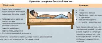
Symptoms
In the early phase of its development, tunnel syndrome is accompanied by trembling, tingling in the limbs and itching. Some symptoms may appear later - after completing a certain wrist movement.
The late stage of the disease is accompanied by:
- numbness;
- pain syndrome;
- heaviness in the hands;
- decreased sensitivity of the hands;
- increasing the severity of paresthesia.
Patients with carpal tunnel syndrome often suffer from insomnia, which is associated with pain and cramps in the affected hand. If the disease progresses to an advanced form, muscle atrophy develops, as a result of which it becomes impossible to clench the hand into a fist. As a result of this process, a person cannot lift weights, hold a phone for a long time, work at a computer, hold the steering wheel of a car for more than 15 minutes, and perform similar manipulations that require rotational movements of the hand. At the same time, fine motor skills of the hands are impaired.
It is noteworthy that such symptoms most often appear during night sleep. At the very beginning of the development of pathology, such ailments quickly pass - for example, after shaking the hand or changing its position. But as the disease progresses, such symptoms appear more and more often, and the previously mentioned measures aimed at eliminating them lose their effectiveness.
Prevention of carpal tunnel syndrome
The incidence of carpal tunnel syndrome has increased significantly in recent years. Doctors attribute this fact to the fact that computers have appeared in human life. People work and spend their leisure time at the computer. If the workplace is not organized correctly and the hand is in an awkward position while using equipment, the prerequisites are created for the development of carpal tunnel syndrome.
To prevent the occurrence of the disease, you should adhere to the following recommendations:
- Set up your workspace correctly. The table should not be too high. While working at the computer, your hand should not sag, but lie comfortably on the table or armrest of a chair. The brush should be straight.
- Choose the right keyboard and mouse. The mouse should fit comfortably in your palm. This way the brush will be more relaxed. There is even a special joystick mouse created for people with carpal tunnel syndrome. No less useful can be special mouse pads equipped with a bolster at wrist level. This will ensure the correct position of the brush. Also, pay attention to the angled keyboard.
- Take breaks every thirty to forty minutes.
- Do hand exercises: shaking your hands, rotating your wrists, clenching and unclenching your fingers.
Grigorova Valeria, medical observer
13, total, today
( 184 votes, average: 4.63 out of 5)
Shoulder blade hurts: what to do?
Cervical spondylosis: symptoms, treatment, prevention
Related Posts
Diagnostics
To undergo diagnostics if you suspect carpal tunnel syndrome, you must contact a neurologist. At the first appointment, the doctor collects anamnesis and conducts an examination to determine the sensitivity of the fingers. Next, dynamometry is performed to assess the muscle strength of the injured arm.
To clarify the diagnosis, the doctor must also carry out:
- Phalen test. With carpal tunnel syndrome, pain occurs when raising the arms up.
- Opposition test. The diagnosis is confirmed if the patient is unable to connect the ring and little fingers.
- Tinel test. When tapping the wrist of the affected hand, the patient feels pain.
- Durkan test. When clenching his fingers into a fist, the patient complains of shooting pain in his hand.
Sometimes, in parallel with the tests described above, patients with suspected carpal tunnel syndrome are prescribed:
- Electromyography. The purpose of this diagnostic method is to determine the bioelectrical muscle and nervous activity at rest and physical stress. Using this procedure, damaged muscle areas can be identified.
- Nerve conduction analysis. This test aims to determine the speed and strength of electrical signals transmitted by nerves. If a slowing of the impulse is detected, this means that the median nerve is pinched.
For differential diagnosis, radiography is performed.
LiveInternetLiveInternet
Cattus_13
all posts by the author
CARPAL TUNAL SYNDROME is manifested by pain, numbness, paresthesia and weakness in the arm and hand. Pain and numbness spread to the palmar surface of the thumb, index, middle and half of the ring finger, as well as to the dorsum of the index and middle finger. Initially, symptoms occur when performing any activities using a brush (working on a computer, drawing, driving), then numbness and pain appear at rest, sometimes occurring at night.
The following tests will help verify the diagnosis:
- Tinel test: tapping the wrist (over the median nerve passage) with a neurological hammer causes a tingling sensation in the fingers or pain radiating (electrical shooting) to the fingers. Pain may also be felt in the tapping area. A positive Tinel sign is found in 26–73% of patients with carpal tunnel syndrome.
- Durkan test: compression of the wrist in the area of the median nerve causes numbness and/or pain in the 1st–3rd and half of the 4th fingers (as with Tinel’s sign).
- Phalen Test: Wrist flexion (or extension) 90 degrees produces numbness, tingling, or pain in less than 60 seconds. A healthy person may also develop similar sensations, but not earlier than after 1 minute.
- Opposition test: With severe thenar weakness (which occurs at a later stage), the patient cannot connect the thumb and little finger. Or, due to less pronounced weakness, the fingers close, but the researcher can easily separate them.
Differential diagnosis of carpal tunnel syndrome is carried out with:
- arthritis of the carpo-metacarpal joint of the thumb (X-ray diagnostics will help)
- cervical radiculopathy (reflex and sensory changes are also associated with neck pain)
- diabetic polyneuropathy (bilateral symmetrical process, but a combination of polyneuropathy and carpal tunnel syndrome in diabetes mellitus is not excluded)
We discussed the principles of therapy The following points deserve special attention:
- Wrist immobilization. There are special devices (splints, orthoses) that immobilize the wrist and are convenient to use. Immobilization should be carried out at least overnight, and preferably for 24 hours (at least in the acute period).
- Surgical treatment - in case of ineffectiveness of conservative treatment and a significant increase in symptoms during therapy - consists of partial or complete resection of the transverse ligament and releasing the median nerve from compression. Recently, endoscopic surgical methods have been successfully used in the treatment of carpal syndrome.
PRONATOR TERESUS SYNDROME or Seyfarth's syndrome is a compression of the median nerve in the proximal part of the forearm between the pronator teres fascicles. The provoking factor is usually a significant muscle load combined with prolonged tissue compression, for many hours, involving the pronator and flexor digitorum muscles. Such types of activities are often found among musicians (pianists, violinists, flutists, and especially often among guitarists), dentists, and athletes. The syndrome can also develop during deep sleep with prolonged compression of the partner’s forearm or shoulder (sleeping in an embrace, when one person’s head lies on the other’s arm). In this case, the median nerve in the pronator snuffbox is compressed, or the compression affects the radial nerve in the spiral canal when the partner’s head is positioned on the outer surface of the shoulder. In this regard, to designate this syndrome in foreign literature, the terms “honeymoon paralysis” (honeymoon paralysis, newlywed paralysis) and “lovers paralysis” (lovers paralysis) have been adopted. Pronator teres syndrome sometimes occurs in nursing mothers, when the baby's head lies on the forearm during feeding and remains in this position for a long time due to the baby's sleep. Clinical manifestations: pain and burning 4–5 cm below the elbow joint, along the anterior surface of the forearm and pain radiating to the first to fourth fingers and palm. Tinel's sign. In case of pronator teres syndrome, Tinel's sign will be positive when tapping with a neurological hammer in the area of the pronator snuff box (on the inside of the forearm). Pronator-flexor test. Pronating the forearm with a tightly clenched fist while creating resistance to this movement (counteraction) leads to increased pain. Increased pain can also be observed when writing (prototype of this test). When studying sensitivity, a violation of sensitivity is revealed on the palmar surface of the first three and a half fourth fingers and palm. The sensory branch of the median nerve, innervating the palmar surface of the hand, usually passes above the transverse carpal ligament. The occurrence of sensory disturbances on the palmar surface of the first finger, the dorsal and palmar surfaces of the second to fourth fingers, with preservation of sensitivity in the palm, allows one to confidently differentiate carpal tunnel syndrome from pronator teres syndrome. Thenar atrophy in pronator teres syndrome is usually not as severe as in progressive carpal tunnel syndromes.
CUBITAL TUNAL SYNDROME is compression of the ulnar nerve in the cubital canal (Mouchet's canal) in the area of the elbow joint between the internal epicondyle of the humerus and the ulna bone and is the second most common condition after carpal tunnel syndrome.
Common reasons:
- repetitive elbow flexion (accumulated traumatic disorder, overuse disorder i.e. the disorder may occur with normal repetitive motions, most often associated with a particular occupational activity, in the absence of obvious traumatic injury)
- injury
- metabolic disorders
Clinical manifestations are pain, numbness. Pain and paresthesia are felt in the lateral part of the shoulder and radiate to the little finger and half of the fourth finger. At first, discomfort and pain occur only when pressure is applied to the elbow or after prolonged bending. In a more severe stage, pain and numbness are felt constantly. Another sign of the disease is weakness in the arm. It is manifested by a loss of “confidence” in the hand, a sudden loss of objects during habitual actions. For example, it becomes difficult for a person to pour water from a kettle. In advanced stages, the hand on the sore arm begins to lose weight, and pits appear between the bones due to muscle atrophy. Diagnosis In the early stages of the disease, the only manifestation (besides weakness of the forearm muscles) may be loss of sensation on the ulnar side of the little finger. If the clinical picture is blurred, the following tests can help verify the diagnosis of Cubital Tunnel Syndrome: Tinel test - the occurrence of pain in the lateral part of the shoulder, radiating to the ring finger and little finger when tapping with a hammer over the area where the nerve passes in the area of the medial epicondyle.
Equivalent to Phalen's sign , sudden flexion of the elbow will cause paresthesia in the ring and little fingers. Frohman's test. Due to weakness of m. abductor policis brevis and m. flexor policis brevis, excessive flexion of the interphalangeal joint of the thumb on the affected hand may be detected in response to a request to hold a paper between the thumb and index finger.
Wartenberg test. Patients with more severe muscle weakness may complain that when putting their hand into a pocket, the little finger is pulled to the side, remaining outside the pocket. Therapy. Changing the load on the elbow and eliminating elbow flexion as much as possible can significantly reduce pressure on the nerve. It is recommended to fix the elbow joint in an extension position at night with the help of orthoses, hold the car steering wheel with your arms straightened at the elbows, straighten the elbow when using a computer mouse, etc. If the use of traditional drugs (NSAIDs, COX-2 inhibitors, splinting) for 1 week does not have a positive effect, an injection of an anesthetic with hydrocortisone is recommended. If the effectiveness of these measures is insufficient, then an operation is performed. There are several techniques for surgical release of the nerve, but all of them in one way or another involve moving the nerve anteriorly from the internal epicondyle. After surgery, treatment is prescribed aimed at quickly restoring nerve conduction. SUPRACONDYLARY PROCESS SYNDROME OF THE SHOULDER (Strother's band syndrome, Coulomb, Lord and Bedosier syndrome) In the population, up to 1% of cases there is a variant of the development of the humerus, in which a “spur” or supracondylar process (apophysis) is found on its distal anteromedial surface. Due to the accessory process, the median nerve is displaced and stretched like a string, making it vulnerable to damage.
This tunnel syndrome, described in 1963 by Coulomb, Lord and Bedossier, has almost complete similarities with the clinical manifestations of pronator teres syndrome: pain, paresthesia, and decreased flexion strength of the hand and fingers are detected in the zone of innervation of the median nerve. In contrast to pronator teres syndrome, when the median nerve is damaged under Strather's ligament, mechanical compression of the brachial artery with corresponding vascular disorders is possible, as well as severe weakness of the pronator teres (teres and minor). in diagnosing supracondylar process syndrome . When extending the forearm and pronation in combination with formed flexion of the fingers, painful sensations are provoked with localization characteristic of compression of the median nerve. If it is suspected that the compression is caused by a “spur” of the humerus, an x-ray examination is indicated. Treatment involves resection of the supracondylar process (“spur”) of the humerus and ligament.
GUYON'S CANAL TUNNEL SYNDROME (Tunnel ulnar syndrome) develops due to compression of the deep branch of the ulnar nerve in the canal formed by the pisiform bone, the hook of the hamate, the palmar metacarpal ligament and the palmaris brevis muscle. There are burning pains and sensitivity disorders in the fourth and fifth fingers, difficulties in pinch movements, adduction and extension of the fingers.
Tunnel ulnar syndrome is very often the result of prolonged pressure from working tools, for example, vibrating tools, screwdrivers, pliers, and therefore occurs more often in representatives of certain professions (gardeners, leather cutters, tailors, violinists, people working with jackhammers). Sometimes the syndrome develops after using a cane or crutch. Pathological factors that can cause compression also include enlarged lymphatic ganglia, fractures, arthrosis, arthritis, ulnar artery aneurysm, tumors and anatomical formations around Guyon's canal.
Differential diagnosis. The difference between Guyon's canal syndrome and ulnar canal syndrome is indicated by the fact that when a nerve is damaged in the hand area, pain occurs in the hypothenar and base of the hand, as well as intensification and irradiation in the distal direction during provoking tests. In this case, sensitivity disorders occupy only the palmar surface of the fourth–fifth fingers. On the back of the hand, sensitivity is not impaired, since it is provided by the dorsal branch of the ulnar nerve, which arises from the main trunk at the level of the distal third of the forearm. When making a differential diagnosis with radicular syndrome (C8), it should be taken into account that paresthesia and sensitivity disorders can also appear along the ulnar edge of the hand. Paresis and hypotrophy of the hypothenar muscles are possible. But with C8 radicular syndrome, the zone of sensory disorders is much larger than with Guyon’s canal, and there is no hypotrophy and paresis of the interosseous muscles.
How to treat carpal tunnel syndrome
Treatment for carpal tunnel syndrome involves preventing further entrapment of the median nerve. Anti-inflammatory and decongestant therapy is required to eliminate pain and discomfort. An important aspect of treatment is the complete elimination of the underlying disease that provoked the development of this syndrome.
Drug treatment
If carpal tunnel syndrome was diagnosed at an early stage of its development, then you can get rid of it without surgery. Modern pharmacology has a wide selection of drugs that will be very effective for this disease.
Drug therapy is carried out on an outpatient basis, that is, at home, but according to a scheme developed by a doctor. Usually prescribed for this:
- NSAIDs. Non-steroidal anti-inflammatory drugs will help eliminate pain and swelling: Ibuprofen, Nimesulide, Analgin, etc. However, you should not pin your hopes on a complete recovery with the help of these medications.
- Hormonal drugs from the GCS group. Corticosteroids have a pronounced anti-inflammatory and anti-edematous effect. They also reduce pressure on the median nerve. Such drugs are administered only by injection, and exclusively into the median canal. Oral corticosteroid hormones do not have the desired effect.
Some patients experienced significant improvement in their condition when taking pyridoxine, a vitamin B6 supplement. The vitamin has an anti-inflammatory effect and also eliminates swelling.
However, pharmacotherapy is only a way to relieve symptoms, therefore, it does not fight the problem itself. That is why, in parallel with taking medications, it is necessary to wear a splint on the sore arm. It fixes and immobilizes the hand, which is especially important and necessary during night sleep. In addition, such fixation is the key to the absence of such unpleasant symptoms as “lumbago” and numbness in the sore wrist.
The main advantage of this technique is that it is not contraindicated for pregnant women and young children.
Folk remedies
Alternative medicine recipes can eliminate an unpleasant symptom, but are not the main means of treating the disease.
Therefore, they can be used as adjuncts in the complex treatment of carpal tunnel syndrome.
The following recipes are considered the most effective:
- Compress of ammonia and sea salt - apply a little ammonia to a clean bandage, sprinkling sea salt on top. The compress is applied to the wrist area, tightly bandaging the entire hand.
- Massage with sea buckthorn oil – apply sea buckthorn oil to the wrist and palm, and rub the hand with soft massaging movements, starting from the fingertips.
- Lingonberry decoction - the leaves of the berry are steamed as indicated in the instructions, after which 2 tablespoons are taken before each meal. The decoction helps relieve swelling and inflammation, as well as speed up the removal of fluid from the body.
Every person has experienced leg cramps at least once in their life. But what to do if your legs cramp with a certain consistency? Let's try to understand the causes of these symptoms and consider treatment methods.
Types of drugs for the treatment of inflammation of the sciatic nerve are presented in this review.
To summarize, we can say that carpal tunnel syndrome increases in intensity as the disease progresses. There is no need to endure and hope that everything will go away on its own. Early diagnosis and comprehensive treatment will make it possible to get rid of the disease forever, with minimal losses to health.
Alternative Treatments
For symptomatic therapy, alternative procedures can be included in the main treatment regimen. But this can be done only after agreement with the doctor, since auxiliary treatment is selected strictly individually.
The following techniques have a beneficial effect on the affected bone:
- Yoga. Yoga poses help strengthen and stretch every joint in the human body. Such exercises for carpal tunnel syndrome significantly reduce the intensity of symptoms of the disease, and also have a beneficial effect on the musculoskeletal system as a whole.
- Manual therapy. To date, research on the effects of manual therapy on affected joints in carpal tunnel syndrome and other similar diseases is still being studied. But recent results show that this approach actually improves the condition of patients with joint pathologies.
- Ultrasound therapy. The use of high frequency ultrasound increases tissue temperature in the treatment area. This helps eliminate pain and launch an active healing and regeneration process. When using this technique to treat patients with carpal tunnel syndrome, the severity of symptoms is significantly reduced, which helps alleviate their condition and improve their well-being.
Causes of the disease
The nerve, which runs in a canal made of hard tissue, is reliably protected from external influences. But at the same time, it can suffer from deformations of the canal, the walls of which surround it. Deformations are caused by overstrain of ligaments and tendons, causing a temporary deterioration in the blood supply to the tissues and a deficiency of nutrients in them. With constant loads on this area, the changes are consolidated and become permanent: the tissues of the tunnel thicken, loosen or swell. As a result, there is no free space left in the tunnel and the pressure on the nerve trunk increases, after which disturbances in its functions begin to develop - the conduction of motor signals.
Much less commonly, tunnel syndrome can be caused by swelling of the nerve itself. This condition can develop due to general intoxication of the body with salts of heavy metals, derivatives of arsenic and mercury and other toxic substances. The prolonged course of someone's disease requiring the use of antibiotics, diuretics and vasodilators can also lead to the development of tunnel neuropathy.
Surgery
If drug therapy does not produce results, surgery is performed. During surgery, the surgeon removes the ligaments that are pressing on the median nerve. This procedure is carried out in several ways:
- Endoscopic surgery. It is low-traumatic and does not leave scars or scars. During the procedure, a small incision is made on the skin through which a device with a camera at the end is inserted. Next, the doctor uses a special tool to destroy the ligaments in the canal. After the operation, a plaster splint is applied to the incision site.
- Open surgery. With this manipulation, a deep incision is made along the median canal. Next, the ligaments pressing on the nerve are removed, and the incision site is sutured. After such an intervention, the patient must undergo a long course of rehabilitation.
The day after surgery, the patient should move his fingers a little. After 1.5 months, the patient is prescribed a course of physiotherapy. In parallel, occupational therapy is indicated.
During the recovery period, massage sessions are performed and a course of exercise therapy is prescribed. To fully restore hand function, the patient must move it regularly. If pain occurs, you must resort to taking painkillers.
When the disease worsens, you can try to slowly bend and straighten the fingers of the affected hand, rotate your hand, squeeze rubber objects, spread your elbows to the sides, clenching the fingers of both hands into fists. Pressing one palm on the other also helps relieve pain.
Treatment and prognosis of the disease
Carpal tunnel syndrome is treated both conservatively and surgically. However, doctors will only recommend surgery if the symptoms are particularly severe and the disease cannot be cured with simple remedies.
Therefore, the first stage of treatment is conservative, that is, treatment without surgery. Exception: in case of accidents, bleeding or acute inflammation of the tissues in the wrist area, emergency surgery is performed.
Conservative therapy
If carpal tunnel syndrome is causing mild to moderate discomfort, immobilizing (immobilizing) the wrist for a period of time is often sufficient. It is recommended to wear a therapeutic splint at night, which prevents the arm from bending at the joint. During the day, the patient should avoid mechanical overload.
In addition, the following remedies can be used against pain:
- Cortisone in the form of tablets or injections into the carpal tunnel area;
- Anti-inflammatory analgesics (non-steroidal anti-inflammatory drugs).
Operation
The prognosis for the development of the disease and the prognosis for treatment of carpal tunnel syndrome depend on two factors: the time during which the narrowing is detected and treated, and the type of treatment. If the operation is performed correctly, the disease can be completely cured. For carpal syndrome, 2 types of operations are used:
- Classical (more frequent) open surgery with conduction anesthesia or general anesthesia;
- A minimally invasive (endoscopic) operation, which is performed through two tubes inserted into the canal cavity through small incisions. This surgical technique leaves only small scars.
Complications
Most often, tunnel neuropathy becomes a chronic state, when exacerbations of the disease alternate with periods of remission (asymptomatic course of the disease).
The good news for people suffering from carpal tunnel syndrome is that the pathology rarely extends beyond the affected area and the worst that can happen is an increase in symptoms and pain.
Therefore, this condition is not life-threatening. But it can greatly disrupt its quality. The pain, which becomes longer and stronger over time, can cause disturbances in sleep, appetite, extreme irritability and ultimately lead to other diseases of the nervous system, such as chronic insomnia, anorexia, bulimia, etc.
Risk factors
Carpal tunnel syndrome usually develops in areas exposed to constant or regular stress in the form of monotonous, repetitive movements. But besides mechanical irritation of the nerve and surrounding tissues, other factors can lead to the disease.
The risk group for carpal tunnel syndrome includes the following categories of the population:
- people whose professional or everyday activities include the same type of flexion-extension movements (hairdressers, typists, tennis players, sign language interpreters, musicians - most often violinists, guitarists, painters, etc.);
- people over 50 years of age (age-related changes that occur throughout the body invariably affect bone tissue);
- people suffering from endocrine diseases (diabetes mellitus, dysfunction of the thyroid gland, pituitary gland), which significantly impair the ability of tissues to recover;
- people who have a family history of musculoskeletal diseases or suffer from these diseases (arthritis, osteochondrosis, etc.);
- people who are often exposed to microtrauma of joints and ligaments (loaders, bodybuilders, masons, etc.);
- people with autoimmune diseases (systemic lupus erythematosus, HIV, etc.)
Treatment of carpal tunnel syndrome
Treatment of median nerve tunnel syndrome - first of all, an attempt is made to correct the leading pathogenetic background, in particular involutive endocrinopathy, or treatment of collagenosis with joint damage. All patients undergo therapeutic measures to reduce tissue edema, improve blood microcirculation and provide nerve decompression.
Reducing tissue edema is achieved by unloading the fingers and hand (temporary cessation of professional activity, immobilization of the hand in a physiologically favorable position - using a splint). Decongestant drugs are prescribed: the first 2-3 days have a general dehydrating effect (hypothiazide, furosemide, Lasix, veroshpiron), then a local effect - paraneural injection of glucocorticoids into the canal (tunnel), applications of dimethyl sulfoxide (Dimexide).
Hydrocortisone for carpal tunnel syndrome
The most effective are paraneural injections of hydrocortisone. The anti-edematous and anti-inflammatory effect of this drug and its beneficial effect on carpal tunnel syndromes are known.
Indications for topical use of hydrocortisone are tunnel syndromes with clearly expressed night or daytime paresthesias, pain with mild paresis of the corresponding muscles and their wasting. The best effect occurs with the inflammatory nature of the disease (rheumatism) and post-traumatic edema (after a fracture of the bones of the upper limb). This method of treatment is contraindicated for carpal tunnel syndromes in pregnant women (during the trimester), purulent processes in the skin, tuberculous tendovaginitis, concomitant oncological diseases, diabetes mellitus with severe glycemia.
Method of administering hydrocortisone for carpal tunnel syndrome. This drug is injected into the affected tunnel area. The most common location along the median nerve is the carpal tunnel. Let us outline the manipulation technique for precisely this syndrome. All aseptic and antiseptic conditions must be observed. The doctor performs surgical treatment of hands and may wear sterile rubber gloves. The skin of the hand and the distal third of the palmar surface of the patient’s forearm is treated with 70° alcohol and a 5% alcohol solution of iodine. The injection field is covered with sterile material.
A standard needle for intramuscular injections is used. After shaking the hydrocortisone suspension, 1 ml of the drug (25 mg) is drawn into the syringe. The injection site is determined as follows. The patient bends the hand at the wrist joint at an angle of 50-60°, providing some resistance to his movement with the other hand. In this case, the tendons of the flexor carpi radialis and the palmaris longus muscle are clearly visible. The injection is made from the ulnar side of the tendon at the level of the distal transverse skin fold, which approximately corresponds to the line separating the first and second rows of the carpal bones.
After accurately determining the injection site, the patient is asked to relax muscle tension and is injected, while avoiding injury to the superficial skin veins. The needle is inserted at an angle of 35-45°, directed to the junction of the II and III interdigital cleft of the fingers. The depth of needle insertion is determined by the sensation of the needle passing through the flexor retinaculum. After passing the ligament, the needle is advanced 0.5 cm, and 25 mg of hydrocortisone is injected.
The needle should be directed distally at an angle of approximately 45° and at the same angle radially. This ensures that the needle enters the carpal tunnel, with its tip passing deeper than the median nerve, and usually it enters one of the flexor tendons or their synovium. This can be demonstrated by transmitting the movement of the needle as the fingers slowly flex and extend. To avoid injection into the tendon itself, the needle is slowly withdrawn until bending the finger no longer causes it to move. The fingers are then extended and ease of insertion confirms that the tip of the needle is in the carpal tunnel and not in the median nerve or flexor tendons.
For the technique to be successful, it is essential that the patient is fully aware of what is required of him. It is considered useful to recommend that the patient rehearse slow, smooth flexion and extension of the fingers before the injection itself. They use positioning of the brush on a suitable surface and then cleansing the skin with an alcohol solution. The authors recommend that the patient fully extend the fingers, including the thumb, and then bend slowly and smoothly until the tip of the needle touches the palm. The fingers are then slowly extended in the same controlled smooth movement. After the rehearsal, the authors injected 40 ml of triamsinolone acetate (kenalog) with a 21-gauge needle in the manner described, without adding local anesthetic to the preparation, since they did not consider it necessary to infiltrate the skin with local anesthetic before performing the injection.
When inserting a needle, you can use the projection onto the skin of the pisiform bone as a guide. The skin is punctured near the distal fold of the palm, 1 cm lateral to the pisiform bone. The intervals between individual administrations of hydrocortisone are 5-7 days, it must be administered 2-3 times.
Complications when inserting a needle can include puncture of the tendon or median nerve. When the needle initially enters the tendon, it is removed to the subcutaneous tissue, and after correcting the direction of needle advancement, the intended dose is administered. A puncture of the median nerve is indicated by pain in fingers I-IV. Then you should refrain from administering the medicine. Occasionally, a hematoma is observed due to injury to the arterial vessel of the wrist. In this case, it is also recommended to refrain from administering the drug. A rare late complication of manipulation is synovitis of the flexor tendon sheaths (increasing pain in the wrist, stiffness and pain when moving the fingers). The wrist joint is immobilized, physiotherapy and salicylates are prescribed.
Strict adherence to asepsis is required to prevent purulent synovitis of the flexor tendon sheaths with the formation of phlegmon of the hand. The administration of glucocorticoids is strictly contraindicated for active purulent inflammation in the hand area.
Under the influence of 2-3 injections of hydrocortisone into the affected tunnel, 86.7% of our patients achieved a good therapeutic effect (practical recovery and significant improvement). No effect was observed in 4.8% of patients, in the rest (8.5%) there was a slight improvement.
Conservative treatment of mild to moderate forms of carpal tunnel syndrome varies. In the last decade, non-operative treatment has been reduced to nightly (less often also daytime) immobilization of the wrist with a splint (splint) or strips of adhesive tape applied on both sides, systemic (oral) administration of glucocorticoids or their injections into the carpal tunnel.
Surgery
For complex forms of tunnel syndromes, including carpal tunnel syndrome, manifested by severe sensory loss, paresis and/or muscle atrophy, surgical treatment (surgical decompression of the corresponding tunnel) is usually recommended. If there is no effect from medication and acupuncture, and especially if the disease is traumatic in nature, surgical treatment of carpal tunnel syndrome is used.
Surgical treatment of carpal tunnel syndrome was developed by Zachary (1945). It consists of cutting the flexor retinaculum retinaculum flexorum.
Indications for surgical treatment are:
- lack of therapeutic effect after repeated injections of glucocorticoids paraneurally at the level of the affected tunnel;
- persistent recurrence of carpal tunnel syndrome;
- the presence of severe symptoms of loss of function of nerve fibers (paresis and atrophy of the corresponding muscles, anesthesia);
- with the bone-traumatic nature of tunnel syndrome (bone fracture with dislocation of bone fragments).
Surgery technique for carpal tunnel syndrome
The operation should be performed in a hospital under local anesthesia with 0.5% novocaine (30-50 ml of anesthetic). The surgical field is treated with 70° ethyl alcohol, followed by lubrication with 5% tincture of iodine. An incision line is marked on the skin; the best is a straight longitudinal incision 6-8 cm long above the wrist joint. The incision is made 0.5 cm ulnar to the projection of the nerve onto the skin. Other skin incision options are also used.
After crossing the skin and tissue, the palmar aponeurosis is exposed, which is crossed approaching the proximal edge of the flexor retinaculum. A groove-shaped probe is placed under the ligament, over which the ligament is crossed with a scalpel. Partial preservation of the ligament may cause failure of surgical treatment. After crossing the ligament, its ends diverge by 1-1.5 cm and the contents of the canal are exposed. The contents of the canal are reviewed and compression of the median nerve is eliminated. The wound is sutured, the affected arm is immobilized and held vertically for the first 24 hours. Treatment in the hospital continues until the stitches are removed (8-10 days).
Section modifications
In surgical treatment of carpal tunnel syndrome, the outcome is sometimes unfavorable due to moderate scarring or recurrent scarring at the incision site. Several incisions have been described for surgical decompression of the median nerve. The following have been proposed: the longitudinal incision, the Z incision, the Lazy S incision (Mackinnon and Deffon, 1988), the multiple Z or zig-zag incision (Eversman, 1983, 1992), and the two-line incision (Gainor, 1990). Mackinnon and Dellon (1988) noted that “a much more cosmetic transverse incision will allow adequate release of the proximal and distal structures.”
Tsai and Syed (1994) used a transverse incision and surgical technique that allows satisfactory decompression of the structures involved in pronator teres syndrome. A verbal and photographic description of this technique is given.
A 6–8 cm long transverse incision is made on the volar side of the forearm, 4 cm distal to the ulnar flexor ligament. The deep fascia and lacertus fibrosus are exposed and incised, preserving the medial and lateral cutaneous nerves and the cubital vein. Small vessels are cauterized using bipolar diathermy. The skin edges with subcutaneous tissue are dissected and retracted proximally. The median nerve is examined before extending this examination toward the deep head of the pronator teres muscle. The median nerve is also exposed distal to the deep ulnar head of the pronator teres, then the deep head is divided. The distal aponeurotic fibers of the pronator teres and the arcade of the superficial flexor digitorum are also released using a diathermy knife.
Since 1985, this technique has been used in 21 patients. All patients had positive signs, including Tinel's sign, pronator compression test and pronator stress test. Comparisons were made with long-term postoperative results from 6 months to 7 years and 8 months. Outcome was assessed as excellent when patients returned to their normal activities with complete freedom from symptoms. Good - was defined as a return to work easier than before, with complete or almost complete relief from symptoms. A satisfactory rating was given when the patient was able to work part of the time or there was only partial improvement in arm function and residual or residual pain. A poor rating was given to those patients who did not improve. Of the 21 patients, the result was excellent in 7, good in 5, fair in 6, and poor in 3. Of these, 2 later developed compression syndromes in other locations (carpal tunnel and thoracic outlet). The third developed rheumatoid arthritis of the carpal and metacarpal bones and joints. All patients were satisfied with the cosmetic results.
Complications after surgery
A complication of carpal tunnel syndrome in the early period is the formation of a hematoma in the area of the surgical bed. Its prevention consists of careful hemostasis during surgery, the use of a pressure bandage on the wound immediately after surgery, and good immobilization of the arm. It is advisable to prescribe a short course of antibiotics to prevent inflammatory complications.
In the late postoperative period, cicatricial adhesions at the surgical site are possible. Therefore, it is advisable to prescribe courses of absorbable drugs (lidase, aloe, vitreous).
The effectiveness of such decompressive operations is good. Only in isolated cases was it not possible to obtain a good effect - in case of surgical complications and severe irreversible damage to nerve fibers.
Carpal tunnel syndrome is treated promptly, more often in severe cases with severe sensory loss, paresis (less commonly, muscle wasting), as well as with little success or short-term effectiveness of conservative treatment (night, less often daytime hand immobilization, mainly in the wrists), systemic glucocorticoids ( orally) or their injections into the carpal tunnel and other less effective methods of treating carpal tunnel syndromes, including injections of vitamin B6, taking nonspecific steroid drugs, diuretics, estrogens, FTL and practically ineffective use of analgesics.
A significant portion of operations for carpal syndromes are performed by highly specialized orthopedists—the so-called hand surgeons—“hand surgery,” but many such operations are performed by neurosurgeons, general surgeons, pediatric surgeons, etc. In the 1990s, the number of operations increased in the United States for carpal tunnel decompression. The authors refer to data from Einhorn, Leddy (1996) - 463-467 operations in the country per year. With the use of expensive endoscopic technology for carpal tunnel syndrome, the total costs have increased, including the fee for surgery per patient up to 10 thousand dollars. The total direct annual cost was $1 billion, and taking into account the losses in the country from decreased labor productivity - $7.5 billion.
According to Krom et at., in the population, carpal tunnel syndrome occurs in 5.8% of women and 0.6% of men and is very common in a number of occupational groups, up to 35% in subgroups where work requires repeated movements of the wrist, in particular among computer operators, carpenters and some other industrial workers.