So, ischemic lacunar stroke is a type of cerebral infarction that can be caused by damage to relatively small and often long-term perforating arteries.
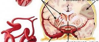
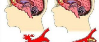
In the vast majority of cases, lacunar stroke is localized as deeply as possible in one or another hemisphere of the brain. Although, of course, there are cases when a lacunar stroke, or rather, the focus of its lesion is located in the so-called pontomesencephalic region of the brain.
Impact on the lacunar type
With this type of brain stroke, the affected area usually cannot exceed 15-20 millimeters in diameter, and such indicators of the lesion are already considered gigantic. The reason why lacunar stroke usually develops can be called a cardiogenic embolism or occlusion of a small artery.
Most often, ischemic lacunar stroke develops in patients over sixty years of age, who already have some health problems in their anamnesis:
- Some or other heart defects of a rheumatic nature.
- Some disturbances in heart rhythm or vascular conduction.
- Emergency conditions such as myocardial infarction.
- Systemic diseases and in particular diabetes mellitus.
The dominant role in the occurrence of ischemic lacunar stroke can be played by certain violations of the so-called rheological properties of our blood, certain pathologies affecting the main arteries.
This type of stroke pathology of the brain is characterized by development during sleep (or at night) in the absence of an extremely acute onset and sudden deep loss of consciousness.
Clinical syndromes of lacunar disease
Clinical manifestations observed during the development of lacunae are referred to as lacunar syndromes. Lacunar cerebral infarction is often accompanied by lacunar transient ischemic attacks (TIA, micro-strokes). Transient ischemic attacks (TIA, micro-strokes) can occur in the patient several times a day and last only a few minutes.
When a stroke occurs, the brain is deprived of oxygen, causing brain cells to die within minutes. However, there are differences between the United States and European countries. The incidence of lacunar stroke increases with age, and it tends to affect more men than women. Several studies have also found higher rates of lacunar stroke in blacks, Mexican Americans, and Hong Kong Chinese.
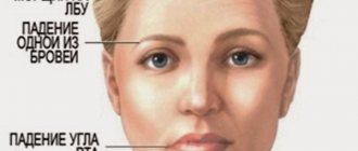
Because lacunar strokes can affect deep brain structures, the damaging effects can occur in several different ways. It often presents with weakness in all or some aspects of voluntary movements, such as walking or simple body movements. This occurs because a stroke damages or destroys brain cells in this area of the brain used to send signals to control muscles. It usually occurs on one side of the body and is often a combination of weakness in the arms and legs, sparing face; or a combination of arms, legs, and facial weakness.
The development of cerebral infarction (ischemic stroke) may be accompanied by a sudden onset or increasing neurological deficit over several days. A few hours or days after the development of a cerebral infarction (ischemic stroke), the patient's condition improves, although some patients become disabled. The patient's recovery may be complete over a period of several weeks to months, or there may be minimal residual neurological deficit.
Causes of lacunar stroke
This type of stroke involves the loss of sensory aspects of the body, representing numbness or unusual perception of pain, temperature, or pressure.
A pure sensory stroke results from damaged or destroyed areas of the brain that are responsible for controlling these sensations, usually the thalamus. It affects sensory functions such as touch, pain, temperature, vision, hearing and taste. They are usually isolated to the side of the body controlled by the affected cerebral hemisphere. Neurological manifestations of many lacunar syndromes are known. Some of these neurological syndromes require confirmation. The most common neurological syndromes with lacunar disease are:
- Pure motor hemiparesis with an infarction in the area of the posterior thigh of the internal capsule or the base of the bridge. The face, arm, leg, foot and fingers are almost always involved. Muscle weakness can be intermittent during a transient ischemic attack (TIA, microstroke), gradually increasing or sudden. Muscle weakness can progress to paralysis (plegia) and then often regress. In many cases of such syndromes, recovery is complete;
- Syndromes with purely sensory disorders of the hemitype in thalamic infarctions;
- True ataxic hemiparesis due to infarction at the base of the pons and dysarthria with awkwardness in the hand or arm due to infarction at the base of the pons or knee of the internal capsule;
- Pure motor hemiparesis with “motor aphasia”, caused by thrombotic blockage of the lenticulostriate branch of the artery of the lentiform nucleus and striatum, supplying blood to the knee and anterior thigh of the internal capsule with the adjacent white matter of the corona radiata.
Before treatment for arterial hypertension, multiple lacunae often cause patients to develop pseudobulbar palsy with emotional lability, a state of lethargy, abulia and bilateral pyramidal symptoms. Currently, this syndrome is rare.
It includes aspects of both pure sensory and pure motor strokes. They occur in the same way as the previously mentioned types of stroke, but affect areas of the brain that control motor and sensory innervation. This type of stroke occurs due to a blockage of blood flow to one of the following areas: internal capsule, corona radiation, or pon. A stroke in these areas can cause imbalance in walking and weakness in the arm or leg on the affected side of the body.
Dysarthria Syndrome
This condition results in speech problems and a clumsy hand as a result of a blow that impacts the anterior portion of the internal capsule. Individuals have difficulty pronouncing or forming words due to inadequate muscle movements in the voice box or larynx, as well as in their tongue and other mouth muscles. They also have difficulty spelling, tying a shoelace, or playing the piano.
There are other lacunar syndromes that have been associated with observed arterial pathology:
- Pseudobulbar syndrome with loss of the ability to form speech sounds (anarthria), caused by bilateral infarctions in the area of the internal capsule, can develop with damage to the lenticular nucleus and striatum.
- Syndromes caused by narrowing of the lumen (occlusion) of the penetrating branches of the underlying portion of the posterior cerebral artery (listed above).
- Syndromes observed with possible narrowing of the lumen (occlusion) of penetrating arteries emanating from the main artery. These lacunar syndromes include ipsilateral ataxia and paresis of the lower extremity, pure motor hemiparesis with horizontal gaze palsy, and hemiparesis with crossed abducens (VI cranial) nerve palsy.
- Downstream basilar artery syndromes include sudden nuclear ophthalmoplegia, horizontal gaze palsy, and appendicular cerebellar ataxia.
- Syndromes that develop with possible narrowing of the lumen (occlusion) of the branches of the vertebral artery include pure motor hemiparesis (while the facial muscles remain intact) due to the involvement of the pyramid of the medulla oblongata, as well as the syndrome of damage to the lateral parts of the pons and medulla oblongata, accompanied by dizziness, vomiting, weakness facial muscles, Horner's syndrome, ipsilateral numbness in the area of innervation of the trigeminal nerve and contralateral loss of sensitivity due to damage to the spinothalamic tract (partial lateral medulla oblongata syndrome).
Classification
This cerebrovascular accident is divided depending on the vessels in which blood supply to the brain the catastrophe occurred. There are two vascular beds: carotid and vertebrobasilar. Thus, there are two types of cerebral infarction:
- lacunar infarction in the area of basal structures - occurs when blood flow in the vertebrobasilar region is disrupted;
- infarction in the cerebral hemispheres - appears when the carotid circulation is impaired.
Clinical classification is based on clinical symptoms and is discussed in the section on symptoms of lacunar infarction.
In the development of the disease there are:
- the most acute period is the first 3 days;
- acute – 28 days;
- early recovery – the next 6 months after suffering a circulatory disorder;
- late recovery – for 2 years.
Due to mild symptoms and the absence of pronounced motor, speech and sensory disorders, it is very difficult to describe the features of each interval. Therefore, this division is of secondary importance.
Instrumental examination for the diagnosis of lacunar disease
Computed tomography (CT) of the brain can detect most supratentorial lacunar infarcts. Magnetic resonance imaging (MRI) of the brain clearly reveals both supra- and subtentorial infarcts (lacunae measuring 7 mm or more), as well as extension into the gray matter of the cortical surface of a small infarction in the white matter of the brain. This spread is predominantly a consequence of embolism rather than narrowing of the lumen (occlusion) of small penetrating vessels. Therefore, in such situations, a diagnosis of lacunar infarction should not be made.
Causes of lacunar stroke and risk factors
Like other types of strokes, lacunar stroke results from insufficient blood flow to the brain through the lacunar arteries.
The biggest risk factor for lacunar stroke is high blood pressure, as well as adulthood, smoking, excessive alcohol consumption, drug abuse, pregnancy, birth control pill use, sedentary lifestyle, poor diet and obstructive sleep apnea. Research has shown that in almost all cases of lacunar stroke, hypertension is a major contributing factor. Having high blood pressure for long periods of time can lead to the development of microatheroma and lipohyalinosis.
Many infarctions larger than 2 cm, combined not only with pure motor hemiparesis, are incorrectly called lacunae in the literature. They are too large to represent the result of occlusion of a single penetrating branch. These are likely embolic infarcts in which brain computed tomography (CT) is unable to demonstrate cortical involvement. The diagnosis of lacunar infarction should be made only when the size of the infarction is less than 2 cm and its localization can be explained by occlusion of a small penetrating branch of one of the large arteries of the base of the brain. Larger deep infarcts in the white matter and middle cerebral artery basin are probably caused by emboli.
What to do for prevention?
Diabetes is widespread in the Western world and is a recognized risk factor for the development of small vessel disease throughout the body.
Therefore, the risk of stroke is exceptionally high for those who have diabetes. Embolism, which is a condition where blood clots travel from other places in the body, is not uncommon when there is an underlying cardiogenetic abnormality that causes the abnormal blood clot to form. These may include atrial fibrillation and ipsilateral carotid stenosis. Electroencephalography (EEG) is usually normal, in contrast to that seen in infarcts involving the cerebral cortex. If normal results are obtained during an EEG study soon after the onset of symptoms, this gives reason to think about a deep infarction in the white matter of the cerebral hemisphere.
It is important to recognize the symptoms of a lacunar stroke so you can get medical help right away. Symptoms of a lacunar stroke are similar to those that accompany other types of stroke, including: slurred speech, inability to raise arms, one side of the face drooping, numbness on one side of the body, difficulty walking or moving arms, confusion, problems with memory when trying to speak or understand tongue, headache and loss of consciousness. Symptoms will depend on the area of the brain that is damaged by the lacunar stroke, with different areas of the brain controlling different aspects of the body, respectively.
Complications and prognosis
The prognosis for a single lacunar stroke is favorable. As a rule, all functions are restored; sometimes partial residual motor or sensory symptoms are observed.
Dementia is a common complication of recurrent lacunar infarctions
If a lacunar stroke often recurs, then there is a high probability of developing a complication such as a lacunar state of the brain. Among patients with vascular dementia, this complication occurs in almost 65-70% of cases.
Treatment of lacunar disease of the brain
The best treatment for small vascular disease is prevention, namely careful control of hypertension. However, a drop in blood pressure during the development of a stroke contributes to an increase in neurological symptoms. Lowering blood pressure begins after the patient's neurological symptoms have stabilized.
It is also interesting to note that the right hemisphere of the brain controls all sensation and motor function for the left side of the body, while the left hemisphere of the brain controls all these processes for the right side. It is for this reason that you may see damage to one side of the brain affecting activities on the opposite side of the body.
For most people, the left hemisphere is dominant and has additional responsibilities, including processing language and behavior. Lesions on this side of the brain can also lead to speech problems, personality changes, or even dementia.
The effectiveness of anticoagulants and antiplatelet agents in the treatment of patients with lacunar transient ischemic attacks (TIA, micro-strokes) and fluctuating strokes has not been clarified. According to some experts, thalamic lacunae caused by lipohyalinosis can be combined with minor hemorrhages (hemorrhages). At autopsy, macrophages loaded with hemosiderin are sometimes found in such infarcts.
Additional symptoms of lacunar infarction include: Gap in speech Inability to raise the arm upward Surface of the face drooping on one side Numbness on one side of the body Problems related to confusion Confusion and altered awareness Memory problems Problems with conversational fluency Persistent headaches Loss of consciousness. Doppler ultrasound can also be used to measure blood flow through veins and arteries.
Your doctor may also run tests to measure your heart function. Kidney and various blood tests may also be requested. The main deciding factor in the prognosis of lacunar stroke is the speed of initiation of treatment. As the brain is depleted of oxygen-rich blood, the longer it takes for this blockage to be relieved, the more brain cells will die. This is why quickly recognizing the symptoms of a stroke is crucial to starting a trip to the nearest hospital. Early treatment within three hours reduces brain damage.
The possibility of using heparin in this condition is also doubtful. But, on the other hand, in some patients with fluctuating hemiparesis in the area of atherothrombotic lesions of the branch of the basilar artery or the arteries of the lentiform nucleus and striatum arising from the trunk of the middle cerebral artery, an improvement in the condition may be observed with the administration of heparin.
Upon admission to the hospital, supportive measures will be provided. This often includes helping with breathing and heart function. Depending on the severity of the lacunar infarction and if current hospital facilities allow it, the patient may be given this anticoagulant medication directly on the side of the blockage in the brain.
Causes and symptoms
Aspirin is also given for 48 hours to reduce the chance of additional blood clotting. Once the worst has passed and the lacunar stroke patient is in the recovery phase, a physical therapy program is usually discussed to help improve any lost or compromised abilities.
Long-term treatment with anticoagulants is not indicated for patients with lacunar stroke. At the same time, it is necessary to carefully control arterial hypertension in order to prevent the progression of vascular damage in patients with a history of hypertension.
The life of a modern person is arranged in such a way that many people simply stopped paying attention to their health. Their rhythm is so fast that people began to get rid of various symptoms, such as headaches, dizziness of varying degrees, by taking painkillers. When they are not trying to understand what is happening to them, they actually receive a diagnosis of lacunar stroke.
Dysarthria and clumsy upper limb
Physical therapy may include rethinking speech, language, and motor skills.
It usually requires a lot of time and effort, with patience from the test patient as it may take months or years to regain lost skills. The best way to prevent lacunar stroke is to prevent stroke risk factors. This involves leading a healthy lifestyle, such as eating regularly, eating healthy, not smoking, reducing stress, maintaining a healthy weight, controlling alcohol consumption and managing other health conditions that may increase the risk of stroke.
According to historical information, people first started talking about lacunar stroke in 1838, when a doctor from France named Decambre raised the problem of hemorrhage in the brain. A pathology that he designated as a lacuna, that is, that affected area of the hemispheres that is not located outside, but in the deeper layers of the cerebral cortex. However, softening of the affected areas began only after a little more than one century, namely in the 80s of the 20th century.
Information for medical professionals
Diseases of the circulatory system Cerebrovascular diseases Cerebral infarction. Also called: Embolic stroke, thrombotic stroke. Stroke is a medical emergency. There are two types - ischemic and hemorrhagic. Ischemic stroke is the most common type. This is usually caused by a blood clot that blocks or occludes a blood vessel to the brain. This causes blood to flow to the brain. After a few minutes, the brain cells begin to die. Another cause is stenosis, or narrowing of the artery.
Causes of ischemic foci
Doctors began to find out the causes of lacunar cerebral infarction in the 19th century. To date, doctors have been able to identify risk factors and specific changes in the vascular system of the brain that provoke the appearance of pathology . These include:
- Arterial hypertension not controlled by the patient. It is characterized by strong pressure surges and crises
. Against this background, hyalinosis is formed, which impairs the distensibility of the arteries and ultimately causes minor hemorrhages. - Disorders of carbohydrate metabolism.
- Atherosclerosis. This pathology usually does not affect small vessels, but, affecting large arteries of the head and neck, they inevitably cause disruption of blood flow in the brain.
- Diabetes.
- Cardioembolic circulatory disorders.
- Tendency to thrombosis.
- Problems with hematopoiesis.
- Arteritis.
- Anomalies in the structure of the vascular system.
- Vasculitis.
In some cases, LI is provoked not by ischemia, but by minor hemorrhages into adjacent tissues, which lead to compaction of the feeding arteries and their subsequent gluing. This causes the formation of a lacuna.
Features of the occurrence of lacunar stroke
A frequent sign of a lacunar stroke involves the formation of a lesion in the deeper subcortical region of the brain, which in area can develop no more than 1.5-2 centimeters. Although such a hemorrhage occupies the smallest piece, despite this, the pathology can greatly affect the patient’s well-being. A feature of this disease is the formation of gradual or sudden blockage of the lumen of small blood vessels.
This can happen due to atherosclerosis, a disease in which plaque builds up inside your arteries. Transient ischemic attacks occur when the blood supply to the brain is briefly interrupted. Sudden numbness or weakness of the face, arm or leg Sudden confusion, trouble speaking or understanding speech Sudden trouble seen in one or both eyes Sudden discomfort, dizziness, loss of balance or coordination Sudden severe headache without any known cause. It is important to treat strokes as quickly as possible.
For rehabilitation after a stroke, our readers recommend Monastic tea. Monastic tea consists exclusively of medicinal herbs, is a completely natural product and does not contain chemicals. Improves the process of restoration of damaged cells in the brain, helps restore speech and hearing functions in the victim, and prevents the occurrence of a recurrent stroke. Doctors' opinion..."
Blood thinners can be used to stop a stroke when it occurs by quickly dissolving the blood clot. Stroke rehabilitation can help people overcome the disabilities caused by stroke damage. Stroke prevention Outline - unloading Thrombolytic therapy. . Lacunar strokes occur due to occlusion of small branches of both the anterior and posterior circulation vessels. Lacunar syndromes are useful in identifying the disease process, but are often less useful in determining the exact location of the lesion.
Lacunar syndromes are as follows. Pure motor hemiparesis: weakness of the face, arms and legs without other deficits. Usually internal damage to the capsule. Ataxic hemiparesis: motor hemiparesis with cerebellar-type ataxia on that side. The affected area is variable: posterior internal capsule, midbrain or pon.
It is also worth noting that lacunar stroke is most often detected when examining elderly patients, mainly those people who have previously had the following health problems:
- Disturbances in the functioning of the heart due to the formation of narrowing of the artery of the cardiovascular system.
- Failure of heart rhythm, low blood flow.
- Due to the formation of atherosclerotic plaques.
- Obesity, signs of high cholesterol and blood sugar.
There is no difference between ischemic and lacunar stroke, since the second one is considered one of the types of illness related to the type of cerebral ischemia. What is especially worth knowing about this pathology is that the symptoms can begin with mild sensations, as a result of which the patient can suddenly lose consciousness during deep sleep.
Rehabilitation period
Treatment with exercise therapy and robotic techniques is of great importance for recovery after a stroke. Rehabilitation begins on the third to fifth day in the hospital department where the patient was admitted.
After the end of the acute period of lacunar stroke, treatment continues in the rehabilitation department of the same hospital. The best option is to undergo recovery at a medical center or specialized sanatorium.
Therapeutic gymnastics, physical education, massage under the supervision of a doctor eliminate the residual effects of a heart attack. After discharge, the patient performs the prescribed set of exercises at home. In addition, it is necessary to periodically consult a rehabilitation doctor.
In case of a stroke, it is necessary to restore not only the movement of the limbs, but also the psycho-emotional state. To do this, it is recommended to visit a neurologist and psychologist.
Lacunar stroke threatens a person’s health and life. But at the first sign of it, you shouldn’t panic or call your relatives and ask what to do. The best decision is to call emergency help without wasting precious time. This is the only way to prevent complications, and in many cases save your own life.
Probability of recovery
In most cases, people who have suffered a stroke are rarely able to fully restore their health. That sometimes even experienced specialists in the field of treating brain diseases cannot always stop the development. Since although it is said that doctors can do anything, in this case one cannot rely only on them; much will also depend on the responsibility of loved ones.
After all, without paying attention and delaying just half an hour, the patient may not be able to help anything. And there is also a chance that in 60% of cases, after all the necessary measures have been taken, he will end up disabled for life, unable to live a full life. In connection with this, it is extremely difficult to determine how positive or negative the forecast will be.
Therefore, in order to take any measures to eliminate or alleviate some of the consequences after a stroke, the doctor must first establish an accurate picture of the course of the disease. Even if the symptoms of two patients are exactly the same, the program of medical procedures for the rehabilitation of each patient will be individual.
However, if the consequences are not so extensive, which especially most often happens when doctors manage to provide medical assistance for the first time 10-15 minutes after the disaster. It is this factor that can influence the successful prognosis that the patient will soon feel better and his life will return to normal. The care and attentiveness of relatives in this case will be the biggest and most influential moment for the patient. Which will also give him confidence in his pursuit of recovery.
About neuroprotectors
To maintain collateral blood flow, treat:
- Cavinton.
- Niceroglin.
- Cynarizine.
- Eufillin.
But there is an opinion among experts that the above drugs will increase blood flow through dilated collateral vessels, and this can increase the ischemic focus because the blood flow goes to a new channel.
To enhance metabolic processes in brain cells where there is damage, they are treated with neuroprotectors and antioxidants. Treatment:
- Glycine.
- Semax.
- Cerebrolysin.
- Nootropil.
- Mexidol.
- Cortexin.
Of course, there is no evidence base for positive treatment results. But many experts are confident that the use of the above remedies is effective and justified.
Symptoms of manifestation
- Refusal of the harmful effects of addictions, such as smoking and alcohol.
- Moderate consumption of fast food.
- Nutrition based on fresh herbs, fruits and vegetables. Lean meats.
- During the day, especially in old age, monitor your blood pressure levels.
- Daily walks, trips to nature, performing light, simple physical exercises.
- If necessary, or preferably 1-2 times a year, undergo MRI diagnostics.
- Less worries and stress.
Prevention
- Avoid stress and overexertion at work. Learn yoga and breathing exercises. Use valerian or motherwort in cases of severe anxiety that you cannot cope with;
- Play sports or lead an active lifestyle. But people with hypertension should not exercise too intensely. Exhausting physical labor is also contraindicated;
- Eat healthy foods. If your weight is above normal, immediately switch to healthy, low-calorie foods;
- Measure your blood pressure with a tonometer; if it deviates from the norm, contact a cardiologist. Follow your doctor's instructions strictly. Sometimes blood pressure medications need to be taken even if it is normal;
- Undergo general medical examination.

