Diseases of the hearing organ cause a person no less discomfort than any other ailment. It is unlikely that many of us can answer the question of what cochlear neuritis is. It is commonly understood as an inflammatory process that affects the nerve located in the area of the inner ear.
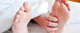
The main complaint that occurs in people with a similar diagnosis is progressive deafness. Elderly men over 50 years of age are most susceptible to this disease. Since cochlear neuritis occurs at an age when many important organs begin to fail, most patients do not dare to consult a doctor for help, thinking that it is age-related and nothing can be done about it.
Treatment of the disease
The main goal of treatment of acute cochlear neuritis is to restore hearing. Treatment of the chronic form of the disease is aimed at stabilizing auditory function.
Acute cochlear neuritis and some cases of progressive chronic cochlear neuritis are an indication for treatment in a hospital. Therapy for cochlear neuritis should be carried out in conjunction with the correction of disorders that could cause it. First of all, this is the elimination of arterial hypertension and hormonal dysfunction, eliminating the effects of ototoxic factors (medicines and other substances, noise, vibration, radioactive radiation).
Drug therapy for cochlear neuritis is carried out with vasodilators, antiplatelet agents, venotonics, neuroprotectors, and detoxification solutions. Combination therapy with trental, vinpocytine, piracetam, Mexidol and Cerebrolysin in the first 2 weeks is carried out by intravenous administration, then proceeds to intramuscular injections and oral administration of drugs. The use of ginkgo biloba in the treatment of cochlear neuritis has a good effect. In the treatment of sudden onset cochlear neuritis, glucocorticoids are additionally used. To relieve dizziness, histamine-like drugs (betagistine) are used.
Physiotherapeutic treatment methods have a positive stimulating effect:
- reflexology (electropuncture, laser puncture, acupuncture);
- electrical stimulation;
- phonophoresis of drugs;
- oxygen barotherapy.
Bilateral hearing loss of up to 40 dB complicates the patient’s speech communication and is an indication for hearing aids. The prelingual form of cochlear neuritis is an indication for wearing a hearing aid at a hearing threshold of 25 dB, since it has been proven that such hearing loss causes disturbances in the development of speech in a child. For the purpose of hearing protection for cochlear neuritis, analog, digital and linear hearing aids can be used. The selection and adjustment of the device is carried out by a hearing therapist.
Surgical treatment of cochlear neuritis is carried out to perform stem or cochlear implantation, removal of acoustic neuroma, hematoma or brain tumor. The need for surgical treatment may be due to painful ear noise or attacks of intense dizziness. In such cases, removal of the stellate ganglion, resection of the tympanic plexus or cervical sympathectomy is performed; in case of deafness or IV degree hearing loss, destructive operations on the cochlea are performed.
Symptoms
Cochleovestibular syndrome consists of:
- Sensorineural hearing loss.
- Systemic dizziness.
- Balance imbalance.
Since cochleovestibular syndrome does not occur in isolation, patients complain of neurological disorders:
- frequent headaches;
- memory impairment, absent-mindedness;
- sleep disturbance;
- slowing down the pace of thought;
- neurasthenia: weakness, irritability, gloomy mood;
- decreased visual acuity, double vision, pain when moving the eyeballs;
- decreased sense of taste and smell.
The first signs are hearing loss and dizziness. Then the balance is upset and complaints of discirculatory encephalopathy appear.
Dizziness is the main symptom of the disease. An attack of dizziness is accompanied by vegetative manifestations: sweating, increased heart rate, shortness of breath, pale skin, nausea and vomiting. Dizziness is paroxysmal: there is a feeling that objects around are moving towards the ear. Nystagmus is directed towards the movements of objects - rapid and rhythmic turns of the eyes. An attack of dizziness can last from a few minutes to several days.
Cochleovestibular syndrome can be complicated by Meniere's disease, when endolymph accumulates in the inner ear and the pressure inside the labyrinth increases. It manifests itself as deafness in one ear, tinnitus and autonomic disorders.
Diagnosis of the disease
The main diagnostic method is an audiogram. Sometimes impedance measurements and Dopplerography of the vessels of the head and neck are additionally performed.
On otoscopic examination of the ear, the eardrum is usually intact.
The study of hearing in whispered and spoken speech reveals a decrease in the perception of high-pitched sounds. However, with pronounced atrophic-degenerative changes, the perception of low sounds may decrease. A positive Rinne test is noted in the initial manifestations of neuritis and a negative one - with a developed process. Research according to Weber, when there is bilateral neuritis, does not lateralize the sound, when there is unilateral neuritis, the lateralization of the sound will be in the healthy direction. The Schwabach test is sharply shortened, the Jelly test is positive.
An audiometric study reveals lesions of the sound-receiving apparatus, characterized by the same impairment of hearing both by air and bone conduction (the bone and air conduction curves are at the same level). In some cases, a paradoxical increase in volume is observed.
It is necessary to differentiate between acoustic neuritis and otosclerosis, in which bone conduction is well preserved.
Causes of cochlear neuritis
Cochlear neuritis is an inflammatory lesion of the auditory nerve in the internal structures of the ear. Accompanied by a significant hearing loss due to impaired conduction of the sound signal from the vestibulocochlear nerve to the cerebral cortex.
Most often it is localized in one ear, much less often it affects both at once. People suffer from the disease mainly in adulthood, but a congenital form is also possible.
The pathology also has other medical names - cochleoneuritis, neurosensory, sensorineural, perceptual hearing loss, auditory neuritis.
A disease such as cochleoneuritis usually occurs acutely with a gradual transition to a chronic form. The reasons lie in many provoking factors:
| Cause | Provocateurs |
| Infectious pathologies | Flu, rubella, measles, herpes, ARVI, meningitis |
| Disorders of the cardiovascular system | Ischemia, hypertension, atherosclerosis, thrombosis, stroke |
| Toxic damage | Potent drugs from the group of antibiotics, cytostatics, salicylates, as well as intoxication with gasoline, phosphorus, and heavy metal salts |
| Mechanical damage | Traumatic brain injuries, surgery, acoustic and barotrauma |
| Oncology | Benign and malignant neoplasms in the brain |
| Chronic systemic diseases | Diabetes mellitus, hyperthyroidism, anemia |
| Genetic disorders and birth trauma | Heredity, Waardenburg syndrome, biotinidase deficiency, Pendred syndrome, fetal hypoxia, premature birth |
| Age | Men and women after 50-60 years against the background of general aging of the body |
According to statistics, a third of cases of neuritis are of infectious origin, the same proportion is due to age-related changes, and other factors are less common.
Prices
| Disease | Approximate price, $ |
| Prices for diagnosis and treatment of sinus pathologies | 10 370 — 17 560 |
| Prices for the treatment of thyrotoxic goiter | 22 590 — 22 670 |
| Prices for treatment of laryngeal cancer | 6 170 — 77 000 |
| Disease | Approximate price, $ |
| Prices for diagnosing migraine | 7 060 — 8 260 |
| Prices for diagnosing childhood epilepsy | 3 100 — 4 900 |
| Prices for brain shunting for hydrocephalus | 33 180 |
| Prices for treatment of Parkinson's disease | 58 600 |
| Prices for migraine treatment | 9 680 |
| Prices for the diagnosis of amyotrophic lateral sclerosis | 6 550 |
| Prices for diagnosing epilepsy | 3 520 |
| Prices for rehabilitation after a stroke | 78 300 — 82 170 |
| Prices for treatment of childhood epilepsy | 3 750 — 5 450 |
| Prices for treatment of multiple sclerosis | 4 990 — 17 300 |
Otolaryngology in Israel receives extremely positive reviews, and the reason for this is not only the high effectiveness of the treatment, but also its affordable cost for most medical tourists. Prices for diagnostic and treatment procedures in Israeli hospitals are at least a third lower than in similar medical institutions in Western Europe, the USA, Canada and other countries with developed medicine.
The cost of treatment for an ENT disease in Israel depends on its nature, location, stage of development, medications used, the number and type of diagnostic procedures required, therapeutic methods, as well as the individual characteristics of the patient. You can find out the approximate cost of solving a particular ENT problem from a medical representative of the clinic during a free consultation. The final cost will be clear on the spot, after all examinations have been completed and the treatment plan has been approved by the doctor.
Possible complications
Patients who seek medical help at the initial stage of development of the syndrome most often manage to restore hearing sensitivity. Although, to assess the likelihood of such an outcome, it is necessary to take into account the cause of the neuritis.
If the cause of the development of the syndrome was trauma, an infectious disease or toxic poisoning, then such patients have a very high chance of regaining their hearing.
If the neuritis managed to move to the third stage or the patient turned to the doctor for help too late, when a lot of time had passed since the first symptoms appeared, then he will have to come to terms with complete hearing loss.
The only solution for such patients may be surgery to install implants. For this reason, you need to consult a doctor as soon as the first symptoms of the disease are detected.
Causes
There are direct injuries and damage to the spinal column, damage to structures associated with it. The causes of cervical artery syndrome are conventionally divided as follows:
- traumatic - falling on your back, trauma to the ribs, severe fear and diaphragmatic spasm;
- myofascial - muscle disorders provoke changes in the position of the ribs and vertebrae;
- visceral – diseases of internal organs are the culprits of reflex problems in the spinal motion segment;
- neurogenic pain - peripheral nerves are affected.
The risk of developing the syndrome increases with muscle overload, prolonged stay in static positions, stress, depression, alcohol and fat abuse.
From a vertebrological position, only circumstances that are official diagnoses of orthopedics are recognized:
- radicular syndromes or compression of nerves due to hernial growths between the vertebrae;
- compression of the spinal canal during listhesis;
- Bekhterev's disease;
- traumatic injuries of the spinal column;
- osteoporosis;
- muscle spasms due to compression of nerves;
- tumor processes;
- muscle inflammation;
- scoliosis;
- osteochondrosis;
- disruption of blood supply to the vertebrae or muscles.
Painful sensations are sometimes reflexive due to inflammation of internal organs: pancreatitis, ulcers, diseases of the genitourinary system. Circumstances are interconnected and form pathological chains. An inflammatory focus in the kidney is fraught with fixation of the ribs, rotation of the vertebrae of the sternum from 10 to 12, weakening of the associated muscles, biomechanical disorders, and the formation of pain. Usually the syndrome occurs due to osteochondrosis, which is detected using x-rays.
Diagnostics
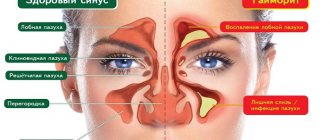
The diagnosis is made on the basis of complaints received, examination and study of the condition of the brain and hearing organs. First, hearing function should be checked, which requires an audiometry procedure, and acoustic impedance testing can also be performed instead. For diagnostic purposes, hardware tests are prescribed to rule out other diseases, including the presence of a foreign body, wax plug in the ear canal, otitis media, Meniere's disease and similar pathologies.
Patients with hearing diseases are advised to undergo an examination by an otolaryngologist, who will write a referral for electrocochleography, prescribe otoscopy, microotoscopy. To exclude concomitant diseases, you need to do an X-ray of the skull, MRI, CT, and ultrasound diagnostics of the vessels of the head.
Often, a hearing problem means there are problems with the spine, which is why X-rays of the cervical spine are indicated. If necessary, the patient will visit a neurologist.
Cochleovestibular syndromes in vertebrobasilar insufficiency
N. S. Alekseeva
The purpose of the study was to determine the main, objective vestibulometric characteristics of peripheral cochleovestibular syndrome caused by arterial hypertension, atherosclerosis and vegetative-vascular dystonia, to clarify the pathogenetic features of its formation, and the therapeutic effectiveness of betaserc in the treatment of peripheral dizziness. We examined 85 patients with PCVS; all of them underwent an ultrasound examination of the main arteries of the head, a study of blood pressure levels and central hemodynamics, and structural changes in the brain.
It has been reliably revealed that an important role in the development of PCVS belongs to the presence of vascular anomalies of the vertebrobasilar system; in isolated cases, stenosis of the vertebral and internal carotid arteries was found. The presence of arterial hypertension and central hemodynamic disturbances in this group of patients was confirmed. It is concluded that in the pathogenesis of ischemic PCVS, an important role belongs to the hemodynamic mechanism of circulatory failure in the inner ear at the microcirculatory level. A comparison of peripheral cochleovestibular syndromes with structural changes in the brain showed that PCVS is not accompanied by ischemic foci in the brain. Pharmacotherapy of peripheral dizziness of vascular etiology consists of the correct choice of drugs that optimize cerebral blood flow in combination with vestibulolytics. The therapeutic effectiveness of betaserc in reducing dizziness and vestibular activity under its influence has been proven when taking the drug over a 2-month course of treatment.
Dizziness most often occurs against the background of vascular pathology of the brain in patients with arterial hypertension, atherosclerosis, and vegetative-vascular dystonia [3, 7, 8, 19, 21, 22, 25]. Acute and chronic circulatory disorders in the vertebrobasilar system (VBS) occupy a significant place in the structure of cerebrovascular diseases [2, 4-6, 11, 18, 20]. Dizziness in these cases usually occurs in response to ischemia in various areas of the VBS and with damage to the vestibular analyzer - from the labyrinth to its cortical section [10, 12, 14, 15, 19]. In clinical practice, both from the standpoint of accurate diagnosis [9, 13, 16, 17, 24], and the most effective approaches to treatment [1, 23], it is important to distinguish between peripheral dizziness caused by ischemic damage to the inner ear (labyrinth), the 8th nerve root and central dizziness caused by ischemia of the vestibular nuclei and pathways.
Peripheral dizziness occurs much more often than central dizziness. In terms of its intensity and the resulting discomfort, it is more pronounced and occurs more often in young people, which leads to their permanent disability. Peripheral and central cochleovestibular syndromes (PCVS and CCWS) have not been sufficiently studied to date. Their connection with the level of blood pressure, the state of cerebral and central hemodynamics has not been established, the pathogenetic mechanisms of their formation have not been revealed, and the problem of effective treatment of heterogeneous dizziness remains unresolved.
The purpose of this work was:
1) determination of the main characteristics of PCVS caused by arterial hypertension and atherosclerosis, vegetative-vascular dystonia;
2) elucidation of the pathogenetic features of its formation;
3) assessment of the therapeutic effectiveness of betaserc in the treatment of peripheral vertigo.
Material and methods
We observed 85 patients with PCVS, of whom 36% were men and 64% were women. The age of the patients ranged from 44.0 to 68.5 years. The otoneurological study was carried out using an expanded technique that included a number of special methods for studying the function of the cochleovestibular analyzer (computer electronystagmography, auditory evoked potentials), and also used impedance tachooscillography, which allowed for multiple registration of arterial blood pressure. pressure before and after experimental vestibular loads, determine the main parameters of central hemodynamics.
Structural changes in the main arteries of the head and hemodynamic indicators of blood flow through the vertebral and internal carotid arteries were studied using Doppler ultrasound (USD), duplex scanning (DS) and, in some cases, using angiography. Structural changes in the brain and cerebrospinal fluid spaces were studied using magnetic resonance imaging tomography (MRI).
Results and discussion
Clinical and pathophysiological features of PCVS in the main group: vestibular disorders in circulatory failure in the VBS were manifested by a wide range of different PCVS. Attacks of systemic rotational dizziness occurred in elderly patients against the background of arterial hypertension in combination with atherosclerosis (65%) and in young patients against the background of vegetative-vascular dystonia (35%), accompanied by acute unilateral sensorineural hearing loss, occurring as an infarction of the inner ear. The attacks of dizziness were isolated or combined with other otoneurological manifestations and hearing loss, and sometimes proceeded like an attack of Meniere's disease.
The onset of the disease was characterized by the development of an acute attack of systemic dizziness, which is accompanied by nausea, vomiting, falling, sometimes with short-term loss of consciousness, both in combination with simultaneous auditory symptoms and without them. Before the development of an attack of dizziness, some patients note the appearance of noise and hearing loss (usually more pronounced in one ear) (see figure). In a number of cases, auditory disturbances were mild and were characterized by patients as impaired speech intelligibility. Recurrences of dizziness attacks are associated with increases or fluctuations in blood pressure, turns of the head and torso, and changes in body position.
It is known that the occurrence of vascular PCVS can be caused by pathology of the vertebral arteries and intracranial vessels of the vertebrobasilar system. However, to date there are no methods that make it possible to visualize pathological changes in blood flow in small arteries supplying blood to the roots of the 8th nerve and the labyrinth. We were the first to identify a connection between the presence of PCVS and the pathological condition of the vertebral artery (p = 0.047), while cochleovestibular syndromes developed both on the side of the altered vertebral artery and on the opposite side.
Analysis of our own observations allowed us to conclude that there are anatomical and physiological prerequisites, in the form of 4 developmental anomalies, against the background of which PCVS is formed.
These anomalies in the development of the vertebral arteries were as follows: asymmetry of diameters in 36% of patients; hypoplasia on the right in 25%; left hypoplasia in 22%; absence of posterior communicating arteries in 9%. To confirm the connection between PCVS and the presence of insufficiency of blood flow in the vertebrobasilar system, we studied the condition of the vertebral and internal carotid arteries according to ultrasound and DS data (Table 1).
As can be seen from table. 1, structural changes in the vertebral arteries were characterized by deformations, including bilateral ones, hypoplasia and asymmetry of diameters, and much less often - stenosis and occlusion. The identified changes in the structure of these arteries were the reason for the presence of chronic insufficiency of blood flow in the vertebrobasilar system in patients. Considering the fact that insufficiency of the blood flow of this system develops against the background of steal syndrome with damage to the internal carotid arteries, the data presented in the same table are of interest. Deformations and stenoses of the internal carotid arteries also occurred in a significant number of cases, which indicates the frequency of a combination of lesions of the vertebral and internal carotid arteries in the studied group of patients. Due to the fact that our patients suffered from arterial hypertension and atherosclerosis, it was important to study the condition of the central hemodynamics (Table 2).
As can be seen from table. 2, all patients with PCVS had a “mild” form of arterial hypertension and relatively stable central hemodynamics, while a decrease in stroke and minute blood volumes was detected. The diagnosis of PCVS of vascular origin is based on the features of cochleovestibular disorders. In the majority of the patients we examined, bilateral spontaneous nystagmus was detected, and only in isolated cases (19 patients) was it unilateral. Unilateral nystagmus was combined with a harmonious deviation of the arms and body towards the slow component of nystagmus, which is characteristic of PCVS in the acute period of the disease. The presence of bilateral nystagmus with PCVS indicates simultaneous ischemic damage to the peripheral and central vestibular structures.
As can be seen from table. 3, in 80% of cases, PCVS was combined with symptoms of damage to the pons, which is due to a single source of blood supply to the peripheral vestibular structures and central pathways and nuclei from the branches of the anterior inferior cerebellar artery and the penetrating arteries of the brain stem.
During experimental vestibular tests (Table 4), the majority of patients showed bilateral hyperreflexia (acute period) and, less often, bilateral hyporeflexia, which corresponded to the period of remission of the disease. Asymmetry along the labyrinth was detected in 56% of cases, unilateral vestibular hyperreflexia was combined with unilateral hearing loss, which formed the clinical basis of PCVS. Asymmetry of nystagmus in direction (a sign of central damage) was identified in isolated cases - 7 patients (16%), indicating the benefit of simultaneous ischemia of the anterolateral parts of the pons and peripheral cochleovestibular structures. The assessment of all components of the vestibular reaction (nystagmus, autonomic and sensory manifestations) was characterized by their harmonious correspondence. No disturbances of optokinetic nystagmus were identified in patients with PCVS. Impairments of auditory function, varying in clinical manifestations, were identified in all patients with PCVS (Table 5).
As can be seen from table. 5, a feature of hearing impairment in this group was the presence of sensorineural hearing loss, and in most patients it was bilateral. Conductive hearing loss was identified in patients with concomitant damage to the middle ear, not purulent, but of a scar-adhesive nature. To support the diagnosis of conductive hearing loss, we used sound lateralization tests in Weber’s experiment (towards the worse-hearing ear) and otoscopy data (scarring of the eardrum).
In 17 patients, unilateral deafness was detected, which corresponded to the diagnosis of acute ischemia of the inner ear. The figure shows an audiogram and vestibulogram of a patient with peripheral dizziness caused by circulatory failure. The main symptom of acute vascular pathology of the inner ear was sudden hearing loss in one ear, rarely both, which was accompanied by paroxysmal systemic rotational dizziness and imbalance. Rarely was there no dizziness.
Acute ischemia of the labyrinth developed against the background of increased blood pressure in combination with hypoplasia and atherostenosis of the VA, an anomaly in the origin of the VA from the aortic arch, cardiac arrhythmia (paroxysmal tachycardia), impaired venous outflow and increased blood viscosity. A comparison of PCVS with structural changes in the brain was also carried out - in the brainstem, cerebellum and hemispheres (Table 6).
From this table it can be seen that the development of PCVS occurs against the background of organic changes in the brain, of which the most common is the expansion of the subarachnoid space. Focal changes in the hemispheres most often corresponded to periventricular changes of the type of leukoaraiosis, characteristic of patients with arterial hypertension. The identified small lesions in the brainstem and cerebellum confirmed the otoneurological diagnosis of simultaneous ischemia in the arteries supplying blood to the brainstem and inner ear.
Based on the data obtained on the hemodynamic features underlying the pathogenesis of peripheral ischemic cochleovestibular syndromes, it was concluded that it is advisable to use a combination of drugs for the treatment of such patients that improve cerebral hemodynamics and have an effect on eliminating vestibular disorders at the peripheral level.
Results of treatment with betaserc
The pharmacological effect of betaserc is based on the fact that it contains betahistine dihydrochloride, which activates microcirculation and increases blood flow in the system of the basilar artery and arteries of the inner ear. In addition, betahistine is an agonist of H1 receptors involved in the stimulation of neurons in the vestibular nuclei. It blocks H3 receptors and stimulates postsynaptic histamine receptors, both in the inner ear and in the brain stem structures. 85 patients (36% of men and 64% of women) under the age of 74 years with peripheral vestibular syndrome were treated with Betaserc. The drug (Germany) was prescribed in tablets of 16 mg 3 times a day for 1 month, then 8 mg 3 times a day for another 1 month against the background of vascular and metabolic therapy. No intolerance to the drug was noted.
The effectiveness of betaserc is presented in table. 7. From the presented data it is clear that dizziness stopped in 20% of cases and decreased in 64%; only 10% of patients had no effect from treatment within a month. After a 2-month course of treatment, dizziness stopped or decreased significantly in almost all patients. Tinnitus decreased in 24% of patients, hearing improvement occurred in 44% of patients. Thus, the study showed a fairly high effectiveness of betaserc in patients with complaints of dizziness and noise in the head that arose against the background of vertebrobasilar insufficiency.
The effectiveness of the treatment was confirmed by the positive results of the dynamics of vestibular function before and after treatment, obtained by computer electronystagmography. During the treatment, a vestibular test was performed with bithermal calorization of the labyrinths. These data are presented in table. 8.
As can be seen from table. 8, the speed of the slow phase (the main parameter of experimental nystagmus) decreased in intensity after 30 days of taking the drug, and vestibular reactions had a clear tendency not only to reduce the speed of the slow phase of nystagmus, but also to their symmetry, which was accompanied by the clinical effect of reducing dizziness. B In this article, we focused on the treatment of PCVS with betaserc, but we also have experience in therapy with vasobral. This drug is known to have a vasodilator, anti-migraine and stimulating metabolism in the central nervous system effect, which is realized due to the blocking effect of β-dihydrocriptine on 1- and 2-adrenergic receptors of vascular smooth muscle cells. Comparison of the effects of betaserc and vasobral allows us to note a more pronounced and faster effect in the treatment of dizziness in the case of betaserc and the advantage of vasobral in relation to the treatment of cochlear disorders. The more pronounced effect of treatment with betaserc is due to the fact that this drug has two mechanisms of action, vasodilator and neuromodulatory. Peripheral vestibular syndrome, caused by circulatory insufficiency in the vertebrobasilar system, responds well to treatment with betaserc, but subject to its long-term use.
Literature
1. Amelin A.V., Skoromets A.A., Gonchar M.A. et al. Comparative effectiveness of betaserc and cinnarizine in the treatment of dizziness in patients with migraine. Journal of neurol and psychiat 2003; 103:5:43-48.
2. Antonov I.P., Gitkina L.S., Sklyut I.A. Some controversial and unresolved issues of vertebrobasilar insufficiency. In the book: Problems of modern neuropathology. Minsk: Belarus 1976.
3. Babiyak V.I., Lilenko S.V., Vavilova A.A. The influence of vertebrogenic pathology on the state of vestibular function in patients with otosclerosis. International Symposium “Modern problems of physiology and pathology of hearing”, 4th: Materials. M 2001; 30-32.
4. Vereshchagin N.V. Diagnosis of circulatory disorders in the vertebrobasilar system. Klin Med 1983; 9:3-9.
5. Vereshchagin N.V. Circulatory insufficiency in the vertebrobasilar system. Consilium medicum 2001; 15-18.
6. Vereshchagin N.V. Pathology of the vertebral arteries. In the book: Vascular diseases of the nervous system. Ed. E.V. Schmidt. M: Medicine 1975; 398-416.
7. Vereshchagin N.V. Structural and functional levels of the vascular system and brain pathology from the perspective of a systems approach. All-Russian Congress of Neurologists, 8th: Proceedings. Kazan 2001; 211.
8. Vereshchagin N.V., Morgunov V.A., Gulevskaya T.S. Brain pathology in arterial hypertension and atherosclerosis. M: Medicine 1997.
9. Gusev E.I. Research methods in neurology and neurosurgery: A guide for doctors. M: Knowledge 2000.
10. Ivanov I. Otoneurological symptoms and syndromes in vertebrobasilar insufficiency. J. neurol and psychiat 1969; 3: 365-369.
11. Kamchatnov P.R. Vertebrobasilar insufficiency: abstract. dis. ...Dr. med. Sci. M 2001.
12. Kekhaiov Atanas. Differential diagnostic problems in otoneurology. Sofia: Medicine and Physical Education 1972: 237.
13. Lilenko S.V. Nystagmometry in the diagnosis of vertebrogenic vertigo: Abstract of thesis. dis. ...cand. honey. Sci. St. Petersburg 2000.
14. Yakovleva I.Ya., Alekseeva N.S. Modern methods of comprehensive otoneurological examination in the diagnosis and pathogenesis of ischemic circulatory disorders of the inner ear and brain. All-Russian Congress of Neurologists, 4th: Proceedings. Kazan 2001; 191.
15. Arai M., Ishida N. Sudden bilateral hearing loss with vertigo due to vertebral artery occlusion. ORL 2000; 40: 844-847.
16. Baloh RW Advances in neuro-otology. Curr Opin Neurol 1998; 11:1:1-3.
17. Baloh RW Approach to the evaluation of the dizzy patient. Otolaryngol Head Neck Surg 1995; 112:1:3-7.
18. Baloh RW Vertebrobasilar insufficiency and stroke. Otolaryngol Head Neck Surg 1995; 112: 114-117.
19. Baloh RW Vertigo. Lancet 1998; 352: 1841-1846.
20. Caplan LR Posterior Circulation Ischemia: Then, Now, and Tomorrow. The Thomas Willis Lecture. Stroke 2000; 31: 2011-2023.
21. Caplan LR Management of acute peripheral vestibular disorders. Eur Neurol 1993; 33: 337-344.
22. Fife TD, Baloh RW, Duckwiler GR Isolated dizziness in vertebral insufficiency. Clinical features, angiography and follow-up. J Stroke Cerebrovasc Dis 1994; 4:4-12.
23. Pyykko I., Magnusson M., Schalen.L., Enbom H. Pharmacological treatment of acute vertigo. Acta Otolaryngol (Stockolm) 1988; Suppl 1:455:77-81.
24. Schick B., Brors D., Koch O. et al. Magnetic resonance imaging in patients with sudden hearing loss, tinnitus and vertigo. Otol Neurootol 2001; 22: 6: 808-812.
25. Troost BD Dizzines and Vertigo in Vertebrobasilar Disease. Stroke 1980; 11: 413-415. Received 06/11/04
Source Journal of Neurology and Psychiatry named after. S.S. Korsakov No. 10 | 2004
What to do if acoustic neuritis is detected in a child?
If a symptom of hearing change (a mild reaction to whispering) appears, parents should consult a doctor.
See also
Causes, symptoms and treatment of ear barotrauma (barotraumatic otitis)
Read
During the examination, the baby is diagnosed in a playful way:
- external examination of the ear;
- audiometry;
- acoustic tests and tests aimed at checking the functioning of the vestibular apparatus.
To accurately determine the causes of the pathology, urine, blood, and smear tests from the site of inflammation are prescribed. Based on the results of the examination and tests, treatment of cochlear neuritis with vasodilators is prescribed.
In addition, parents are advised to adjust the diet and prescribe a set of physical exercises for the child. The baby is given daily drops for auditory neuritis.
Children's hearing is restored using physiotherapeutic procedures:
- acupuncture;
- reflexology;
- laser
With an integrated approach, the source of infection is eliminated, after which the spasm goes away, drainage of the ear canal improves, and hearing is restored.
Treatment methods
Therapy of vertebrogenic pain syndrome involves relief of the inflammatory focus. The patient is prescribed bed rest for 5-9 days, a firm mattress is used and the position is alleviated with the help of pillows under the neck. If a person is unable to reduce physical activity, orthopedic corsets are prescribed to relieve the axial load on the back.
Conservative methods
It is necessary to resort to drug treatment of vertebral artery syndrome when all pathological links that lead to the development of pathology are discovered. The basic principles of conservative therapy include:
- unloading of the spinal column;
- symptomatic treatment;
- correction of psychological state;
- strengthening the spinal muscles.
You might be interested in the article - How to treat muscular-tonic syndrome?
Taking medications helps relieve inflammation, relax muscle spasms and normalize the tone of the nervous system. Below are the drugs included in the complex treatment.
- Non-steroidal anti-inflammatory drugs are also used to relieve pain. Diclofenac is widely prescribed in the form of intramuscular injections or tablets with a maximum dosage of 150 mg per day. Sometimes Analgin is prescribed.
- Muscle relaxants are used in injection or tablet form, depending on home or hospital treatment. Relanium in solution and Baclofen in tablets are shown.
- Tricyclic antidepressants complement therapy for severe pain. Amitriptyline is used in small dosages.
Among anti-inflammatory medications, selective inhibitors of COX, mediators of the inflammatory process, are used: Nimesulide, Piroxicam. For persistent pain, blockades are made with corticosteroids, which reduce immune function to block inflammation.
Read about what analogues of Berlition exist and when their use is justified in this article.
Exercise therapy and physiotherapy
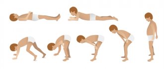
To eliminate compression of the vertebral artery, muscle tissue should be strengthened and physical activity on the spine should be performed. In the form of exercise therapy, the doctor selects a set of exercises. They help the patient return to the usual rhythm of life. The load should be moderate. The training complex is based on the following exercises:
- circular rotations of the head, tilting it in different directions;
- raising your shoulders as high as possible, trying to reach your ears;
- turns the body in different directions;
- circular rotation of the body clockwise and back.
If pain occurs after exercise, stop exercising and consult a doctor. He will make adjustments to the training scheme and reduce their load.
The main goal of physiotherapeutic manipulations is to accelerate tissue restoration and blood circulation. Effective methods include:
- Magnetic therapy – a pulsed low-frequency or constant field is applied to the affected area;
- electrophoresis - tissue is affected by direct current in tandem with analgesics in the form of ointments and creams;
- phonophoresis - manipulation is carried out using ultrasonic waves in combination with Analgin and Hydrocortisone.
In addition to drug therapy and physiotherapeutic measures, it is worth wearing a Shants collar. It will help fix the neck and compensate for the load on it.
Surgery
If drug treatment is not beneficial, the pain does not subside within 3-4 months, surgical intervention is resorted to. Indications for surgical manipulation also include decreased sensitivity of the legs and disruption of the functioning of the pelvic organs. Then urgent surgery is needed. In vertebrology and neurosurgical centers, interventions on the arteries are performed using minimally invasive techniques using an endoscope. A small incision is made, less than 2 centimeters, this reduces trauma to the body, prevents damage to nearby organs, and shortens the rehabilitation period. During the operation, pathological bone growths are removed, the compressed artery at the site of narrowing is excised, and plastic surgery is performed. The effectiveness of the procedure is 90%.
Diagnosis of vertebral artery syndrome
In order to begin treatment of the disease, it is necessary to conduct a broad diagnosis of this problem. As already mentioned, cochlear neuritis exists in a wide variety of types and types and can be classified and defined into groups. Therefore, the doctor needs to conduct many tests, analyzes and procedures in order to fully describe the disease, identifying all aspects of cochlear neuritis and determining its genesis, causes, symptoms, period of occurrence, etc. This is a meticulous process, difficult at an early stage both if there is unilateral and bilateral cochlear neuritis.
The degree of damage to the auditory nerve is determined using audiometry or acoustic impedance testing. Additionally, the ability of the vestibular apparatus to respond to stimuli and load is studied. To determine the nature of the disease, various tests and analyzes are carried out; separately, using computed tomography, x-rays or magnetic resonance imaging, the vascular system, cranial bones, and human brain are studied.
To clarify the diagnosis and check its truth, manipulations are carried out aimed at detecting other diseases. Thus, the patient can be checked for otitis media, cerumen impaction, and otosclerosis using endoscopy or otoscopy.
After other possible options have been excluded from the diagnostic field, the doctor narrows the search and not only records the disease, but also characterizes it, describing the type, variety, and possible cause that caused it.
Vertebral artery syndrome is diagnosed by a neurologist, and the patient can additionally be consulted by an otolaryngologist, ophthalmologist, or vestibulologist. During examination, signs of autonomic disorders may be revealed, in the neurological status - instability in the Romberg position, slight symmetrical discoordination when performing coordination tests. X-rays of the spine in the cervical region are performed with functional tests in 2 projections. It determines a variety of vertebral pathologies: spondylosis, osteochondrosis, hypermobility, subluxation of articular processes, instability, structural abnormalities. If it is necessary to obtain more accurate information, a CT scan of the spine is performed, and an MRI of the spine is performed to assess the condition of the spinal cord and its roots.
In order to study vascular disorders accompanying SPA, rheoencephalography with functional tests is performed. As a rule, it diagnoses a decrease in blood flow in the VSB, which occurs or intensifies during rotational tests. Currently, REG is giving way to more modern studies of blood flow - duplex scanning and ultrasound scanning of the vessels of the head. MRI of the brain allows us to establish the nature of the morphological changes in cerebral tissues that arose as a result of stroke in the organic stage of SPA. According to indications, visimetry, perimetry, ophthalmoscopy, audiometry, caloric test and other studies are performed.
It is important to make an accurate diagnosis, then the effectiveness of therapy can be guaranteed. Diagnostics is carried out in two stages.
On the first:
- The doctor listens to the patient's complaints and clarifies the symptoms.
- Anamnesis is studied to identify diseases that can lead to vertebral artery syndrome.
- A survey on the presence of genetic abnormalities in the structure of blood vessels and the musculoskeletal system.
- Conversation about lifestyle, professional activities.
At the second stage of diagnosis, various methods are used:
- MRI. The resulting image reveals the cause of the circulatory disorder.
- CT scan. Allows you to study the pathological area in detail.
- X-ray of the cervical spine. The picture is taken in different projections.
- Doppler ultrasound. The vertebral artery is analyzed after the injection of a special substance to detect an area with problematic blood circulation.
After making an accurate diagnosis, the specialist prescribes therapy.
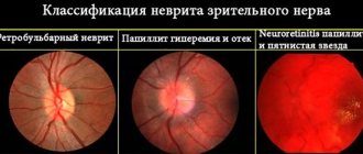
Neuritis - symptoms, causes, types and treatment of neuritis
Good day, dear readers!
In today's article we will look at the disease neuritis and everything connected with it.
What is neuritis?
Neuritis is an inflammatory disease of peripheral nerves, characterized by a decrease or complete loss of sensitivity, as well as motor disorders of the tissue innervated by this nerve.
It should immediately be noted that innervation is the supply of various tissues and organs with nerves, through which the central nervous system (CNS) provides them with sensitivity and motor function.
Neuritis can also cause the development of partial (paresis) or complete paralysis.
Most often, the optic, auditory, facial, trigeminal, radial and sciatic nerves are affected by the disease.
If the inflammatory process develops in one place, the disease is called neuritis, while damage to nerves in several places is called polyneuritis.
The main symptoms of neuritis (manifested at the site of inflammation) are decreased sensitivity, numbness, partial or complete impairment of motor function, and pain.
The main causes of neuritis are infections, trauma, tumors, hypothermia, poisoning, various diseases (osteochondrosis, arthritis, diphtheria and others).
Development of neuritis
Thanks to the nervous system, we can see, hear, smell, move, breathe, etc.
The collection of nerves in the body form the peripheral nervous system.
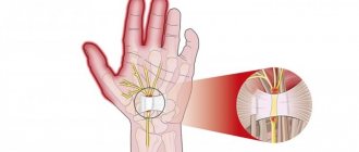
A nerve is a part of the nervous system, which consists of bundles of nerve fibers covered with a sheath, providing communication between the brain (cerebral, spinal) and other parts of the body, organs, and tissues.
There are also blood vessels inside the nerve.
The largest nerves are called nerve trunks, after which they branch significantly, and at the end points, control of the tissue/organ by the nervous system can be achieved with just one nerve fiber. The structure of the nerve may differ depending on its location.
The mechanism of development of neuritis is quite complex, but is caused mainly by disturbances in the nerves - metabolic and vascular processes, their injury, tumors, and infection.
These factors lead to the destruction of myelin and Schwann cells, which are involved in the transmission of nerve impulses along the fibers. With severe pathology, the axial cylinder is also destroyed. At the same time, the nerve fibers are not able to perform the function of transmitting nerve impulses from the brain to the tissues, which is why the latter are not able to perform their functions.
Neuritis, neuralgia and neuropathy (neuropathy) - difference
This is also an interesting question, since various sources combine these concepts, indicating the sameness of this disease. However, in clinical practice these concepts are separated, since the causes, localization, symptoms and further treatment regimen may differ slightly.
Let's consider the distinctive features of these concepts.
Neuritis is characterized by inflammation of the peripheral nerve itself, in which pronounced changes also occur. The inflammatory process involves the myelin sheath (contains myelin and is located inside the glial sheath of the nerve) and the axial cylinder.
Neuropathy (neuropathy) is a disease of peripheral nerves (most often nerve trunks) of a non-inflammatory nature, in which degenerative and metabolic damage to the nerves occurs.
The causes of neuropathy are usually impaired blood supply, injury, and metabolic disorders. Symptoms of neuropathy are decreased sensitivity, decreased reflexes, and decreased strength.
Neuropathy in psychiatry is diagnosed in the case of increased excitability of the nervous system with increased fatigue.
Neuralgia is characterized by inflammation of the peripheral nerves, but loss of sensitivity, paresis, paralysis and impaired motor activity in the innervation zone are not observed, nor are there any (or minimal) structural changes in the nerve itself. The main symptom of neuralgia is pain (often severe) at the site of innervation, decreased sensitivity. There may be autonomic disorders.
Neuritis statistics
According to medical statistics, neuritis most often occurs in older people, especially women.
Neuritis - ICD
Neuritis: ICD-10 - M79.2, ICD-9: 729.2; Neuropathy: ICD-10 - G60-G64;
The first signs of neuritis:
- Feeling of pain at the site of the inflammatory process;
- Numbness of the innervated area;
- Tingling sensation.
Main symptoms of neuritis:
The main symptoms of neuritis depend on the type of nerve fibers affected - sensory, motor and autonomic, as well as on the cause and severity of the inflammatory process:
Inflammation of sensory fibers causes paresthesia (decreased sensitivity, goosebumps, numbness and tingling in the innervation zone), and a feeling of pain.
Inflammation of motor fibers is caused by impaired motor function (paresis - partial, paralysis - complete), weakening and/or atrophy of muscles, decreased or loss of tendon reflexes.
Inflammation of vegetative fibers is caused by local hair loss, skin depigmentation (the appearance of vitiligo), xeroderma, thinning and swelling of the skin, fragility of the nail plate, the appearance of trophic ulcers, increased sweating and others.
Additional symptoms of neuritis
The following symptoms are characteristic of different types of neuritis, and depend mainly on the location of the innervated area/organ/tissue:
Neuritis of the facial nerve (Bell's palsy) is an inflammation of the nerve responsible for the work of the facial muscles of one half of the face.
Symptoms of facial neuritis are the appearance of weakness in the facial muscles, which manifests itself in the form of partial or complete absence of facial movements, as well as facial asymmetry, similar to the consequences of a stroke.
With neuritis of the facial nerve, on the affected side, wrinkles on the forehead are also smoothed out, the eyelid is drooping, and the corner of the mouth is drooping.
Source: https://medicina.dobro-est.com/nevrit-simptomyi-prichinyi-vidyi-i-lechenie-nevrita.html