Provoking factors
Visual impairment with VSD has distinctive features. Usually these are severe pain in the head, discomfort in the sternum, and the deterioration of vision occurs gradually, so it is quite difficult to determine the cause of its impairment. The image looks blurry, darkening occurs in the eyes during an attack, which may be mild or accompanied by complete loss of vision. For the purpose of prevention, at the first symptoms you should consult a doctor and undergo an annual examination with an ophthalmologist.
During the period of VSD, many disorders occur in the body. Any system can be a weak link, including the one responsible for vision
What causes vision problems:
- Insufficient amount of healthy ingredients in the daily diet. With an unbalanced diet, nutrients do not reach the brain, and vision deteriorates. In order for the information coming from the eyes to the central nervous system to be processed promptly and on time, many vitamins and microelements are needed.
- Circulatory disorders. If there is insufficient blood supply to the brain and eyes, visual distortion of the image occurs. VSD can have such an effect on the eyes, as the capillaries and blood vessels become thinner.
- Panic state. Feelings of fear and anxiety negatively affect the functioning of the central nervous system, disrupting the perception of images by the visual system.
- Connection with consciousness and subconscious. In stressful situations and accumulated problems, the world looks unattractive. The optic nerves perceive information from the environment, but consciousness does not record it. This condition is characterized by unclear, dim images, quiet words, red eyes with prolonged depletion of the body by stress.
Stress and negative emotions prevent you from thinking soberly, this is typical for any person, not only those with VSD, and vision deteriorates.
Visual impairment can be caused by inadequate supply of nutrients to the brain and retina
Why does visual impairment occur with VSD?
It is known that a person whose psyche is practically defenseless in the face of stressful situations constantly suffers from the manifestation of many unpleasant symptoms.
Regular unrest leads to excessive stimulation of the nervous system, which provokes overstrain of the eye muscles, changes in the shape of the eyeball, narrowing of blood vessels and a lack of blood in the tissues. As a result, pathological processes may begin to develop in the organs of vision. However, vision problems with VSD do not always arise due to nervous shock. For example, the basis for the occurrence of a symptom may be a disorder of the vestibular apparatus. Diseases such as glaucoma, cataracts, myopia, retinal detachment or conjunctivitis should not be excluded. Vegetative-vascular dystonia is not a reason to forget about possible eye pathologies that can develop in any person.
VSD activates the stress response. The stress response immediately causes physiological, psychological and emotional changes that increase the body's ability to cope with a threat - either to fight it or to flee from it - which is why the stress response is often called the "fight or flight" response.
Part of the changes to the stress response involves stimulation of the nervous system. The sympathetic system releases hormones that enhance the functioning of sensory organs, including the eyes.
When stress reactions occur infrequently, the body can quickly recover from the physiological, psychological and emotional changes caused by the response. However, when the stress response occurs too frequently or abruptly, the body takes longer to recover, which can cause the body to remain in a semi-solid state (called stress overstimulation). In an overstimulated body, vision problems and other sensory disturbances occur.
Because stress reactions have a profound effect on the nervous system and sensory organs, persistent stress causes sensory abnormalities. The eye problems and anxiety symptoms listed above are a good example of how the eyes and vision are affected by constant increased stress, such as from excessive worry.
Clinical manifestations
The patient does not always associate pain in the eyes and impaired visual function with dystonia. Poor vision with VSD is not as pronounced as numbness of the legs and arms or hemicrania. This symptom can be mistaken for another disease.
If you are concerned about darkening of your eyes, this is the main sign of dystonia. As the patient's condition worsens, pressure surges become more frequent. For this reason, a person is worried about dizziness and fainting. The central nervous system is overloaded and can no longer cope with all tasks. In addition to blood pressure, intracranial pressure increases. If you notice that your vision is blurred, be prepared to lose consciousness.
In addition to the main symptom, a person may notice other clinical manifestations:
- Flash Light. The symptom appears during the transition from bright to dark lighting or vice versa. Possible loss of balance.
- Floaters in the eyes. A characteristic symptom after overwork, prolonged concentration on one object, or a change in starting position.
Deterioration of blood supply to the visual system (eyes, occipital part of the cerebral hemispheres, pathways) can lead to incorrect visual perception of the world
- Blurring of the background within the visible space. Sometimes an object is clearly visible, but the background around it appears blurry. VSD can also have such an effect on the eyes.
- Eyes hurt. Discomfort can occur in different conditions - bright lighting, touch, blinking.
- Double vision. Objects may become distorted. The patient does not see a clear image.
If you experience any of these symptoms, seek medical help immediately.
A different picture is observed during an acute attack of visual impairment. The patient complains of sparkling ripples, lack of understanding of what is happening, and panic attacks. This condition can occur with physical fatigue, acute or chronic illness. At first, the ripples are weakly expressed, but gradually the attack increases and can be felt unevenly by the eyes. At first, the patient cannot distinguish small objects, write, or read. Then the ripples intensify and sparks appear. The surrounding space is blurry. The man is disoriented. An attack while driving a car is especially dangerous.
If the slightest symptoms appear, you need to stop working for a short period of time. No special therapy is required; medications are useless. It is necessary to lie down so that the blood supply to the brain improves. Close your eyes, overcome the panic. After 10–20 minutes, everything will gradually begin to recover.
If vision is impaired during vegetative-vascular dystonia, the patient may have the following symptoms: distorted image
Objective signs of vegetative-vascular dystonia
Upon examination, the main symptoms are identified:
- changes in the fundus of the eye;
- red eye syndrome;
- bruises and swelling under the eyes;
- different pupil sizes (functional anisocoria);
- deterioration of vision, detected using an ophthalmological table.
Fundus examination allows you to assess the condition of the blood vessels supplying the retina. During instrumental examination, spasm or hypotonicity of blood vessels is noted.
Visually, you can determine the size of a possible pathological area, as well as assess the degree of influence of the dystonic process on visual perception.
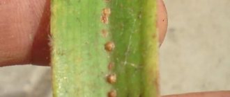
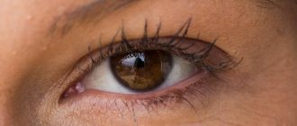
Red eyes – with vegetative-vascular dystonia, this symptom often appears.
It's easy to discover on your own. Redness is caused by various factors:
- Bruising occurs against the background of prolonged spasm of small vessels, which is visually manifested by redness of the protein membrane (sclera).
- The appearance of swelling and bruising under the eyes is associated with excessive decompensatory overstrain of the eye muscles.
When diagnosing visual impairments in VSD, visual impairment such as myopia is often detected. Its characteristic feature is its transient nature. After the smooth muscle function is restored, restoring the normal curvature of the lens, vision improves.
Different pupil sizes indicate a disruption in the functioning of the sympathetic and parasympathetic nervous system.
To determine the size of the pupils in both eyes, an examination is carried out with uniform illumination of the eyes, testing the pupils for constriction or dilation using light exposure.
What to do?
The main thing is not to panic and clearly understand that such a condition may be temporary. To confirm that this was due to VSD, laboratory tests are necessary. Then the doctor makes a conclusion. To prevent such manifestations, it is important to adhere to the norms of prevention (do not overload the central nervous system, refrain from worries, stress), and establish the cause.
At the initial stage, when clinical manifestations are short-term, visual impairment does not harm health. But an alarming symptom should be a reason to seek medical help in order to prevent serious complications.
Necessary treatment
Any visual impairment requires the attention of a medical specialist. If a person experiences darkening or ripples in the eyes, he needs to consult an ophthalmologist. After carrying out diagnostic procedures and diagnosing VSD, the doctor draws up the optimal course of treatment, taking into account the individual characteristics of the patient’s body.
If the cause of visual impairment is explained by low or high blood pressure, the course of treatment is complex. To normalize indicators, different techniques are used. Among them are drug therapy, psychophysical techniques and many others.
The choice of treatment method is made by a medical specialist; it is dangerous to independently determine the course of therapy. This may make the situation worse. To improve the functioning of the visual apparatus, a special breathing technique was developed, which is carried out with energy replenishment. Medical experts recommend doing it daily; even after half an hour of exercise, the first improvements in the functioning of the visual apparatus are noticeable.
To improve the patient's condition, the attending physician may prescribe courses of physical therapy. It is better to give up bad habits at least until recovery. This will increase the effectiveness of the treatment course. Some doctors prescribe psychological trainings that help normalize the energy of thought activity.
Particular attention must also be paid to sleep patterns. Overwork and stressful situations have a detrimental effect on the entire body, including visual acuity. An active lifestyle plays an important role; almost all patients with vegetative-vascular dystonia led a sedentary lifestyle. An excellent way to prevent vision problems is to exercise in the morning.
Since VSD is a psychovegetative syndrome, the course of therapy must necessarily include psychotropic medications. The effectiveness of the entire treatment directly depends on the choice of medications.
If the drugs were chosen incorrectly, the patient’s well-being may worsen, and in addition to the clinical manifestations of the disease, side effects from the use of drugs may occur.
One of the most effective treatment methods is reflexology. These include massage treatments, manual therapies, acupuncture and much more. The impact is on reflex areas.
Psychotherapy is often included in a comprehensive course of treatment. Qualified specialists recommend psychoregulation techniques. In other words, self-hypnosis. To achieve maximum effectiveness when using this technique, you must adhere to the recommendations of a neurologist.
To treat a sharp deterioration in vision due to VSD, the doctor prescribes medications:
- sedatives;
- psychotropic drugs;
- vasodilator tablets.
For the treatment of retinal angiopathy of both eyes, Okumetil drops are used. Improved visual function can be achieved with the help of the drug Halidor. If the symptom of dry eye develops, the antioxidant agent Mexidol and Taufon drops are prescribed. To stabilize the nervous system, effective sedatives are used:
- Novo-Passit;
- Persen;
- Cardiplant;
- Bromcamphar;
- Corvalol;
- Valocordin;
- Validol.
They give a long-lasting and lasting effect. If the patient is worried about blurred vision, it is necessary to visit a doctor to choose the right medications for treatment. In many cases, he prescribes medications that eliminate neurotic disorders.
To prevent the disease from progressing, the patient is recommended to take tranquilizers and antidepressants. The drug Eltacin prevents the appearance of flickering glare during a panic attack.
The doctor recommends that the patient take vitamin complexes. For the treatment of vascular disorders, the medicine Picamilon and Ocuvait eye drops are prescribed. To strengthen blood vessels, a course of Mildronate is required.
The doctor recommends eye exercises to the patient. Exercises are performed regularly, then the eyeballs are massaged. To prevent vision from deteriorating, the patient is advised to rinse his eyes with weak brewed black tea or boiled water at night.
You can make lotions with herbal infusion:
- dill inflorescences;
- chamomile flowers;
- peppermint leaves.
If tension appears in the eyes, massage the earlobes and stimulate biologically active points that improve visual acuity during VSD.
These simple techniques keep the patient healthy.
Visual impairment with VSD requires immediate medical attention. You should consult with a therapist and ophthalmologist and undergo the prescribed examination. Only after a diagnosis is made, comprehensive treatment is prescribed. If vision deterioration occurs due to hypo- or hypertension, medications are used to normalize blood pressure.
If a patient has a vegetative disorder, neurotropic and psychotropic drugs are prescribed. This allows you to quickly get rid of fear and anxiety. An ophthalmologist prescribes medications that normalize blood circulation in the organs of vision. The course of therapy is selected individually. Attempts at self-medication will lead to aggravation of the patient's condition.
MORE ABOUT: Congenital cataracts in a kitten - Eye diseases
First of all, whether you are dystonic or not, if you have any vision problems, you must consult a doctor and undergo the necessary examination. Treatment should only be prescribed by a qualified specialist. At home, you can perform special gymnastics for the eyes.
- Close your eyes, then open them wide. Do this at least four times.
- Move your gaze from right to left, then up and down. First perform the exercise with your eyes open, then with your eyes closed. Do not turn or tilt your head. Do it at least three times.
- Rotate your eyes clockwise, then in the opposite direction. Repeat at least three times with eyes closed and open.
- Blink quickly for a minute.
- Rub your palms until they become warm, then hold them over your eyes for two to three minutes. When performing the exercise, you need to completely relax and think about what calms you down and evokes positive emotions.
You can also try to correct vision impairment with VSD with a nutritious, balanced diet. It will be beneficial to eat fresh blueberries, as well as foods rich in vitamins A, B, D, E. Special eye vitamins and herbal teas can be effective.
It must be remembered that loss of clarity of vision is not always a cause for great concern. Sometimes a blurry picture can be short-term and be the result of ordinary fatigue or excessive physical exertion. To avoid discomfort, you should pay attention to the correct daily routine, sleep at least 7-8 hours and not burden yourself with heavy physical and mental work.
Therapy begins with a visit to an ophthalmologist. After a survey and tests, a specialist can confirm or refute vegetative-vascular dystonia as the main provoking factor.
Some patients complain that there is pressure on their eyes. The reason is eye or blood pressure. To solve the problem, an integrated approach is required. First you need to determine whether the pressure is low or high. Then appropriate drug treatment, physiotherapy, and traditional medicine recipes are prescribed. The duration of the course of therapy is determined by the doctor.
Most people diagnosed with VSD lead a sedentary lifestyle. Therefore, physical therapy occupies a key place in therapy. You can choose an activity “to your liking”: swimming pool, yoga, gymnastics, fitness. Giving up bad habits will make treatment easier for the patient.
A balanced diet plays an important role in therapy. You need to reconsider your diet so that more nutrients enter the bloodstream. Food should contain minerals and vitamins of different groups.
In more advanced stages, treatment without medication is impossible. To improve the result, neurotropic drugs and drugs with psychotropic effects are prescribed. Feelings of anxiety and restlessness will no longer bother you, which means recovery will come sooner. Preventive measures for vision problems:
- stick to the regime;
- do not overwork;
- avoid stress and worries;
- have a full rest.
A favorite hobby will help you take your mind off sad thoughts.
The basic rule to follow is that at the first sign of vision deterioration, you should seek medical advice. But even before the doctor confirms or denies vegetative-vascular dystonia as the cause of visual impairment, the patient should try to bring his mental state back to normal on his own. This will relieve the nervous system from overload and prevent the situation from worsening, and may even improve it. It is possible to use mild sedatives such as herbal teas.
Once the doctor has made a diagnosis, appropriate treatment will be prescribed, which may include:
- psychophysical methods of influence;
- physiotherapy;
- herbal medicine;
- medicines.
If the cause of vision deterioration was problems of a nervous nature, the help of a psychotherapist is needed who can quickly bring the patient’s state of mind back to normal.
- Working with a psychotherapist who identifies situations and thinking patterns in a person that can cause increased stress, anxiety and, ultimately, lead to dysfunction of the autonomic nervous system.
- Physiotherapy methods, primarily massage, physical therapy, and electrosleep, can stabilize brain function and get rid of most neurological symptoms.
- Medicines in the form of sedatives are used in patients of different ages. They help improve the functioning of the central nervous system and cope with the clinical manifestations of VSD.
- Lifestyle changes (diet adjustments, regular physical activity, etc.) are a necessary part of therapy.
Prevention of visual impairment
With VSD, the best methods for preventing visual complications are reducing stress on the central nervous system and maintaining a daily routine. It is useful to engage in physical exercise. The following types of activities have a beneficial effect on health:
- walking;
- moderate running;
- swimming;
- fitness;
- yoga;
- gymnastics.
Along with sports exercises, it is necessary to give up bad habits. It is also necessary to balance the diet. The menu should include food containing a large amount of minerals and vitamins. For VSD, you need to drink freshly squeezed fruit and vegetable juices, compote, green tea, and fruit juice. Eye exercises are useful.
Visual impairment is minimized by following preventive measures. They are aimed at eliminating the influence of provoking factors in the development of dystonia.
Diseases are much easier to prevent than to treat. Timely treatment, preventive measures and early diagnosis of pathologies help to avoid undesirable consequences.
Posts 1 page 10 of 10
Share12012-04-24 00:12:02
- Author: Halluhorsez
- Beginner fly swatter
- From: MO
- Registered: 2012-04-23
- Posts: 12
- Respect: +1
- Positive: +2
- Gender: Male
- Age: 32 [1986-07-21]
- Last visit: 2012-08-05 00:39:08
Hi all! I was very surprised that such a forum existed. 1.5 years ago, dystonia struck and I was surprised, and at the same time breathed a sigh of relief when I found out that I was not the only one. The Internet is full of information on this topic, there are even entire forums. In general, we went through fire and water with all that it entails. Anyone who has had VSD will understand. A full bouquet of various symptoms, visiting doctors. Thank God, I managed to somehow figure it out and cope with everything myself. But the only problem with my eyes still remains. Moreover, the jokes with vision began only about 3 months after the first crisis. Namely, at first the image began to tremble, more precisely where the gaze focused, vessels appeared on the proteins from the center to the edge, then flies with sparks appeared, at the end of the day the picture sometimes turned into interference, like on a TV when there is no signal. Then, after another 2 months, various transparent dots and cobwebs began to appear. When problems with my eyes started, I went to an ophthalmologist, and a month later I repeated the visit. Nothing was found. They said that the blood vessels were narrowed and that’s all. I have had astigmatism since childhood, but my vision has not deteriorated over these 15 years, thank God. Then I was referred to a neurologist, who diagnosed VSD. I explained everything to him. The fact is that before that I went to the gym, worked with fairly heavy weights, slept little, and on weekends I liked to drink and sometimes indulge in drugs. In general, I combined everything, for the time being. until one night there was a good shaking, so much so that my veins began to shake. Naturally, after everything that happened, Running Sports Healthy Lifestyle began. In six months I got rid of half the symptoms, and after another half I almost finished off the remaining ones. Only the eyes remained. And by the end of the 11th year, things started to improve with them, there were fewer flies with sparks, the picture stopped shaking. But here, you understand, NG again, DR, etc. How can I resist here, everything seems to have passed. I drank normally a few times and, voila, the vision symptoms returned, even more severely, especially the next day after drinking. But then another symptom was added after I read a lot on the Internet about retinal detachment. About 3 months ago, a small speck, a sunbeam, came into view. Only in the final stage, when he is about to disappear. And when you start blinking, it becomes clearly visible. Periodically, this spot appears, but quickly disappears, within 30 seconds. Today this bunny sparkled once brighter than usual, it has already really lit up. It was no longer that dark spot. That’s when I decided to write here. Everyone's mood has dropped, apparently it's a total mess (((( .
ps in addition to these symptoms, there are also these: the outlines of objects remain in the field of vision, i.e. if you look at the circle and then look at the wall, then you see this circle on the wall; irritability to very bright light, especially in sunny weather, if you look at the sky; the appearance of single sparks lasting 2-3 seconds; if you sniffle near the monitor, standing sideways to it, then your peripheral vision gets the impression that the monitor is blinking, it’s like when computers appear on TV; Well, at the end of the working day, the eyes get very tired, it hurts, they become mucus, and vision deteriorates, especially in dark lighting.
That's basically all I wanted to write.
I don't know what to do. It’s clear that you’ll have to go to the ophthalmologist, but will this again give any results?
ps2 I also have scoliosis and chest deformation, they say that the spine can also cause such problems. Don't know.
Edited by Halluhorsez (2012-04-24 00:21:30)
Share22012-04-24 14:22:09
- Author: Kosh
- Beginner fly swatter
- Registered: 2010-02-15
- Posts: 12
- Respect: +0
- Positive: +0
- Last visit: 2017-11-01 13:45:37
I don't know what to do. It’s clear that you’ll have to go to the ophthalmologist, but will this again give any results?
The sparks you see may be caused by traction on the vitreous, which can lead to a retinal tear. Be sure to go to an ophthalmologist and check the fundus of the eye if you have not already done so.
Share32012-04-24 21:37:07
- Author: Halluhorsez
- Beginner fly swatter
- From: MO
- Registered: 2012-04-23
- Posts: 12
- Respect: +1
- Positive: +2
- Gender: Male
- Age: 32 [1986-07-21]
- Last visit: 2012-08-05 00:39:08
The sparks you see may be caused by traction on the vitreous, which can lead to a retinal tear. Be sure to go to an ophthalmologist and check the fundus of the eye if you have not already done so.
Well, a year ago they didn’t find anything. Let's see what happens this time. I already signed up for tomorrow, I feel like I got it :/ what if suddenly. How is this generally tolerated? What about sports, running? After all, apparently no loads can be given?
Share42012-04-24 23:03:46
- Author: Victor23
- Advanced fly swatter
- Registered: 2012-01-28
- Posts: 97
- Respect: +4
- Positive: +2
- Last visit: 2018-03-07 11:36:13
what if suddenly... How is this generally tolerated? What about sports, running? After all, apparently no loads can be given?
the doctor will tell you everything) we don’t know what you have there) In general, don’t guess, go to an ophthalmologist and, better yet, not to a clinic, but to a specialized ophthalmological center.
Share52012-04-24 23:33:41
- Author: Admin
- Administrator
- From: Moscow
- Registered: 2005-09-01
- Posts: 2927
- Respect: +266
- Positive: +156
- Skype: musheknet
- Last visit: 2018-10-05 14:39:38
Hello Halluhorsez ! Once again one of the trends in the appearance of flies is confirmed. Everything is just like mine, only I didn’t use drugs.
My friend, what can I say.
First of all, you need to change your psychology, start thinking differently. This is the root cause, which is usually hidden. Second. Body position and mental state are directly interconnected. Simply put, if you are used to looking at your feet, sitting bent over, you are loaded and negative. If you look straight and up at the sky, you radiate positivity and confidence. Always control your body position: your back should be straight and your neck open! But from school we were taught to hide our heads like a turtle and sit in a slavish, bent position. Especially on the computer. VSD is already a consequence, especially if the body is physically overloaded with harmful substances. And only then there is a reflection on the physical condition.
With vegetative-vascular dystonia, a change in the tone of blood vessels is observed, which leads to blurred vision and reduced visual acuity. This fact does not frighten a person suffering from VSD, since vision decreases gradually and slightly, without foreshadowing anything terrible. But there are cases when an attack of vision loss occurs suddenly, resulting in panic and misunderstanding of what is happening. A person with VSD, faced with acute vision loss, may experience panic attacks and stress. To avoid panic and prevent complete loss of vision in the future, it is necessary to understand the causes of the disease, its origin and methods that will help prevent the disorder.
How to get rid of vision problems?
If visual impairment during VSD is caused by an intense panic crisis, then a decrease in anxiety will lead to a reduction in symptoms. As the body recovers from an active stress response, the unpleasant feeling diminishes until it disappears completely. It may take up to 20 minutes or more until the body has fully recovered from a severe stress response. But such a long recovery in medicine is considered to be the norm and should not alert the patient.
If red eyes or other visual disturbances in VSD are caused by overstimulation, it will take much longer for the body to recover. In some cases, drug intervention is necessary to relieve the adrenergic crisis.
But when the body is fully restored, vision problems disappear. Therefore, they should not cause concern, since they are only related to stress.
The patient can speed up the recovery process by reducing stress, practicing relaxed breathing, increasing rest and relaxation intervals, and not thinking about the sensation. Of course, this can be unsettling and annoying. But as the body recovers from the stress response or chronic stress, the symptom will disappear.
Fundus diagnostics in adults and children
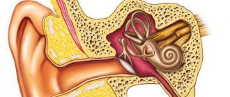
What is the fundus of the eye?
Our readers successfully use Oko-plus to treat joints. Seeing how popular this product is, we decided to bring it to your attention. Read more here...
So, ophthalmologists call the back inner part of the eyeball, which is seen during ophthalmoscopy. Normally, the fundus of the eye looks like this: the retina (inner layer) has a reddish color, against its background a rounded and lighter pale pink optic disc stands out. In the center of the disc, the central vein (darker and thicker) and the central artery are clearly visible; they are divided into lower and upper branches, and then they are divided into smaller vessels, which form the choroid. Against the background of the retina, the oval and darker macula (yellow spot) is also very clearly visible.
Using an ophthalmoscope, you can not only clearly examine the structure of the fundus of the eye, but also study the sizes of veins and arteries, and determine the deviation of their sizes from the norm. And also evaluate the condition of the retina and choroid, the presence of hemorrhages, ruptures, detachments, and tumors. Defects of the eye disc (seals, dystrophy, darkening) and macula are clearly visible here.
Symptoms of fundus pathologies
The following symptoms should alert you and be a reason to visit an eye doctor:
- the appearance of flies and lightning before the eyes,
- distortion of color perception,
- rapid (sharp) decrease in visual acuity,
- narrowing of visual fields,
- decreased twilight vision acuity,
- blurred picture (veil before the eyes).
How often and why should a fundus examination be performed?
Ophthalmologists recommend that healthy adult patients undergo fundus examinations at least once a year. If there are visual impairments or eye diseases, at least once every 6 months. And for newborns, children and adolescents at least once every six months. This is especially true in light of the following figures: from 70 to 80% of the world's population experience some form of vision problems.
Ophthalmological diseases of the posterior part of the eye are dangerous due to loss of vision, which is unlikely to be restored. These eye diseases cannot be cured at home; they require hospital treatment, and in many cases, surgery.
If the pathology is caused by a systemic disease, then the treatment will be complex and most often urgent. The fact is that the fundus of the eye provides very extensive information about the state of the structures and systems of the body as a whole and about other systemic diseases.
Thus, enlarged veins and arterial atrophy may indicate hypertension, slight bleeding in the fundus indicates the possibility of diabetes mellitus, the presence of retinopathy, and some types of it warn of leukemia (and this symptom appears the very first). High fundus pressure will indicate glaucoma.
Fundus diseases
Fundus diagnostics will provide comprehensive information about such ophthalmological diseases as:
- retinal detachment,
- retinal tears,
- retinal hemorrhages,
- optic nerve retinopathy,
- atrophy of the blood vessels of the eye,
- narrowing (dilation of veins and arteries),
- optic nerve atrophy,
- glaucoma.
Examination of the retina and eye structures will help detect systemic diseases of the endocrine, vascular, cardiac, body systems, neurological problems, and brain tumors. Thus, examination of the fundus can help in diagnosing:
- diabetes mellitus type II,
- brain tumors,
- vegetative-vascular dystonia,
- multiple sclerosis,
- rheumatoid arthritis,
- blood cancer,
- breast cancer,
- cardiovascular diseases,
- hypertension.
Very often, changes in the fundus appear before other symptoms of the disease. In these cases, this examination will help identify the problem at an early stage, which will significantly increase the patient's chances of recovery.
The ophthalmologist will write out a referral to a cardiologist, neurologist, or therapist. Additional examinations and consultations will be required to confirm (or establish) the diagnosis.
Methods for examining the fundus of the eye in adults
A fundus examination can be performed in any ophthalmology office, where the doctor will perform an ophthalmoscopy using a special ophthalmological device - an ophthalmoscope. However, today modern equipment is used for more accurate studies: electric microscopes (slit lamps) and laser ophthalmoscopes.
In examination rooms, doctors use two types of examinations: direct and reverse ophthalmoscopy. In some cases, to obtain greater viewing opportunities, mydriatics (drops that dilate the pupils) are instilled into the eyes.
Sometimes, for complex pathologies, a more complex and modern examination using a laser ophthalmoscope may be recommended (it is also more expensive).
Direct ophthalmoscopy is carried out using a conventional hand-held or head-mounted ophthalmoscope, a Skepens ophthalmoscope and allows you to see the fundus of the eye. To do this, the patient is placed in a dark room and a light source is installed behind him. A beam of light is reflected from the concave mirror of the ophthalmoscope and falls through the pupil onto the fundus.
Now, with the help of a special magnifying lens, you can see vessels, veins, retina, macula (macula), and optic nerve enlarged 12-20 times. With direct ophthalmoscopy, changes in the fundus of the eye, or more precisely, in its center, are clearly visible. Thus, direct ophthalmoscopy makes it possible to detect optic nerve dystrophy, vein pathologies, and tumors in the eye.
Reverse ophthalmoscopy is carried out using an additional lens; here the image will be upside down, but the area being examined increases. Here not only the center of the retina, but also the periphery will be clearly visible. In this case, you can clearly see retinal detachments and tears, peripheral vascular abnormalities, and tumors in the periphery.
Slit lamp (electric ophthalmoscope) - this device was invented a long time ago, but today it has been significantly improved, allowing the ophthalmologist to obtain a three-dimensional image of the internal structures of the eye. Higher magnification is obtained when examining the posterior part of the eye using a slit lamp.
The laser ophthalmoscope is the gold standard for examining the back of the eye. When conducting a fundus examination using a laser, you can obtain not only a picture, but also record the examination, measure the size of veins and arteries, and obtain the size of the optic disc and macula. The results will be presented in the form of tables and graphs. However, this survey will only be in black and white.
Fundus examination in children
Fundus examination in children requires preparation. In order to be able to conduct ophthalmoscopy in young children, parents need to have a preparatory conversation with them. You will need to tell the child how the procedure will take place, what the doctor will do, explain that the examination will not cause pain to the baby and tell why it is necessary.
Children's eyes are constantly growing, and in order not to miss the disease, children undergo ophthalmoscopy more often than adults.
For newborns, this procedure can also be performed, but most often it is prescribed for suspected congenital pathologies: congenital cataracts, retinal dystrophy and others.
After ophthalmoscopy, newborns are often prescribed an ultrasound of the brain based on the results of the examination.
Features of the examination in children
Fundus examinations for young children and older children are carried out using the same method as for adults: in a dark room using a light source and an ophthalmoscope.
For particularly active young children, the doctor may use a toy (this distracts attention and forces the child to look in the right direction).
The examination of newborns follows the same scheme, with the active participation of the mother.
Gymnastics for the eyes
1. Close your eyes tightly and open them wide. Repeat 5-6 times at intervals of 30 seconds.
2. Look up, down, to the sides, without rotating your head, 3 times with an interval of 1-2 minutes. Do the same with your eyes closed.
3. Rotate your eyeballs in a circle: down, right, up, left and in the opposite direction. Repeat 3 times with an interval of 1-2 minutes.
Do the same with your eyes closed.
4. Close your eyes tightly for 3-5 seconds, then open them for 3-5 seconds. Repeat 6-8 times.
5. Blink quickly for a minute.
6. It is also useful to hang a bright calendar, photograph or painting at a distance of 1-2 m from the desktop (this place should be well lit) so that during classes you can look at it from time to time.
7. Extend your hand in front of you and look at the tip of your finger at a distance of 20-30 cm for 3-5 seconds. Repeat 10-12 times.
8. This exercise also has a good effect on the eyes: standing at the window, look for some point or scratch on the glass (you can glue a small circle of dark plaster), then turn your gaze, for example, to the television antenna of a neighboring house or a branch of a tree growing in the distance.
What are the symptoms of panic disorder?
Symptoms of panic disorder often appear in teenagers and young adults under the age of 25. If he has had four or more panic attacks, or lives in fear of another panic attack, he has panic disorder.
Panic attacks cause intense fear that begins suddenly. The attack lasts from 10 to 20 minutes, and in extreme cases more than an hour. The experience is different for everyone, and symptoms often vary.
Common symptoms associated with a panic attack include:
- Tachycardia.
- Hesitant breathing.
- Suffocation.
- Paroxysmal vertigo.
- Nausea.
- Sweating or chills.
- Changes in mental status: feelings of derealization (unreality) or depersonalization (dissociation from oneself).
- Numbness or tingling in the arms/legs.
- Poor coordination.
- Redness of the eyes.
- Problems with movement.
- Cardialgia.
- Fear of dying or going crazy.
Symptoms of panic attacks occur for unknown reasons and are disproportionate to the level of danger. Since attacks cannot be predicted, they significantly affect the functioning of the patient. In rare cases, patients suffer from visual disturbances due to attacks of VSD. They may have red eyes or spots in front of their eyes.