Varieties
With a disease such as myopathy, the muscle structures of the shoulder girdle and pelvic girdle are predominantly affected. But other muscles can also be affected, so depending on the severity of the symptoms, several forms of this disease are distinguished.
The most common form is Duchenne myopathy . This form of pathology is otherwise called pseudohypertrophic, since it is characterized by an increase in muscle mass due to the accumulation of fat, which causes the muscles to become large but weak. Duchenne myopathy is the most malignant form of pathology - it is characterized by a rapid course and severe consequences. Most patients with Duchenne atrophy become disabled and even die due to respiratory or heart failure. It must be said that Duchenne myopathy manifests itself already in the first years of life, and it mainly affects boys. In addition, the earlier it begins, the more severe the pathology.
The second form is also no less common - this is Erb's myopathy or the juvenile form of the pathology. The disease develops in men and women aged 20–30 years, and is manifested by atrophy of the muscles of the hips and pelvic girdle. Patients develop a “duck” gait and develop atrophy of the oral muscles, which is characterized by the inability to purse their lips and whistle, which also causes problems with the pronunciation of certain sounds. Early onset of the disease leads to immobility and disability, but if the disease begins later, its course is less aggressive.
Another common form is Becker myopathy . It is considered the mildest pathology of all varieties. It begins in young people at the age of 20 and is manifested by hypertrophy of the calf muscles. There are no mental abnormalities with this form.
The next form of pathology is glenohumeral-facial . Both men and women are affected by this variant, and the disease manifests itself between the ages of 10 and 20 years. The initial symptom of the disease is weakness of the facial muscles, after which atrophy spreads to the muscles of the shoulder girdle, affecting the shoulder blades.
and this disease affects the muscles of the mouth and eyes, which leads to their hypertrophy. Very rarely the process reaches the pelvic girdle. The course of this type of myopathy is slow, so patients can maintain mobility and performance for a long time. The later the disease begins, the easier it is, and disability in most cases does not develop in this form.
There is also a type of disease called eye myopathy . Most often, due to damage to the eye muscles, a person develops myopia and this is the main and only symptom of this pathology. No other disorders are found in eye myopathy, so this form of the disease can be considered the mildest.
In medical practice, there are some other types of myopathies, for example, distal, filiform, mitochondrial, Oppenheim myopathy. These forms of the disease are less common and do not have pronounced manifestations, so they are often not even diagnosed.
As mentioned above, the causes of the disease lie in gene mutations, and scientists have still not been able to figure out why these mutations occur. The consensus is that myopathy develops as a result of metabolic disorders in muscle tissue.
Where did the problem come from?
It has not yet been possible to identify why children develop myotonic syndrome. Presumably, the tendency to it is inherited, and a metabolic failure in the mother’s body plays a role, if it occurred during pregnancy.
Acquired myotonic syndrome can form if the child is inactive, has had rickets, or suffers from metabolic failures. Various forms of the pathological condition are observed in tetanus and neuroleptic syndrome.
Symptoms similar to myopathy may include Schwartz-Jampel disease, encephalomyelitis, or fever. The so-called “rigid person” condition is associated with similar risks. Similar symptoms appear after a Black Widow spider bite.
Symptoms
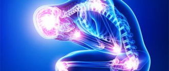
Of course, the main symptom of the disease is weakness and subsequent atrophy of the body muscles. However, each type of pathology has its own typical symptoms, which allow doctors to make the correct diagnosis.
The general symptoms of myopathy inherent in each form of the disease are as follows:
- increased fatigue;
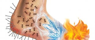
- pain in those muscles that are affected;
- reduced or, conversely, increased joint mobility;
- a feeling of “ache” in the muscles, as with colds;
- decreased muscle strength.
Other symptoms of the disease are characteristic of certain varieties. Thus, Erb's myopathy is characterized by:
- muscular dystrophy of the hips;
- atrophy of the back muscles and curvature of the spine;
- the appearance of a “wasp” waist;
- the appearance of a “duck” gait;
- atrophy of the muscles around the mouth.
Duchenne myopathy has its own characteristic symptoms, including:
- the growth of adipose tissue on the calf muscles, causing them to increase in size;
- inability to stand up independently when the patient is sitting on the floor;
- dystrophy and complete dysfunction of all muscles of the body;
- joint deformities;
- atrophy of the cardiac and respiratory muscles with the development of respiratory or heart failure.
As mentioned above, Duchenne myopathy is the most severe form of pathology.
Becker myopathy is characterized by symptoms such as:
- fatigue and increased fatigue in the legs;
- change in the appearance of the legs;
- atrophy of the muscles of the pelvic girdle.
With glenohumeral-facial muscular dystrophy, there is hypertrophy of the lips, impaired sound pronunciation, atrophy of the eye muscles, which is why a person cannot close them, and changes in facial expressions.
A mild form of this pathology, ocular myopathy, is characterized only by changes in the eye muscles, which leads to blurred vision and difficulty closing and opening the eyes.
How to help?
Treatment of myotonic syndrome in children is mainly palliative; a course of drugs and procedures is selected to improve the general condition of the body. Medicine does not yet know the path to an absolute cure. The symptomatic course allows the patient to lead a normal, adequate life. If the attacks are intense, medications are prescribed to reduce the likelihood of manifestations.
If a child has myotonic syndrome, parents will have to immediately learn special massage - such procedures are considered the most effective in a therapeutic course. The child should be protected from stress, increased physical activity, and exposure to the cold.
It is contraindicated to spend a long time in a relaxed position. The muscles suffering from the syndrome should be strengthened. Hardening, daily walks, and a strictly healthy lifestyle are recommended.
Overview of Myopathy
Neuromuscular diseases in which dystrophic lesions of certain muscles are observed, accompanied by steadily progressive degeneration of muscle tissue, are called myopathies . Pathology develops due to:
- disturbances in the functioning of mitochondria, which ensure the oxidation of organic compounds and use the energy resulting from their breakdown for further actions;
- destructive changes in the structure of myofibrils, which provide contraction of muscle fibers;
- disturbances in the production of proteins and enzymes that regulate metabolism in muscles and contribute to the formation of muscle fibers;
- changes in the functioning of the autonomic nervous system, which regulates the functioning of internal organs, endocrine glands, lymphatic and blood vessels, and is responsible for adaptive reactions.
Such disorders cause degenerative changes in muscle fibers, atrophy of myofibrils, which are replaced by connective and adipose tissue. The muscles lose their ability to contract, weaken and stop actively moving. Physical activity is unable to restore the strength of atrophied muscles, since their weakness is not due to “underdevelopment”, but due to systemic changes at the molecular level, which have led to biochemical processes in muscle tissue being disrupted and certain connections between cells being weakened or absent.
Muscles with myopathy are weakened unevenly, so weaker areas of muscle tissue are not used during physical stress, which leads to accelerated atrophy. At the same time, stronger muscles take on the entire load. At first, after physical exercise, a person is able to feel an improvement, but then the tone of the “pumped up” muscles decreases, and the condition worsens. Sometimes complete immobilization occurs.
Malignant processes
Sometimes myopathic syndrome and muscular dystrophy are observed in sarcoidosis. The cause of the pathological condition is granulomatous neoplasms localized in the patient’s muscles. A distinctive feature of the underlying disease is the proliferation of lymph nodes on the periphery of this system, enlargement of the lymph nodes in the mediastinum.
The described clinical picture of the myopathic syndrome is accompanied by specific tissue changes identified during histological examination. Muscle biopsies are obtained for analysis. In laboratory conditions, it is possible to detect an infiltrate formed by lymphocytes, histiocytes, and blood serum cells.
The structure of granulomas is similar to those formed during tuberculosis, but they are not characterized by caseous decomposition. Dystrophic processes are localized in muscle tissue. To alleviate the patient's condition, corticosteroid therapy is indicated.
Types of myopathies
In most cases, the pathology is hereditary in nature (primary), and therefore is diagnosed in young children. Less commonly, the disease is a consequence of some illness (acquired or secondary pathology). There are many types of myopathies, the classification of which is based on the cause that provoked destructive changes in muscle tissue. A common option is the approach according to which the following types of disease are distinguished:
- Hereditary - Erb's disease, pseudohypertrophic form of Duchenne, scapuloperoneal, oculopharyngeal, pathology of the central rod.
- Inflammatory. There are two options: infectious and idiopathic. The first can be provoked by bacterial (streptococci), viral (influenza, enteroviruses, rubella, HIV), parasitic (toxoplasmosis, trichinosis) lesions. Idiopathic inflammation has an unclear origin - dermatomyositis, myositis, scleroderma, Sjögren's syndrome, collagenosis.
- Metabolic. Myopathic syndrome is caused by a disorder of lipid metabolism in muscle tissue, glycogen and purine metabolism. The group includes one type - mitochondrial myopathies (genetic pathologies based on dysfunction of mitochondria).
- Membrane. Dystrophy is associated with the loss of amino acids and enzymes in muscle fibers due to defects in cell structure. This group includes myotonia congenita and myotonic dystrophy.
- Paraneoplastic. The development of the disease is caused by the growth of a malignant tumor. For example, lung cancer leads to Eaton-Lambert syndrome, in which neuromuscular signal transmission is disrupted.
- Toxic. It develops under the influence of chemicals and drugs that are injected into the muscle (drugs, lipid-lowering drugs, glucocorticoids, etc.). Numerous injections lead to muscle hardening, suppuration, ulceration and further degeneration.
Based on the location of the lesion, myopathy is divided into three types. Distal muscular dystrophy is characterized by damage to the muscles of the arms and legs. In the proximal form, the muscle tissue closer to the center, the torso, is affected. The third option is mixed, when muscles located at different distances are affected. Another type of classification is by location:
- facioscapulohumeral muscular dystrophy;
- limb-girdle (Erb-Roth hip disease);
- ocular myopathy – bulbar-ophthalmoplegic form;
- distal myopathy is a disease of the final parts of the arms and legs (hands, feet).
Literature
- Belenikin M.S., Zhilina S.S., Barinov A.A., Sharina M.Yu., Bryukhanova N.O., Magomedova R.M., Meshcheryakova T.I., Petrin A.N., Demidova I .A., Prokopyev G.G., Mutovin G.R., Prityko A.G.
[npcmed.ru/wp-content/uploads/2016/06/Miopatiya-Saliha.pdf Allelic variant of Saliha congenital myopathy] // Russian Bulletin of Perinatology and Pediatrics. - 2020. - No. 3. - P. 89-93. — ISSN [www.sigla.ru/table.jsp?f=8&t=3&v0=1027-4065&f=1003&t=1&v1=&f=4&t=2&v2=&f=21&t=3&v3=&f=1016&t=3&v4=&f=1016&t=3&v5 =&bf=4&b=&d=0&ys=&ye=&lng=&ft=&mt=&dt=&vol=&pt=&iss=&ps=&pe=&tr=&tro=&cc=UNION&i=1&v=tagged&s=0&ss=0&st=0&i18n=ru&rlf=&psz =20&bs=20&ce=hJfuypee8JzzufeGmImYYIpZKRJeeOeeWGJIZRrRRrdmtdeee88NJJJJpeeefTJ3peKJJ3UWWPtzzzzzzzzzzzzzzzzbzzvzzpy5zzjzzzzzzzzzzzzzzzzzzzzzzzzz zzzzztzzzzzzzbzzzzzzzzzzzzzzzzzzzzzzzzzzzzzzzzzyeyTjkDnyHzTuueKZePz9decyzzLzzzL*.c8.NzrGJJvufeeeeeJheeyzjeeeeJh*peeeeKJJJJJJJJJmjHvOJJJJJJJJfe eeieeeeSJJJJSJJJ3TeIJJJJ3..E.UEAcyhxD.eeeeeeuzzzLJJJJ5.e8JJJheeeeeeeeeeeeyeyeK3JJJJJJJJ*s7defeeeeeeeeeeeeeeeeeeeeeeeeSJJJJJJJZIJJzzz1..6LJJJJJJtJJZ4….EK*&deb ug=false 1027-4065].
Reasons for the development of the disease
Myodystrophy is another name for genetic myopathy. The defective gene can be either recessive or dominant. The development of pathology can be triggered by external factors:
- infections – influenza, ARVI, pyelonephritis, bacterial pneumonia;
- severe injuries - multiple tissue and organ damage, pelvic fracture, traumatic brain injury;
- poisoning;
- strong physical activity.
An acquired disease can develop due to problems with the endocrine system (hypothyroidism, thyrotoxicosis, hyperaldosteronism, diabetes mellitus). The cause of secondary myopathy can be:
- severe chronic disease (heart, kidney, liver failure, pyelonephritis);
- malignant or benign neoplasms;
- avitaminosis;
- malabsorption (digestive disorder in the small intestine);
- pregnancy (Becker myopathy);
- pelvic fracture;
- bronchitis;
- scleroderma (a systemic disease based on impaired microcirculation, manifested by thickening and hardening of connective tissue and skin, damage to internal organs);
- constant depression;
- alcoholism, drug addiction, substance abuse, hazardous production and other factors under the influence of which constant intoxication of the body occurs;
- salmonellosis (intestinal infection).
How to notice?
Myopathic syndrome is accompanied by the inability of the myocardium to contract normally. The patient feels aching pain in the chest, which intensifies with physical activity. The nature is not similar to ischemia; it does not tend to spread to other tissues and organs.
Gradually, the pain subsides on its own - no special medications are needed. Then the condition worsens, shortness of breath becomes stronger, swelling is bothersome, the patient feels weak and lethargic. You feel dizzy, arrhythmia and tachycardia develop.
Symptoms of myopathies
Almost all types of myopathies develop gradually. At first, the disease makes itself felt by slight muscle weakness in the arms and legs, pain, body aches, and rapid fatigue after a short walk or other minor exertion. Over the course of several years, the muscles weaken significantly, which makes it difficult for patients to rise from a chair, up stairs, run, jump, and a duck's gait appears. Dystrophic changes in the limbs occur symmetrically, changing them in size, highlighting them against the background of other parts of the body.
Simultaneously with the loss of strength, tendon reflexes fade, muscle tone decreases - peripheral flaccid paralysis develops, which over time can lead to complete immobilization. Lack of active movements leads to joints losing mobility. Curvature of the spine is possible due to the inability of the muscles to support the body in the desired position.
Signs of some forms
The most common form of myopathy is Duchenne-Becker disease, characterized by severe disease and high mortality . This is a hereditary pathology, the initial symptoms of which often appear in the first three years of life. The disease begins with atrophy of the muscles of the pelvis and proximal legs, resulting in pseudohypertrophy of the calf muscles and curvature of the spine. Possible oligophrenia. In 90% of cases, the respiratory muscles and cardiovascular system are affected, which can cause death.
Erb's myopathy makes itself felt at the age of twenty to thirty years. Destructive processes first affect the muscles of the thigh, pelvic girdle, and waist, then quickly move to the shoulders and torso. The limbs lose mobility, become thin, a duck's gait appears, and the appearance of the legs changes. If the deformity appears at a young age, early immobility is possible. Older people tolerate the disease more easily and maintain physical activity for a long time. Other complications are respiratory failure, intervertebral hernia, which can lead to death.
Landouzi Dejerine myopathy is known as glenohumeral-facial pathology. The first signs of the disease appear at the age of ten to twenty years in the form of damage to the muscles around the eyes and mouth. Over time, dystrophy spreads to the shoulders, upper arms, chest, legs, and abdominal muscles. Fixation of the joints in one position, slight hearing loss, and pathological processes in the retina may be observed. The patient remains able to work for a long time, although problems with the heart and breathing are possible.
Ocular myopathy is a drooping eyelid, limited mobility of the eyeballs, and pigmentary degeneration of the retina. The pathology leads to vision problems, difficulties opening and closing the eyes. After a few years, degenerative processes can spread to the face and shoulder girdle, and affect the muscles of the pharynx. In most cases, the disease develops after forty years of age.
Complications
Duchenne myopathy causes severe complications. They are associated with damage to various organ systems:
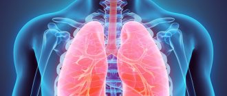
- Respiratory problems. Muscle weakness leads to shallow breathing and the inability to clear the throat normally. As a result, sputum remains in the lungs and bronchi. This leads to frequent respiratory diseases. In order to prevent them, vaccinations are given and treatment begins immediately after the first manifestations of ARVI. In severe cases, mucus is suctioned. Another problem is a decrease in the level of oxygen in the blood, which causes sleep disturbances, headaches, and irritability.
- Cardiomyopathy. Weakening of the heart muscle leads to arrhythmia and heart failure. The patient suffers from shortness of breath, weakness, and swelling.
- Stool disorders. A sedentary lifestyle causes constipation. To prevent it, it is recommended to eat fiber-rich foods and take laxatives.
- Osteoporosis. Immobility and taking hormonal drugs are the main causes of decreased bone density. Prevention of this complication involves additional use of medications that contain calcium and vitamin D. If the disease has already been diagnosed, bisphosphonates are recommended.
- Skeletal pathologies. Progressive atrophy of muscle tissue causes curvature of the spine such as kyphosis and scoliosis. Patients are advised to wear corsets; in severe cases, surgery is performed.
- Increase or decrease in body weight. Taking corticosteroids leads to excess weight. However, there are also opposite situations when patients lose it due to muscle atrophy. In any case, patients are prescribed special nutrition.
- Eating disorder. The patient gradually loses the ability to chew and swallow. To maintain life, intravenous feeding or gastrostomy is performed. In the latter case, patients eat through a special tube.
Diagnostics
Having discovered symptoms of the disease, you need to consult a neurologist. To make a diagnosis, the doctor prescribes the following types of examination:
- general blood analysis;
- biochemical study of plasma for AST, ALT, LDH, CPK, creatinine, the level of which increases with muscle dystrophy;
- biopsy of muscle tissue to determine the type of pathology and degree of damage;
- electroneurography (ENG), electromyography (EMG) - assess the condition of muscles, nerves, signal transmission.
To determine the state of the cardiovascular system, the doctor prescribes a consultation with a cardiologist, electrocardiography, and ultrasound examination of the heart. If you suspect problems with the respiratory system or the development of pneumonia, you need to take an x-ray of your lungs and undergo an examination by a pulmonologist. To clarify the diagnosis, magnetic resonance imaging may be prescribed.
Treatment of myopathy
Therapy for acquired myopathy is aimed at combating the disease that provoked the pathology. The treatment of a hereditary disease is at the stage of study and scientific experiments. In clinical practice, symptomatic therapy is used, aimed at eliminating the symptoms of the disease and improving muscle metabolism. For this purpose, the following drugs are prescribed:
- Vitamins B1, B6, B12, E.
- ATP (adenosine triphosphoric acid) - normalizes metabolic processes in the heart muscle, stimulates energy metabolism, reduces uric acid levels, increases the activity of ion transport systems in cell membranes.
- Glutamic acid is a drug from the group of amino acids. Participates in carbohydrate and protein metabolism, promotes the work of skeletal muscles, activates oxidative processes, neutralizes and removes ammonia from the body.
- Anticholinesterase drugs (Galantamine, Ambenonium, Neostigmine) are inhibitors of the cholinesterase enzyme, which is involved in muscle function at the stage of muscle relaxation.
- Anabolic steroids (methandienone, nandrolone decanoate) - accelerate the renewal and formation of muscle structures, tissues, and cells.
- Calcium and potassium preparations – provide the appearance of electrical potential and nerve impulses in muscle fibers and nerve cells, ensuring muscle contraction.
- Thiamine pyrophosphate – promotes carbohydrate metabolism and is used as part of complex therapy.
In addition to drug treatment, massage, physiotherapy (ultrasound, electrophoresis with neostigmine, iontophoresis with calcium), and special gymnastics are prescribed. Exercise therapy exercises should be gentle so as not to overload weakened muscles. You may need to consult an orthopedist, after which you should select orthopedic correction devices (shoes, corsets).
Nuances of the case
Secondary myopathy occurs with uniformly spreading damage to myocardial tissue. The start of the process is the incorrect operation of enzyme systems that regulate metabolic processes at the cellular level. Metabolic failures make myofibrils inoperative, and the myocardium loses the ability to actively contract. Tests show metabolic instability of the heart muscle.
MORE ABOUT: Treatment of cataracts without surgery with drugs
In childhood, myopathic syndrome is not so common, but myotonic syndrome is quite common. This is also a neuromuscular pathology that brings a lot of concern to both the child and his parents.
Not all doctors recognize the syndrome as a distinct disease, since the term includes various disorders in muscle function. The description of myotonic syndrome in children in different sources is quite different.
Some call for this term to be understood as myotonia that appears from birth, others diagnose as pathology all cases of problems with fiber relaxation, including temporary disorders. It is generally accepted that congenital disease is the classic form of the syndrome.
Etiology and pathogenesis of myopathies
Primary myopathies are based on genetically determined disorders in the functioning of mitochondria and ion channels of myofibrils, in the synthesis of muscle proteins or enzymes that regulate the metabolism of muscle tissue. Inheritance of a defective gene can occur recessively, dominantly and linked to the X chromosome. At the same time, external factors often act as triggers that initiate the development of the disease. Such “trigger” factors can be a variety of infections (chronic tonsillitis, frequent acute respiratory viral infections, bacterial pneumonia, salmonellosis, pyelonephritis, etc.), nutritional dystrophy, severe injuries (pelvic fracture, polytrauma, head injury, etc.), physical overexertion, intoxication.
Acquired myopathies can develop against the background of endocrine disorders (hyperparathyroidism, Itsenko-Cushing's disease, hypothyroidism, hyperthyroidism, hyperaldosteronism), chronic intoxication (substance abuse, drug addiction, alcoholism, occupational hazards), malabsorption and vitamin deficiencies, severe chronic diseases (CKD, chronic liver failure, heart failure, COPD), tumor processes.
The presence of genetically determined or acquired defects in metabolites involved in metabolism and the construction of muscle fibers leads to the occurrence and progression of degenerative changes in the latter. Atrophy of myofibrils develops, and they are replaced by adipose and connective tissue. Muscles lose the ability to contract, which causes muscle weakness and limited ability to perform active movements.
Recent studies have revealed in patients with various forms of myopathies dysfunction of both central (at the diencephalic level) and peripheral parts of the autonomic nervous system, which play an important role in the pathogenesis of the disease. This is precisely what can explain the typical predominant lesion of myopathies in the proximal parts of the extremities, which have richer autonomic innervation.
Endocrine system
Myopathic syndrome is more often detected in children and adults against the background of disorders of the thyroid gland. In some cases, acute myopathy with severe thyrotoxicosis develops. At the same time, muscle weakness actively and aggressively progresses, and paresis of certain muscles is observed.
A complex of symptoms can be provoked by chronic myopathy with thyrotoxicosis. This form is more widespread. Its key differences are gradually progressive atrophy and paresis, which are more characteristic of the proximal parts of the extremities. Possible weakness of the neck muscle tissue.
The basic principle of diagnosing the symptoms of myopathy is accurate differentiation of the case with identification of the triggering factor. The patient is sent for an EMG. With myopathic syndrome, specific deviations in the study results are noticeable.
A therapeutic course aimed at eliminating thyrotoxicosis makes it possible to eliminate the complex of symptoms in question if the patient’s condition is not too severe. In rare cases, myopathy occurs due to hypothyroidism.
Classification of myopathies
Specialists in the field of neurology have developed several classifications of myopathies. The most popular among clinicians is the etiopathogenetic principle of separation, according to which hereditary, inflammatory, metabolic, membrane, paraneoplastic and toxic myopathies are distinguished. Among hereditary myopathies, 3 types are most common: juvenile/adolescent form of Erb, pseudohypertrophic form of Duchenne and humeroscapulofacial form. Less common are scapuloperoneal, oculopharyngeal, distal and other forms. A separate group includes congenital myopathies: central core disease, nemaline and myotubular myopathy, disproportion of myofibril types.
Inflammatory myopathies are classified as infectious - arising as a result of infectious-inflammatory damage to muscle tissue during various infectious processes: bacterial (streptococcal infection), viral (enteroviruses, influenza, rubella, HIV), parasitic (trichinosis, toxoplasmosis) and idiopathic - dermatomyositis, inclusion body myositis , polymyositis, myopathies in Sjogren's syndrome, SLE, scleroderma and other collagenoses.
Metabolic myopathies are divided into those associated with disorders of lipid metabolism in muscles (acetyl-CoA dehydrogenase deficiency, carnitine deficiency), glycogen metabolism (Andersen disease, Pompe disease, glycogenosis type III, McArdle disease, phosphorylase b kinase deficiency, phosphoglyceromutase deficiency), purine metabolism (MADA enzyme deficiency) and mitochondrial myopathies (reductase, ATP, cytochrome b, b1 deficiency).
Forecast
The prognosis is determined by the speed of progression of dystrophic changes in the skeletal muscles, as well as by the age at which the disease begins. In the most malignant form of Duchenne myopathy, complete immobility of patients develops already in childhood. The lethal outcome may be caused by an increase in pulmonary and heart failure, hypoventilation and hypostatic pneumonia. In late forms, beginning after 20–30 years, the course is relatively benign; patients can remain able to work for a long time. Myotubular, nemaline myopathies, “central core disease,” mitochondrial myopathies are usually characterized by slow progression and can become stationary with age.
Bibliography:
Gausmanova-Petrusevich I. Muscular diseases, trans. from Polish, Warsaw, 1971; Kopyeva T.N. and Potomskaya L. 3. Features of skeletal muscle metabolism in progressive dystrophy of the Duchenne form, Arch. pathol. , t. 36, v. 2, p. 35, 1974, bibliogr.; Kryzhanovsky G. N., Pozdnyakov O. M. and Polgar A. A. Pathology of the synaptic apparatus of the muscle, M., 1974; Myopathies, ed. S. Bozhinova et al., trans. from Bulgarian, Sofia, 1977, bibliogr.; Hereditary diseases of the neuromuscular system, ed. L. O. Badalyan, M., 1974; Adams R. Diseases of muscle, NY, 1975; Bethlem J. Muscle pathology, Amsterdam -L., 1970; Dubowitz Y. a. Brooke MH Muscle biopsy, a modern approach, L., 1973; Mair WGP a. Tome FMS Atlas of the ultrastructure of diseased human muscle, Edinburgh - L., 1972; New developments in electromyography and clinical neurophysiology, ed. by JE Desmedt, v. 1-3, Basel, 1973.
L. O. Badalyan, T. N. Kopieva (pat. an.).
Symptoms of myopathies
Most myopathies have a gradual onset with the appearance of slight muscle weakness in the extremities, more quickly occurring fatigue from walking and other physical activity. Over the course of several years, weakness increases, muscle atrophy appears and progresses, and limb deformities occur. Due to significant muscle weakness, patients have difficulty getting up from the floor and walking up stairs, and cannot jump or run. In order to get up from a chair, they have to use special techniques. The patient's appearance is characteristic: wing-shaped shoulder blades, drooping shoulders, protruding abdomen and increased lumbar lordosis. A “duck” gait is observed - the patient moves, swaying to the sides.
Pathological changes in myopathies occur symmetrically in the muscles of the limbs and trunk. Typically, muscle atrophy is observed in the proximal arms and legs. In this regard, the muscles of the distal limbs may appear hypertrophied. This myopathic pseudohypertrophy is most noticeable in the muscles of the legs. Along with the increase in muscle weakness, there is a gradual extinction of tendon reflexes and a progressive decrease in muscle tone, i.e., peripheral flaccid paralysis develops and worsens. Over time, the result of a sharp restriction of active movements is joint contractures.
Myopathies may be accompanied by damage to the facial muscles, which is manifested by the inability to stretch out the lips, whistle, frown or smile. Damage to the orbicularis oris muscle leads to dysarthria associated with difficulty pronouncing vowel sounds.
The clinical picture of some myopathies includes damage to the respiratory muscles, leading to congestive pneumonia and the development of respiratory failure. Pathological changes in the heart muscle with the occurrence of cardiomyopathy and heart failure, and in the muscles of the pharynx and larynx with the development of dysphagia and myopathic paresis of the larynx are possible.
What to do?
Secondary myopathic syndrome is a polyetiological pathological condition characterized by a wide variety of manifestations. To manage the case, a committee of doctors of different specializations is assembled. Treatment is chosen in such a way as to normalize metabolic processes, optimize cardiac output and prevent degradation of the heart muscle.
The patient will have to give up smoking and alcohol, significant physical activity, and review his diet. If the manifestations are very pronounced, a medication course is selected to alleviate them, cardiac glycosides, vitamin therapy, and metabolic activators in the myocardium are prescribed.
Features of individual forms of myopathy
Juvenile Erb's myopathy
inherited autosomal recessively. Pathological processes begin to appear at the age of 20-30 years. First of all, they cover the muscles of the pelvic girdle and hips, then quickly spread to other muscle groups. Involvement of the facial muscles is not typical. The onset of myopathy at a younger age leads to early immobility of patients. When the disease develops at an older age, its course is less severe: patients retain the ability to move for a long time.
Pseudohypertrophic Duchenne myopathy
inherited in a sex-linked recessive manner. Only boys are affected. As a rule, it manifests itself during the first 3 years of life, less often - in the period from 5 to 10 years. Typically, it begins with atrophic changes in the muscles of the pelvic girdle and proximal legs, accompanied by pseudohypertrophy of the calf muscles. Contractures and curvature of the spine (kyphosis, scoliosis, hyperlordosis) occur early. Oligophrenia may occur. The disease occurs with damage to the respiratory muscles and heart (cardiomyopathy is observed in 90% of patients with Duchenne myopathy), which causes early death.
Landouzi-Dejerine myopathy
has autosomal dominant inheritance. Manifests at 10-20 years of age with damage to the facial muscles. Gradually, weakness and atrophy cover the muscles of the shoulder girdle, shoulders and chest. The muscles of the pelvic girdle are usually not affected. Characterized by a slow course with long-term preservation of performance, without a reduction in life expectancy.
Scapuloperoneal myopathy
- an autosomal dominant disease. Its peculiarity is the development of atrophy in the muscles of the distal legs and proximal arms, as well as the presence of mild sensory disturbances in the distal parts of both lower and upper extremities.
Oculopharyngeal myopathy
characterized by a combination of damage to the extraocular muscles with weakness of the muscles of the tongue and pharynx. Usually manifests with bilateral ptosis, followed by swallowing disorders. A feature of this myopathy is its late onset - in the 4th-6th decade of life.
Distal late myopathy
inherited in an autosomal dominant manner. It is characterized by the development of weakness and atrophy in the distal parts of the extremities: first in the feet and hands, and then in the legs and forearms. Characterized by a slow flow.
Features of clinical manifestations of various forms of congenital, hereditary and metabolic myopathies are described in independent reviews.
How to identify?
Myotonic syndrome manifests itself with specific deviations in movements: after a contractile act, muscle tissue does not relax, as it should normally. Instead, tonic tension occurs.
It is difficult for the patient to take the first step after a prolonged state of rest, and after several cycles of relaxation and activation, the process proceeds more easily. Typically, the muscle relaxation stage requires about half a minute.
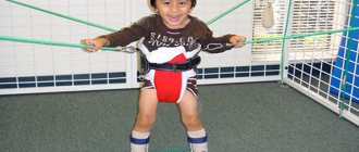
In the congenital form, it is not easy to immediately notice myotonic syndrome. Its symptoms are most obvious if the child is active and tries to make quick movements that require complex coordination. With congenital myotonia, the baby learns to walk and talk later than his peers. Such children tend to have problems with posture.
Diagnosis of myopathies
Electrophysiological examination methods help a neurologist to establish a diagnosis of myopathy: electroneurography (ENG) and electromyography (EMG). They make it possible to exclude damage to the peripheral motor neuron and, thus, differentiate myopathy from infectious myelopathy, cerebrospinal circulatory disorders, myelitis and spinal cord tumors. EMG data indicate changes in muscle potentials characteristic of myopathies—a decrease in their amplitude and a reduction in duration. The presence of a large number of short peaks indicates a progressive process.
A biochemical blood test for myopathy shows an increase in the content of aldolase, CPK, ALT, AST, LDH and other enzymes. In a biochemical analysis of urine, an increase in creatinine concentration is indicative. In establishing the form of myopathy, muscle biopsy is of paramount importance. Morphological examination of muscle tissue samples reveals the presence of randomly scattered atrophied myofibrils among practically intact and hypertrophied muscle fibers, as well as the replacement of areas of muscle tissue with connective or fatty tissue. Making a final diagnosis is possible only after comparing the results of histochemical, immunobiochemical and molecular genetic studies.
In order to diagnose lesions of the heart muscle, a patient with myopathy may be prescribed a consultation with a cardiologist, ECG, or ultrasound of the heart; if pneumonia is suspected, consult a pulmonologist and take an X-ray of the lungs.
Prevention
Duchenne pathology is progressive. The prognosis for its course is extremely unfavorable; most patients do not live to see 30 years of age. The main causes of death are concomitant diseases associated with cardiac activity and breathing.
In many ways, life expectancy depends on care, compliance with treatment, including physical therapy, massage, and exercise therapy, if possible.
Symptoms of myopathy appear gradually. Over the course of several years, a person loses self-care skills and loses the ability to move independently. It is currently impossible to cure the pathology. Supportive symptomatic treatment is carried out. The prognosis is unfavorable.
To prepare the article, the following sources were used: Latysheva V. Ya., Drivotinov B. V., Olizarovich M. V. // Neurology and neurosurgery: textbook. allowance - Minsk, Higher. school 2013.
Team of authors // Nervous diseases - “SpetsLit”, 2011 (Textbook for secondary medical schools).
Kurushina O.V., Andryushchenko F.A., Agarkova O.I., Dvoretskaya Yu.A. Modern approach to the diagnosis and treatment of primary and secondary myopathies // Bulletin of VolgSMU - 2020 Issue 1 (61).
The most unfavorable prognosis are hereditary myopathies that manifest themselves in early childhood. Otherwise, the prognosis depends on the form of myopathy and the involvement of the cardiac and respiratory muscles in the process. The prognosis of secondary myopathies is more favorable provided that the underlying disease is successfully treated.
Prevention of primary myopathies is a thorough collection of family history and mandatory consultation with a geneticist for couples planning pregnancy. Prevention of secondary myopathies is the elimination of toxic effects on the body, timely treatment of infectious and endocrine diseases, and correction of metabolic disorders.