Causes and provoking factors
Radicular osteochondrosis can be caused by a sedentary lifestyle, excessive physical activity, and obesity. Spinal roots often become inflamed during acute syphilis, tuberculosis or spinal meningitis. Their damage is also possible against the background of such pathological conditions:
- spinal cord injuries, severe bruises, subluxations of vertebral bodies;
- congenital and acquired anomalies of the spinal column, including kyphosis and scoliosis;
- benign and malignant neoplasms, localized mainly in the back;
- hormonal imbalances.
The course of osteochondrosis is accompanied by a slow but persistent decrease in the height of the cartilaginous disc and a decrease in the diameter of the intervertebral foramen. The result is pinching, compression of the spinal root. The development of such a process is even more likely with the traditional complication of osteochondrosis - the formation of an intervertebral hernia. Chronic spondylosis also predisposes to radicular syndrome. In this case, the nerve endings are simultaneously pinched by both the bone growths of the deformed vertebrae and the modified facet joints.
Important! With prolonged infringement of the spinal root, numerous neurological disorders occur. The most severe of them cannot be eliminated even by surgery - the patient becomes disabled.
Classification
Radicular syndrome in osteochondrosis is treated with various methods. When choosing therapeutic tactics, the division of pathology into groups is taken into account. It is based on the localization of vertebral segments that have undergone destructive and degenerative changes. The intensity of pain often depends on the location of such structures. Here is the generally accepted classification of radicular syndrome:
- cervical. It is diagnosed very rarely due to the small diameter of the spinal canal in this section and the high strength and elasticity of the ligaments. But this pathology is also considered the most dangerous, since the pinched root is located close to the brain. If its compression is too long, the risk of developing an inflammatory process increases, aggravating the patient’s condition. Due to compression of the vertebral artery, hypoxia (oxygen starvation) occurs, the main symptoms of which are visual and auditory disorders, arterial hypertension;
- chest It is also detected quite rarely, but is distinguished by the most diverse and severe symptoms. After all, the nerves located in the back area also innervate the internal organs. Therefore, pain spreads to the heart, kidneys, and gastrointestinal tract. Patients misinterpret their origin and complain to gastroenterologists, nephrologists, and cardiologists;
- lumbar. The most common pathology in which bending, turning, and walking are significantly difficult. At the stage of remission, osteochondrosis quickly recurs with increased physical activity or hypothermia. Gait and posture change, muscle strength and sensitivity decrease. The pain is so acute that a person freezes in one body position for a long time so as not to provoke its intensification.
Separately, it is worth noting the radicular syndrome in lumbosacral osteochondrosis. If a person does not consult a doctor and prefers to independently get rid of pain radiating to the sides and hips, then the destructive-degenerative process quickly progresses. After several years, the damaged segment of the spine is completely or partially immobilized.
Pain in osteochondrosis with radicular syndrome occurs especially often as a result of compression of the nerve by a bone growth
On a note! A severe course and severe symptoms are also characteristic of cervicothoracic osteochondrosis. With this pathology, the intervertebral discs of the cervical and thoracic spine are simultaneously or sequentially destroyed. Clinically, it manifests itself as pain, aggravated by walking, bending and turning the head, and stiffness.
Osteochondrosis with radicular syndrome - what is it?
The disease osteochondrosis develops due to metabolic disorders in the tissues of the discs located between the vertebrae. The long course of the pathology and the lack of competent therapy lead to the fact that the discs undergoing degenerative changes gradually change their natural structure.
The annulus fibrosus surrounding the inner core of the disc changes, cracks appear and widen, through which a jelly-like substance protrudes. The nucleus, released from its anatomical, normal position, begins to compress the spinal nerves, or rather their roots, causing all the symptoms of radicular syndrome.
The irreversibility of the frequent occurrence of radicular syndrome in patients with osteochondrosis is explained by the fact that due to changes in the discs and all adjacent osteochondral structures, the hole through which the main vessels and nerves pass is reduced.
For some people, this process begins to occur after 2-3 years, while others, thanks to treatment and maintaining a healthy lifestyle, can delay the development of radicular syndrome by decades.
Signs
Symptoms of any osteochondrosis with radicular syndrome appear gradually. At the initial stage of development, the course of the pathology is asymptomatic. From time to time a person experiences mild discomfort. Usually the cause of their appearance is physical activity, so the patient mistakes them for overwork and does not seek medical help. And cartilage tissue continues to slowly deteriorate.
At a certain stage, the spinal root is pinched. As a rule, it is provoked by an awkward, excessively sudden movement. The pain is so acute that the person freezes in place, afraid to breathe. Radicular syndrome is usually complicated by the following symptoms:
Ointments for osteochondrosis
- crunching when walking, bending, turning the body or head;
- decreased sensitivity of the shoulders, hips, buttocks, ankles, and sometimes feet;
- constant muscle tension, provoking an increase in the intensity of pain;
- paresthesia - sensitivity disorders characterized by numbness, tingling sensations, “crawling sensations”;
- by adopting a forced body position, in which the severity of pain is slightly reduced;
- visible curvature of the spinal column.
A person with osteochondrosis complicated by radicular syndrome moves deliberately slowly and avoids making movements that can cause acute pain. Changes in tendon reflexes are especially dangerous in lumbar and lumbosacral pathologies. It manifests itself as urination disorders, decreased sexual desire, and problems with bowel movements.
Helpful information! Lumbar osteochondrosis with radicular syndrome is the most severe due to lumbodynia - prolonged pain in the lower back. Its frequent appearance indicates severe damage to the lumbar segment and the formation of an intervertebral hernia.
Pregnancy and osteochondrosis
This is a special condition of the female body, when several factors that provoke the development of the disease appear in a short period of time.
- A rapid increase in weight and a shift in the center of gravity leads to increased stress on the lumbar region;
- Less movement and lack of physical activity for several months allow salts to accumulate, cartilage tissue becomes stiffer;
- Hormonal changes negatively affect the elasticity of ligaments and cartilage.
Sometimes radicular pain radiating to the lower abdomen can be perceived as premature contractions. Therefore, if any discomfort occurs, it is recommended to visit a doctor and consult a neurologist.
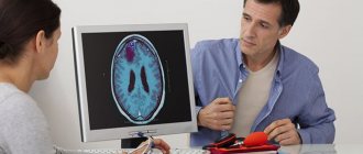
Diagnostics
Osteochondrosis of the lumbar spine, cervical or thoracic segments is first diagnosed based on data from functional tests, which assess sensitivity and range of motion. The doctor palpates the area of pain to detect trigger points located in the area of the spinous processes. As a rule, it immediately determines the degree of tonic-muscular tension. To confirm the location of the damaged spinal root identified during an external examination, electroneuromyography is performed, which also makes it possible to assess the general functional state of the autonomic nervous system and skeletal muscles.
Osteochondrosis with radicular syndrome can be quickly diagnosed using radiographic images
But the main task of diagnosis is to establish the cause of the development of osteochondrosis complicated by radicular syndrome. As well as determining its severity, the rate of involvement of neighboring vertebral segments in the pathological process. For this purpose, a number of instrumental studies are carried out:
- X-rays help directly detect osteochondrosis or spondyloarthrosis, spondylolisthesis, congenital and acquired anomalies of the spinal column;
- MRI and CT are considered the most informative in the diagnosis of intervertebral hernia, traumatic hematoma, hemorrhage in one part of the spinal cord, meningoradiculitis, benign and malignant neoplasms.
If the doctor suspects that the cause of inflammation of the spinal root is an infectious process, then bacterial culture and (or) biochemical examination of the exudate collected by puncture is necessary. Their results make it possible to judge the species of microbes or viruses and their sensitivity to drugs.
Clinical characteristics of symptoms
General signs of the development of radicular syndrome depend on the degree of exposure to the effect of compression of the roots and on what part of the spinal column undergoes changes.
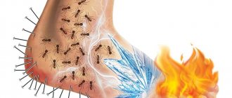
The main complaint is pain, it always spreads along the passage of the nerves and its intensity is quite pronounced. In addition to pain, the patient complains of numbness, burning, paresthesia in the area of localization of pain, in the limbs and along the passage of nerve endings.
The clinical features of the disease depend on the area in which the roots are pinched.
Signs of disease in cervical osteochondrosis
Disturbances in the roots of the cervical spine are observed less frequently compared to the thoracic and lumbar spine.
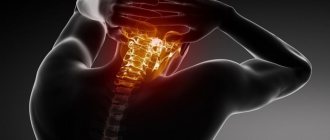
The photo shows the location of pain during the development of radicular syndrome in the cervical spine
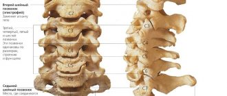
The pain intensifies significantly with movement and coughing, so a person tries to provide rest to the cervical region during an attack. In the cervical spine there are eight pairs of spinal nerve roots; an experienced neurologist, based on the patient’s complaints, can determine between which vertebrae the pathological process is most pronounced.
- If the first root of the cervical spine is affected, the patient complains of pain mainly in the parietal region, and is also bothered by a feeling of numbness in the occipital region and crown.
- When the second root is affected, the nature of the manifestations of the disease is identical to that of the first root. But besides this, you can identify sagging muscles and skin under the chin, which is explained by malnutrition and decreased tone.
- When the third pair of roots is affected, severe pain, numbness of the skin, and swelling of the tongue appear. All these signs appear on the side where the root compression occurs. In addition, the patient exhibits a slight speech impairment when speaking.
- If the 4th root is compressed, the pain is very disturbing in the area of the scapula, shoulder girdle, and collarbone. In some patients, lumbago is recorded in the area of the heart and liver. The patient complains of numbness in the same areas of the body; upon examination, muscle weakness is determined.
- When the 5th root is affected, pain appears in the neck, along the outer part of the shoulder on the affected side. There is numbness and weakness in hand movements. All changes affect the side where the root is compressed.
- If the 6th root is affected, then pain and severe numbness from the neck spread to the shoulder blade, then to the entire upper limb up to the thumb.
- Compression of the 7th root causes pain to spread throughout the neck, scapula area, and the outer and posterior surfaces of the upper limb. All changes are pronounced and affect the area of the second and third fingers of the hand.
- Damage to the 8th root leads to the spread of pain and numbness to the little finger on the side of the compression. There is severe weakness when moving the arm.
All pain during the development of radicular syndrome in the cervical region begins acutely and intensifies significantly if you move your neck.
During breastfeeding
Pain is expressed in the back or chest area. Manifestations of the disease also depend on which of the 12 thoracic roots is affected.
- With pathological compression of the 1st thoracic root, aching pain occurs and a slight decrease in skin sensitivity in the area of the shoulder blades. Similar signs are detected on the inside of the upper limb, starting from the axillary region and ending with the elbow joint.
- If the roots are affected, starting from the 2nd and ending with the sixth, then the patient is bothered by girdling pain. This pain affects the area of the shoulder blades, armpits and lower chest. Severe discomfort of the esophagus and pharynx may occur.
- When the 7th and 8th roots are affected, the nature of the pain corresponds to that described above, but the discomfort extends somewhat lower. There is also some pain in the stomach and heart area.
- When the 9th and 10th vertebrae are compressed, the patient complains of acute pain with localized localization, in front they cover the area from the bottom of the chest and up to the navel. From behind they are observed symmetrically.
- When the 11th-12th vertebrae are affected, pain, numbness and discomfort from the navel reach the groin area.
A pronounced increase in pain occurs at the time of coughing or sharp inhalation. By these same signs, radicular syndrome can be distinguished from an angina attack that manifests itself with similar symptoms.
Symptoms of the syndrome in the lumbar spine
Since the lumbar spine bears the largest, constant load, radicular syndrome occurs most often in this section. Its manifestations also depend on which of the roots is damaged.
- When the 1st to 3rd roots , acute or aching pain is observed in the groin and thigh area. These same parts of the body experience severe numbness.
- Compression of the 4th root leads to the development of aching, dull pain. It begins in the lumbar region and moves to the lower leg and knee area.
- Damage to the 5th root causes acute pain in the thigh and lower leg. The pain reaches the foot and affects the big toe.
The pain decreases significantly with rest, especially if the person lies on the healthy side.
Treatment methods
Only an integrated approach to treatment will allow you to get rid of all the symptoms of osteochondrosis with radicular syndrome. For pathologies of mild severity, conservative methods are used. Osteochondrosis of 3-4 degrees is characterized by the formation of a large hernia. To eliminate the compression of blood vessels and the spinal root, surgical treatment is often required.
Surgical interventions
For lumbar osteochondrosis, a pathology localized in the neck or chest, surgical treatment is necessary if progression of loss of function is detected. It is also required when the spinal root is pinched by a tumor. The surgeon directly removes the compression of the nerve ending itself, and then removes its cause. He excises the hernial protrusion along with the destroyed disc. The operation is performed using one of the following methods:
- open or endoscopic discectomy;
- microdiscectomy.
At the final stage of the surgical intervention, stabilization of the spinal column is performed, including the installation of B-Twin implants. Decompression is also carried out by laminectomy, or removal of the vertebral arch. Intradiscal electrothermal therapy is also in demand in the surgical treatment of osteochondrosis with radicular syndrome.
Important! The most modern and safe way to remove a hernial protrusion is considered to be nucleoplasty. This puncture surgical intervention is aimed at eliminating a specific fragment of the nucleus pulposus of the damaged intervertebral disc.
How are spinal nerves formed?
The spinal nerve is formed by two roots that arise from the spinal cord. The roots consist of neurons (nerve cells) of different types. One root is formed by sensory neurons (sensory or afferent), the second - by motor neurons (motor, efferent). Therefore, the spinal nerve is of a mixed type.
Spinal nerves leave the spinal cord through the bony canal of the corresponding vertebra. Located between adjacent vertebrae. When leaving the spine, they are divided into two branches: anterior and posterior. The posterior branches innervate the back muscles and spine. The anterior ones, intertwining with each other, form nerve plexuses and then go to the corresponding organs and tissues of the body.
There are a total of 31 pairs of spinal nerves. These include:
- Eight neck ones. Innervates the corresponding segments of the cervical spine - C1, C2...C8.
- Twelve chest (T1…T12).
- Five lumbar (L1…L5).
- Five sacral (S1…S5).
- One coccygeal nerve (Co1).
Each spinal nerve is accompanied by vessels (artery and vein), providing it with nutrition and oxygen enrichment.
How does radicular syndrome occur?
In various pathological processes of the spine, compression (squeezing) and/or inflammation of the spinal nerve roots may occur, which leads to the development of the disease. Such pathological processes can be:
- Osteochondrosis.
- Spinal cord herniation.
- Deforming spondyloarthrosis.
- Bruise and other vertebral injuries.
- Osteoporosis and its complications (fractures).
- Spinal cord tumors (neurinomas).
- Infectious process in the vertebrae (osteomyelitis, tuberculosis, syphilis) and other causes.
Whatever the causes of radicular syndrome, the essence of the syndrome is the same - trauma to the spinal nerve.