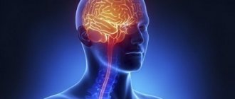
Returning to the issue of encephalopathy in children, let us consider the basic concepts, causes and symptoms of this pathology in children of different ages.
Encephalopathy in children is a group of diseases of the nervous system of non-inflammatory origin that develop as a result of the impact on neurons of the brain of various damaging factors that cause disruption of its functions.
The disease develops as a result of pathological changes directly in nerve cells, and diffuse changes inside the cells are associated with a significant disruption of their blood supply, exposure to hypoxia, toxic or infectious agents.
Causes of perinatal encephalopathy.
Signs of pathology are often vague. To identify the problem, dynamic observation over a long period of time is sometimes required. There is evidence that at the initial appointment with a neurologist, up to 80% of children receive a diagnosis of PPE or “unspecified perinatal encephalopathy.” It is reassuring to know that by the age of one year, most children are cleared of their diagnosis. This can be explained by the lack of clear diagnostic criteria and the mobility of the infant’s neuropsychological structure. Perinatal encephalopathy is an organic and functional disorder of the brain in children during the days of intrauterine development and the first hours of life. The time limits are determined as follows: from the 28th week of pregnancy to the 7th day after birth. In premature babies, the period is extended to 28 days.
The seriousness of the disease and the difficulty of diagnosis are not a death sentence. The main factors that cause the disease still exist:
- Hypoxia is a lack of oxygen and is ischemic in nature. When oxygen supply stops, the blood stops supplying food and fills with carbon dioxide. This compound provokes cell oxidation. As a result, acidosis appears (body oxidation syndrome) - a disorder of the acid-base balance. Its most dangerous consequence is swelling.
Hypoxia can have both destructive and restorative effects. Immediately after the baby leaves the womb, acidosis acts as a means of adaptation to changes in its environment. When its form is prolonged, encephalopathy of newborns manifests itself.
With a complicated birth, asphyxia occurs - oxygen starvation of the children's brain. Then uncompensated acidosis may occur. As a result of a disruption in the blood supply to the brain, the acid-base balance changes (it is measured by pH). If the baby has uncompensated acidosis, the indicator drops below 7.1, which requires immediate resuscitation measures.
- Cranial trauma. It entails post-traumatic encephalopathy. As a result of a difficult birth, the child experiences mental and physical disorders. Depending on the severity of the injury, neonatal encephalopathy can be expressed in different forms.
- Damage to the frontal or temporal lobes of the brain at the time of birth;
- Swelling of the brain, disruption of the blood supply to cells by connecting material: this causes adhesions and scars;
- Autoneurosensitization is a conflict of the immune system: it mistakenly begins to react to its own cells as a foreign object.
- Toxic-metabolic syndrome is the result of the toxic effects of the metabolic activity of unhealthy kidneys and biliary tract in children. Bilirubin encephalopathy (jaundice of newborns), i.e. bilirubin intoxication, is quite widespread. Bilirubin is a neurotoxic poison that leads to damage to the cerebral cortex and subcortical nuclei. Depending on the causes, there are several types of jaundice in newborns: conjugative (low binding capacity of the liver), hemolytic (caused by hemolysis - destruction of red blood cells), obstructive (problems with the outflow of bile), parenchymal.
- Intrauterine infections infect the newborn and provoke inflammatory processes. Infection of the fetus can lead to serious pathology of the nervous system, perinatal encephalopathy, premature birth and even the death of the child.
- Disruption of uteroplacental blood flow (fetoplacental insufficiency) is a failure of blood circulation in the placenta in the late period of gestation. Through this protective shell the fetus is nourished, supplied with oxygen, and hormonal exchange occurs. When the dysfunction is insignificant, the danger is minimal, but with severe fetoplacental insufficiency, hypoxia occurs, followed by encephalopathy.
- Severe chronic or acute illness of the mother also negatively affects the child. Preeclampsia is a complication of the late period of pregnancy, it is called late toxicosis. As a result of the disease, the internal structures of the mother (kidneys, liver, blood circulation) are damaged.
- The threat of termination of pregnancy is fraught with many negative side effects, including the above factors.
Symptoms of the disease
The severity of the symptoms of this pathology and their combination depend on:
- on the age of the child and the degree of maturation of the nervous system;
- on the location and extent of damage to brain tissue and its glial structures;
- on the causative factor and the duration of its effect on brain cells;
- from the presence of concomitant diseases that can aggravate the pathological effect of the main etiological factors (intoxication, metabolic or vascular disorders).
Common signs and symptoms of encephalopathy in children are:
- frequent headaches, anxiety, moodiness, constant screaming (in young children);
- hyperactivity, uncontrollability, disinhibition, obsessive states;
- sleep disorders, which are manifested by drowsiness during the day and insomnia at night;
- neuroses in the form of behavioral disorders or phobias in the form of panic fear of any objects, obvious or imagined, as well as environmental phenomena;
- decreased memory (usually impaired short-term memory for remembering words, numbers or names of objects), cognitive activity and intelligence of the child;
- hearing and vision impairment;
- development of hydrocephalic syndrome (in children of the first year of life).
Parents need to remember that in the absence of diagnosis and timely treatment, there is a progression of diffuse changes in brain cells, the breakdown of connections between neurons and their death lead to significant deviations in the functioning of the central nervous system.
In this case, the symptoms become permanent with worsening signs of encephalopathy in children, the following are noted:
- frequent fainting;
- dizziness;
- asthenia;
- deterioration of attention and memory;
- changes in motor coordination;
- movement disorders (flaccid paralysis, paresis);
- mental and vegetative disorders;
- significant decrease in intelligence;
- behavioral disorders;
- developmental delay.
Diagnosis of encephalopathy.
Symptoms in children do not appear immediately. Immediately establishing the level and nature of the problem in the first days after birth can be difficult.
Sometimes natural manifestations of the nervous organization of a newborn are mistaken for symptoms of encephalopathy, in other cases the disease goes unnoticed. Objective conclusions can often be drawn only after six months. According to the rules, a child is examined by a doctor every month in the first half of his life. The doctor examines the baby’s reflexes and determines the quality of his physical development. In a fairly short period of time, it is possible not only to diagnose an anomaly, but also to successfully correct it. Long-term follow-up may be required so that the diagnosis does not give rise to doubts, and the development of a treatment regimen for the disease becomes possible.
The depth of damage to a child’s brain varies. Depending on the severity of the disease, 3 levels of brain destruction with long-term consequences have been established:
- The initial stage of the disease is characterized by a slight slowdown in the motor development of children for more than 2 weeks. In the first degree, fortunately, the brain substances are not affected.
- Deviations of moderate severity are determined by the presence of convulsions when crying, the baby’s motor skills are 8 weeks behind, hyperexcitability, cyanosis with short crying, “marbling” of the skin, twitching of the arms and legs. Muscle hypertonicity develops, adaptation syndromes are revealed. A year later, the disease “offers” new symptoms (when trying to walk, the child leans on his toe). Lack of treatment in the future threatens serious deviations.
- Deep brain damage results in cerebral palsy (CP) and autism. The help of restorative medicine should be resorted to as early as possible.
Manifestations of asthenic syndrome
Without taking into account the signs of diseases against the background and due to which neuropsychic weakness occurs, a set of signs characterizing the state of emotional exhaustion has diagnostic significance. These are mainly disturbances in the processes of higher nervous activity. These include:
- severe weakness;
- extremely rapid fatigue (even with minor loads) and reduced performance;
- daytime drowsiness and sleep disturbance (prolonged falling asleep, rare or absent dreams, light shallow sleep);
- decreased interest in life, apathy, loss of desire for favorite activities;
- disturbances in memory processes (short-term), difficulties in learning new material, suppression of analytical abilities;
- nervousness, irritability, negativity in views and contradictions, increased criticism of others;
- decreased libido, sexual dysfunction;
- changes in blood pressure and pulse rate, unmotivated shortness of breath, pain in the lower back muscles, chilliness in the legs, disorders of the digestive and urinary system.
The last section of signs is manifestations of autonomic disorders. What brings asthenic syndrome closer to vegetative-vascular and neurocirculatory dystonia.
What should alert parents?
The peculiarity of encephalopathy is the need for the earliest possible start of stabilization and elimination of neurological defects of the fetus.
Emerging syndromes can be stopped and corrected during pregnancy and infancy, but qualified assistance from a neurologist is required.
Particular responsibility falls on parents. One of the features of the child’s behavior or well-being should cause concern:
- sleep disorders;
- muscle hypertonicity;
- poor coordination;
- lethargy of the hands;
- does not turn over;
- legs are not involved in crawling;
- does not sit down or stand up;
- improper crawling;
- increased excitability to various stimuli (frequently cries);
- depressed appearance in the waking phase;
- delayed motor development syndrome (cannot pick up a toy);
- can only do pull-ups with his right arm;
- spitting up like a fountain after eating;
- tremor – fine muscle trembling;
- body weight slowly increases;
Such symptoms force you to go to the clinic. Important preventive measures should not be neglected, even if the child seems to be healthy.
What is important to do in time:
- Thorough examination by a neurologist at the age of 4 weeks.
- Regular monthly examination by a pediatrician, and if necessary, treatment of the disease.
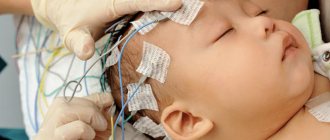
- Do a neurosonography test - NSG (establishes movement disorder syndrome).
If the presence of cerebral palsy or epilepsy has already been established, it is necessary to begin treatment as quickly as possible to prevent further pathology.
Definition and variants of the syndrome
The term “asthenia” is derived from the Greek word “sthenos”, which literally means strength, vital activity. The prefix "a-" means negation. As a result, asthenia is impotence, inactivity in life.
In the general understanding, these are emotional disorders in the form of decreased mood in the hyposthenic type or, on the contrary, manifested by nervousness (irritability) in hypersthenia. Usually this condition occurs against the background of a current or past illness. But it can also manifest itself as a result of severe emotional shock, and also, in the mildest degree, against the background of complete health - asthenia of overwork.
In information sources there are variations of the term: neuro-asthenic syndrome, neuropsychic weakness, asthenic conditions or reactions, chronic fatigue syndrome.
Osteopathy in the correction of neonatal encephalopathy.
Organic disorders of brain development in infants must be identified and treated immediately.
Due to neural disorders, diseases of the child’s internal organs occur. The damaged network begins to control the entire body. The opposite effect also occurs: normalization of the kidneys, liver, and lungs affects neural connections. Therefore, when stabilizing the brain, it is necessary to take into account the overall picture of the body’s development and look for options for complex treatment.
Osteotherapy is one of the branches of restorative medicine. A person is considered as a single holistic scheme. The basic principles of the technique were proclaimed more than 30 years ago. Over several decades, new approaches have been developed, management methods have been found, and practical experience has been acquired. Medications are used with great caution or are not used at all. Treatment occurs through the mobilization of all elements included in recovery. The advantage of this area of medicine is that drugs are practically not used. The child activates his internal reserves. Complex treatment is the basis of the theory.
The following osteopathic directions have been identified:
craniosacral
structural
visceral
fascial
Craniosacral method of treatment.
The craniosacral (cranium-skull, sacrum-sacrum) pathway is based on the theory that the bones of the skull are movable (previously it was believed that only the lower jaw was movable).
The dynamics of their movement are imperceptible and strictly obey the impulses of the brain. If the body as a whole and the main control center are functioning normally, then the rhythm of movement of the elements of the skull is clear and correct. Failure of brain impulses against the background of encephalopathy indicates deviations from the norm. The study of the rhythmic movement of children's cranial areas is used not only for diagnosing and identifying nervous destruction, but also for the reverse process. Through the systematization of impulses, it is possible to influence the damaged area. This method is especially effective in eliminating the consequences of encephalopathy. Treatment of areas of the brain eliminates the symptoms of the disease, normalizes intracranial pressure in children and can lead to restoration of the affected areas.
Structural method.
The musculoskeletal system of the newborn was chosen as the object of structural impact.
Bone and cartilage substances influence the function of the spinal cord. Neuronal fibers and veins connected to the spine are also integrated into the overall circuit. Treatment of the limbs, neck, and spine helps to normalize certain damaged areas of the brain affected by pathology (perinatal encephalopathy).
The structural theory in encephalopathy operates on measures affecting the bones and is rarely used to get rid of problems of fetal formation and their influence.
Fascial method.
Fascial therapy examines soft tissues, mucous membranes, and bloodstreams. Treatment of encephalopathy is based on the development of neural connections between individual parts of the body. The fascial method eliminates vegetative-visceral disorders of the brain of children and their consequence - hyperexcitability. Treatment of encephalopathy with fascial techniques involves correcting muscle dystonia if there are signs of increased muscle tone.
Visceral method for the correction of encephalopathy.
Visceral therapy uses the ability of internal structures to interact and influence each other. Thus, bilirubin encephalopathy requires synchronization of liver function.
Cerebral ischemia syndrome associated with hypoxia manifests itself in abnormal activity of the heart and lungs. Treatment is complex (minimal use of drugs).
Why is it developing?
Factors that provoke encephalopathy in newborns differ from the causes of the disease in older children.
In the first case, the process of intrauterine development of the fetus plays an important role.
Birth injuries can also cause the development of encephalopathy.
In older children, the disease in most cases is a complication of other pathologies or the result of traumatic brain injury.
provoke encephalopathy in children:
- consequences of intrauterine hypoxia;
- complications of birth injuries;
- abuse of bad habits during pregnancy;
- congenital diseases of the cardiovascular and respiratory systems;
- instability of blood pressure;
- complications of infectious and viral diseases;
- disruption of the circulatory system;
- consequences of traumatic brain injuries;
- complications of neonatal jaundice;
- lack of vitamins in the body (especially vitamin B);
- toxic effects on the central nervous system.
Etiology based on age
infancy
Opsoclonus syndrome Myoclonus ataxia
Opsoclonus myoclonus ataxia (OMA) appears as early as 6 months of age. Opsoclonus myoclonus syndrome is a paraneoplastic autoimmune phenomenon characterized by chaotic conjugate high-amplitude eye movements, body spasms, limbal ataxia, and regression and developmental pathology. An underlying neuroblastoma or ganglioneroblastoma is often recognized, although not universally accepted.
Recognition is facilitated when the triad of symptoms occurs in close proximity to each other. However, ataxia itself may precede findings, leading to diagnostic confusion and delays of months to years in initiating investigations.
Find out more Frontal Lobe Syndrome
Metaidobenzylguanidine scintigraphy (MIBG) scans have moderately high sensitivity, but children whose scans are negative should have a high-resolution computed tomography (CT) scan or magnetic resonance imaging (MRI) scan of the chest and abdomen.
Children's age from 1 to 4 years (preschool)
Acute post-infectious cerebral ataxia
Acute postinfectious cerebellar ataxia (APCA) accounts for up to 40% of cases of acute cerebellar ataxia. Usually occurs after febrile illness or immunization. Previous chickenpox was recorded in 26% of patients. A large number of other viruses are involved, including coxsackie B, echoviruses, mumps, Epstein-Barr, influenza A and B.
The pathology is manifested by acute demyelination caused by cross-reacting antibodies to epitopes in the cerebellum. The onset of the disease lasts up to 3 weeks after the systemic disease has subsided. Symptoms take several hours to develop, being most noticeable upon initial presentation with relatively rapid resolution over the next few days.
Mental status remains clear throughout the illness. The presence of extreme irritability should raise suspicion for the diagnosis.
Screening reveals a pure cerebellar syndrome with marked gait involvement and significant ataxia. The child recovers in less than 2 weeks after the onset of the disease. This is a self-limiting condition that does not require any specific intervention.
Cerebrospinal fluid (CSF) analysis is performed without risk of herniation and usually shows mild pleocytosis with negative viral and bacterial cultures. MRI is normal or reveals mild, nonspecific changes.
Acute cerebellitis
The acute syndrome occurs after a systemic illness or is a direct result of infection of the cerebellum. Common agents that cause acute ataxia are rotavirus, mycoplasma, and human herpes virus.
Clinical features are altered sensorium, increased intracranial pressure in addition to the features of pure cerebellar syndrome. CSF analysis will likely reveal pleocytosis and, in rare cases, antibodies against the infectious agent.
It should be noted that performing a spinal puncture in conditions of significant cerebellar edema is life-threatening. MRI shows abnormalities suggestive of edema. Fatalities have been reported from acute cerebellitis.
Essentially, acute cerebellitis is distinguished from APCA by the presence of systemic symptoms such as fever, neck stiffness, symptoms and signs of increased intracranial pressure due to rapid compression of the fourth ventricle, and risk of death; therefore, there is an urgent need for early therapy.
There is significant overlap between postoperative ataxia and acute infectious ataxia, so it may be difficult to distinguish between the two diseases.
Toxic ingestion
Accidental use of medications, medications, and pills in preschool children accounts for up to 30% of cases of acute ataxia. Cerebellar Purkinje cells are particularly susceptible to toxic insults.
Accidental ingestion of anticonvulsants, lead, eucalyptus oil, insecticides such as paraquat, phosphine, dextromethorphan, and shellfish poisoning provoke noticeable cerebellar symptoms.
Clinical features: depressed mood or agitation, seizures, cerebellar signs. The latter is often masked by the overall severity of the situation. Parents should be asked to bring all prescription medications, and a urine test is required.
Benign paroxysmal vertigo
Benign paroxysmal vertigo (BPV) must be differentiated from benign paroxysmal positional vertigo, which is the most common cause of vertigo in adults.
Characterized by brief periods of dizziness and ataxia.
A healthy child suddenly appears fearful, pale, and clings to a parent during episodes of BPV. The symptoms last no more than a few minutes, after which the child does not experience confusion or drowsiness.
The typical age of onset is 1 to 4 years, with resolution occurring between 7 and 10 years. Screening is invariably normal between attacks. In many cases, a family history of migraine is found, and BPV itself is believed to be a precursor to migraine.
Acute disseminated encephalomyelitis
ADEM is an immune-mediated phenomenon that occurs following a viral illness or immunization. It is characterized by encephalopathy (confusion, irritability, drowsiness, personality changes) and the acute onset of multifocal neurological deficits - most often ataxia. Convulsions, cranial nerve palsy, hemiparesis, fever, and meningism are present.
Images show striking lesions in the subcortical white matter, cerebellum, and basal ganglia. CSF is abnormal in about half of the cases, with increased white cells and protein. ADEM is usually a monophasic disease.
Internal disease
One of the rare complications of acute otitis media is vestibular neuritis, which is an extension of a bacterial middle ear infection to the inner ear. Causes sensory hearing loss, tinnitus, nystagmus, dizziness. Dizziness leads to ataxia.
Fever, ear pain, and mastoiditis are key to the diagnosis. The presence of nystagmus in a child with acute otitis media is often a strong clue to the presence of impending vestibular neuritis and should be treated promptly.
The presence of contrast enhancement in the auricular capsule on MRI is a sensitive indicator of the disease process in this area. CT is less reliable.
Children's age from 5 to 16 years (school age)
Concussion
Cerebellar concussion is a clinical syndrome in which head injury is accompanied by transient deficits in cerebellar function with intact consciousness. Patients demonstrate a wide gait, triangular instability, and dysarthria.
Most cases follow severe injuries; however, cerebellar concussion has been noted following relatively minor head trauma.
Learn more Geniuses with Savant syndrome: past, present, future
The pathophysiology involves damage to connections between the cerebellum and the cortex, especially the superior cerebellum, after severe traumatic brain injury. Transient vasospasm is a proposed mechanism for ataxia after mild injuries. No specific intervention is required.
Stroke
Strains in the cerebellar region (posterior circulation) are easily missed because strokes are uncommon in children. They have subtle symptoms that are often nonspecific, without obvious mechanical or sensory abnormalities.
Dizziness about three quarters of the time, nausea, vomiting, gait disturbance is observed about half the time, headache about one third of the time.
The onset of symptoms is abrupt. Examination reveals ataxia with a tendency to fall on the affected side, vertical nystagmus, in addition to other classic cerebellar stigmata. The presence of an altered level of consciousness, hemiplegia, and cranial nerve weakness, if present, will quickly direct the doctor to the appropriate diagnosis.
MRI is a diagnostic test because CT may miss cerebellar and brainstem strokes.
Most cerebellar strokes are caused by arteriopathy that follows infection (eg, chickenpox), vasculitis (Kawasaki disease), or head trauma. Swelling after a stroke is most noticeable in the first 24 hours.
If located in the posterior circulation area, it is especially ominous. Rapid swelling can compress the brain stem. The vast majority of these patients will die without surgery. Therefore, all cerebellar strokes should be closely monitored in an intensive care setting.
Multiple sclerosis
Multiple sclerosis (MS) is an autoimmune demyelinating disease that affects different parts of the nervous system over time. About 50% of children under 5 years of age have multiple sclerosis, and 5% to 15% of adolescents have MS with acute ataxia, making it a relatively common manifestation of the disease.
Children with MS show more frequent disease relapses than adults and experience significant cognitive decline and severe residual disability early in life. MRI with contrast agent and referral to a neurologist to initiate therapy is essential.
Prescription drug poisoning, substance abuse
Adolescents suffering from acute ataxia should be suspected of using psychoactive, therapeutic drugs that are used for medical purposes. Benzodiazepines, phenytoin, and carbamazepine are anticonvulsants that are most likely to cause ataxia after acute intoxication.
In these situations, it is necessary to measure free and total drug concentrations. Antineoplasts such as fluorouracil (5-FU), cytarabine (ara-C), methotrexate cause acute cerebellar damage. Abuse of drugs that provoke ataxia, such as toluene, cocaine, heroin, phencyclidine.
Toluene intoxication occurs due to inhalation in poorly ventilated areas with high levels of the substance. Cocaine use predisposes to cerebellar infarctions, as does recovery from a scorpion sting.
Basilar migraine
Children with basilar migraine with an aura consisting of ataxia, diplopia, ringing in the ears, tingling in the extremities, or changes in consciousness that last from 5 minutes to 1 hour.
A severe, pounding headache follows within 1 hour of these symptoms, usually accompanied by nausea, vomiting, photophobia, and phonophobia. The headache can be one-sided or two-sided and prevents you from falling asleep.
The average duration of a headache ranges from 30 minutes to 3 days. Screening was unremarkable, with no clear evidence of cerebellar pathology other than ataxia. Initial episodes require an MRI.
Guillain-Barre syndrome
Children exhibit symptoms of ataxia due to disease processes in the peripheral nerves that prevent appropriate sensory signals from reaching higher focal centers. A classic example is Guillain-Barre syndrome.
A previous viral infection or gastroenteritis is noted in half of the cases. Children experience severe pain in the lower extremities until weakness sets in. Weakness and ataxia peak at 4 weeks. Rare cases progress and involve the respiratory muscles, quadriparesis.
Diagnostic testing includes imaging of the spinal cord to rule out myelitis and spinal fluid.
In most cases the diagnosis remains clinical, which can be made with confidence due to the triad of ataxia, isflexia and motor weakness.
Nerve conduction studies are performed in atypical cases to confirm the diagnosis. Collaboration with a neurologist is vital for children ready to receive treatment for Guillain-Barré syndrome.
Episodic ataxia
Hereditary episodic ataxia (EA) refers to a group of inherited conditions characterized by periods of cerebellar dysfunction that lead to acute ataxia.
Seven different subtypes are recognized, with EA2 being the most common. Attacks are accelerated by fatigue or strong emotions (anger, sadness) and can last for hours.
In addition to ataxia, patients experience dizziness, nausea, vomiting, and (rarely) convulsions. Patients may have migratory headaches and acute nystagmus between attacks. The mutation responsible for EA2 is located on the CACNA1A gene.